产品: 磷酸化 YAP (Ser127) 抗体
货号: AF3328
描述: Rabbit polyclonal antibody to Phospho-YAP (Ser127)
应用: WB IHC IF/ICC
反应: Human, Mouse, Rat, Monkey
预测: Pig, Zebrafish, Horse, Sheep, Rabbit, Chicken, Xenopus
分子量: 65~78kD; 54kD(Calculated).
蛋白号: P46937
RRID: AB_2810276

浏览相似产品>>

   规格 价格 库存
 50ul RMB¥ 1300 现货
 100ul RMB¥ 2400 现货
 200ul RMB¥ 3200 现货

货期: 当天发货

联系销售

产品描述

来源:
Rabbit
应用:
WB 1:500-1:2000, IHC 1:50-1:200, IF/ICC 1:100-1:500
*The optimal dilutions should be determined by the end user.
*Tips:

WB: 适用于变性蛋白样本的免疫印迹检测. IHC: 适用于组织样本的石蜡(IHC-p)或冰冻(IHC-f)切片样本的免疫组化/荧光检测. IF/ICC: 适用于细胞样本的荧光检测. ELISA(peptide): 适用于抗原肽的ELISA检测.

反应:
Human,Mouse,Rat,Monkey
预测:
Pig(100%), Zebrafish(100%), Horse(100%), Sheep(100%), Rabbit(100%), Chicken(100%), Xenopus(100%)
克隆:
Polyclonal
特异性:
Phospho-YAP (Ser127) Antibody detects endogenous levels of YAP only when phosphorylated at Serine 127.
RRID:
AB_2810276
引用格式: Affinity Biosciences Cat# AF3328, RRID:AB_2810276.
偶联:
Unconjugated.
纯化:
The antibody is from purified rabbit serum by affinity purification via sequential chromatography on phospho-peptide and non-phospho-peptide affinity columns.
保存:
Rabbit IgG in phosphate buffered saline , pH 7.4, 150mM NaCl, 0.02% sodium azide and 50% glycerol. Store at -20 °C. Stable for 12 months from date of receipt.
别名:

展开/折叠

65 kDa Yes associated protein; 65 kDa Yes-associated protein; COB1; YAp 1; YAP 65; YAP; YAP1; YAP1_HUMAN; YAP2; YAP65; yes -associated protein delta; Yes associated protein 1 65kDa; Yes associated protein 1; Yes associated protein 2; yes associated protein beta; YKI; Yorkie homolog;

抗原和靶标

免疫原:
Uniprot:
基因/基因ID:
表达:
P46937 YAP1_HUMAN:

Increased expression seen in some liver and prostate cancers. Isoforms lacking the transactivation domain found in striatal neurons of patients with Huntington disease (at protein level).

描述:
This gene encodes the human ortholog of chicken YAP protein which binds to the SH3 domain of the Yes proto-oncogene product. This protein contains a WW domain that is found in various structural, regulatory and signaling molecules in yeast, nematode, and mammals, and may be involved in protein-protein interaction.
序列:
MDPGQQPPPQPAPQGQGQPPSQPPQGQGPPSGPGQPAPAATQAAPQAPPAGHQIVHVRGDSETDLEALFNAVMNPKTANVPQTVPMRLRKLPDSFFKPPEPKSHSRQASTDAGTAGALTPQHVRAHSSPASLQLGAVSPGTLTPTGVVSGPAATPTAQHLRQSSFEIPDDVPLPAGWEMAKTSSGQRYFLNHIDQTTTWQDPRKAMLSQMNVTAPTSPPVQQNMMNSASGPLPDGWEQAMTQDGEIYYINHKNKTTSWLDPRLDPRFAMNQRISQSAPVKQPPPLAPQSPQGGVMGGSNSNQQQQMRLQQLQMEKERLRLKQQELLRQAMRNINPSTANSPKCQELALRSQLPTLEQDGGTQNPVSSPGMSQELRTMTTNSSDPFLNSGTYHSRDESTDSGLSMSSYSVPRTPDDFLNSVDEMDTGDTINQSTLPSQQNRFPDYLEAIPGTNVDLGTLEGDGMNIEGEELMPSLQEALSSDILNDMESVLAATKLDKESFLTWL

种属预测

种属预测:

score>80的预测可信度较高,可尝试用于WB检测。*预测模型主要基于免疫原序列比对,结果仅作参考,不作为质保凭据。

Species
Results
Score
Pig
100
Horse
100
Sheep
100
Xenopus
100
Zebrafish
100
Chicken
100
Rabbit
100
Bovine
0
Dog
0
Model Confidence:
High(score>80) Medium(80>score>50) Low(score<50) No confidence

翻译修饰 - P46937 作为底物

Site PTM Type Enzyme
S61 Phosphorylation Q15208 (STK38) , Q13131 (PRKAA1)
T63 Phosphorylation
T77 Phosphorylation
T83 Phosphorylation
K90 Ubiquitination
S94 Phosphorylation Q13131 (PRKAA1)
K97 Acetylation
K97 Sumoylation
K97 Ubiquitination
K102 Ubiquitination
S103 Phosphorylation
S105 Phosphorylation
S109 Phosphorylation Q05513 (PRKCZ) , Q15208 (STK38)
T110 Phosphorylation Q05513 (PRKCZ)
T114 Phosphorylation
T119 Phosphorylation P06493 (CDK1) , P45984 (MAPK9)
S127 Phosphorylation Q15208 (STK38) , P31749 (AKT1) , Q9NRM7 (LATS2) , Q13188 (STK3) , Q9Y2H1 (STK38L) , O95835 (LATS1)
S128 Phosphorylation Q9UBE8 (NLK)
S131 Phosphorylation
S138 Phosphorylation P45984 (MAPK9)
T141 Phosphorylation
T143 Phosphorylation
T145 Phosphorylation
S149 Phosphorylation
T154 Phosphorylation P45984 (MAPK9)
T156 Phosphorylation
S163 Phosphorylation
S164 Phosphorylation Q15208 (STK38)
K181 Ubiquitination
Y188 Phosphorylation
S217 Phosphorylation
T241 O-Glycosylation
K254 Ubiquitination
S274 Phosphorylation
S276 Phosphorylation
K280 Sumoylation
K280 Ubiquitination
S289 Phosphorylation P06493 (CDK1)
S300 Phosphorylation
K315 Acetylation
K315 Ubiquitination
K321 Ubiquitination
S340 Phosphorylation
K342 Ubiquitination
T354 Phosphorylation
T361 Phosphorylation
S366 Phosphorylation
S367 Phosphorylation P45984 (MAPK9) , P06493 (CDK1)
S371 Phosphorylation
S381 Phosphorylation
S382 Phosphorylation
S388 Phosphorylation
T390 Phosphorylation
Y391 Phosphorylation
S397 Phosphorylation O95835 (LATS1) , Q9NRM7 (LATS2)
T398 Phosphorylation
S400 Phosphorylation P48730 (CSNK1D) , P49674 (CSNK1E)
S403 Phosphorylation P48730 (CSNK1D) , P49674 (CSNK1E)
S405 Phosphorylation
Y407 Phosphorylation P00519 (ABL1)
T412 Phosphorylation
S419 Phosphorylation
S473 Phosphorylation
K494 Methylation
K497 Methylation
K497 Ubiquitination
S499 Phosphorylation

研究背景

功能:

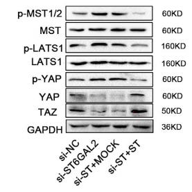
Transcriptional regulator which can act both as a coactivator and a corepressor and is the critical downstream regulatory target in the Hippo signaling pathway that plays a pivotal role in organ size control and tumor suppression by restricting proliferation and promoting apoptosis. The core of this pathway is composed of a kinase cascade wherein STK3/MST2 and STK4/MST1, in complex with its regulatory protein SAV1, phosphorylates and activates LATS1/2 in complex with its regulatory protein MOB1, which in turn phosphorylates and inactivates YAP1 oncoprotein and WWTR1/TAZ. Plays a key role in tissue tension and 3D tissue shape by regulating cortical actomyosin network formation. Acts via ARHGAP18, a Rho GTPase activating protein that suppresses F-actin polymerization. Plays a key role to control cell proliferation in response to cell contact. Phosphorylation of YAP1 by LATS1/2 inhibits its translocation into the nucleus to regulate cellular genes important for cell proliferation, cell death, and cell migration. The presence of TEAD transcription factors are required for it to stimulate gene expression, cell growth, anchorage-independent growth, and epithelial mesenchymal transition (EMT) induction.

Isoform 2 and isoform 3 can activate the C-terminal fragment (CTF) of ERBB4 (isoform 3).

翻译修饰:

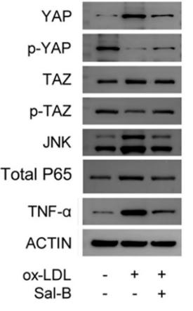
Phosphorylated by LATS1 and LATS2; leading to cytoplasmic translocation and inactivation. Phosphorylated by ABL1; leading to YAP1 stabilization, enhanced interaction with TP73 and recruitment onto proapoptotic genes; in response to DNA damage. Phosphorylation at Ser-400 and Ser-403 by CK1 is triggered by previous phosphorylation at Ser-397 by LATS proteins and leads to YAP1 ubiquitination by SCF(beta-TRCP) E3 ubiquitin ligase and subsequent degradation. Phosphorylated at Thr-119, Ser-138, Thr-154, Ser-367 and Thr-412 by MAPK8/JNK1 and MAPK9/JNK2, which is required for the regulation of apoptosis by YAP1.

Ubiquitinated by SCF(beta-TRCP) E3 ubiquitin ligase.
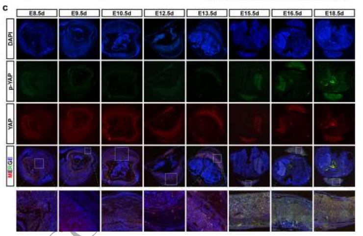
细胞定位:

Cytoplasm. Nucleus.
Note: Both phosphorylation and cell density can regulate its subcellular localization. Phosphorylation sequesters it in the cytoplasm by inhibiting its translocation into the nucleus. At low density, predominantly nuclear and is translocated to the cytoplasm at high density (PubMed:18158288, PubMed:20048001). PTPN14 induces translocation from the nucleus to the cytoplasm (PubMed:22525271).

Extracellular region or secreted Cytosol Plasma membrane Cytoskeleton Lysosome Endosome Peroxisome ER Golgi apparatus Nucleus Mitochondrion Manual annotation Automatic computational assertionSubcellular location
组织特异性:

Increased expression seen in some liver and prostate cancers. Isoforms lacking the transactivation domain found in striatal neurons of patients with Huntington disease (at protein level).

亚基结构:

Binds to the SH3 domain of the YES kinase. Binds to WBP1 and WBP2. Binds, in vitro, through the WW1 domain, to neural isoforms of ENAH that contain the PPSY motif (By similarity). The phosphorylated form interacts with YWHAB. Interacts (via WW domains) with LATS1 (via PPxY motif 2). Interacts with LATS2. Isoform 2 and isoform 3 interact (via WW domain 1) with isoform 3 of ERBB4 (via PPxY motif 2). Interacts with TEAD1, TEAD2, TEAD3 and TEAD4. Interacts with TP73. Interacts with RUNX1. Interacts with HCK. Interacts (via WW domains) with PTPN14 (via PPxY motif 2); this interaction leads to the cytoplasmic sequestration of YAP1 and inhibits its transcriptional coactivator activity.

蛋白家族:

The first coiled-coil region mediates most of the interaction with TEAD transcription factors.

Belongs to the YAP1 family.

研究领域

· Environmental Information Processing > Signal transduction > Hippo signaling pathway.   (View pathway)

· Environmental Information Processing > Signal transduction > Hippo signaling pathway - multiple species.   (View pathway)

文献引用

1). 3D-bioprinted GelMA nerve guidance conduits promoted peripheral nerve regeneration by inducing trans-differentiation of MSCs into SCLCs via PIEZO1/YAP axis. Materials Today Advances [IF=10.0]

Application: WB    Species: Rat    Sample: MSCs

Fig. 4. The correlation between trans-differentiation and PIEZO1/YAP expression in MSCs. (A) Representative immunofluorescent images for the subcellular localization and activating states of YAP/TAZ in MSCs plating on GelMA and hard substrate (HS) before trans-differentiation. Scale bar:100 μm. (B) Representative immunofluorescent images for the activating states of YAP/TAZ and expression of S100β in GelMA and HS groups after trans-differentiation. Scale bar:100 μm. (C) Representative immunofluorescent images for the subcellular localization and activating states of YAP/TAZ and PIEZO1 in MSCs after trans-differentiation. Scale bar:10 μm. (D) Immunoblotted image for PIEZO2, PIEZO1, YAP/TAZ, p-YAP, GFAP, NGF, S100β in MSCs plating on GelMA and HS after co-culture with Ne–4C. (E) Densitometric analysis of blots showing the values of relevant proteins in GelMA and HS groups. (F) Immunoblotted image for PIEZO1, YAP/TAZ, p-YAP and GFAP in MSCs in co-culture system, after being treated with Yoda1 and GsMTx4. (G) Densitometric analysis of blots showing the values of relevant proteins in MSCs after being treated with Yoda1 and GsMTx4. (H) An indirect co-culture system including NE-4C in the upper chamber and MSCs plated on GelMA in the lower chambers.

2). Interaction between laminin-5γ2 and integrin β1 promotes the tumor budding of colorectal cancer via the activation of Yes-associated proteins. ONCOGENE (PubMed: 31676872) [IF=8.0]

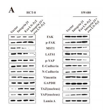
Application: WB    Species: human    Sample: HCT-8 and SW480 cells

Fig. 4| Interaction of Integrin β1 and LN-5γ2 promoted TB and aggressiveness of CRC cells by activation of YAP. a The HCT-8 and SW480 cells were transfected with M02-ITGB1 or treated with exogenous recombinant LN-5γ2 (2 μg/mL) or both. The expression of FAK, p-FAK(Y397), MST1, LATS1, p-YAP, E-cadherin, N-Cadherin, and Vimentin was then examined by western blot analysis of the whole cell lysates, and the expression of YAP and TAZ in the nucleus was also examined.

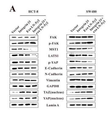
Application: WB    Species: Human    Sample: HCT-8 and SW480 cells

Fig. 4 Interaction of Integrin β1 and LN-5γ2 promoted TB and aggressiveness of CRC cells by activation of YAP. a The HCT-8 and SW480 cells were transfected with M02-ITGB1 or treated with exogenous recombinant LN-5γ2 (2 μg/mL) or both. The expression of FAK, p-FAK(Y397), MST1, LATS1, p-YAP, E-cadherin, N-Cadherin, and Vimentin was then examined by western blot analysis of the whole cell lysates, and the expression of YAP and TAZ in the nucleus was also examined. b In vitro inverse Matrigel invasion model showed that treatment with LN-5γ2 or overexpression of integrin β1 or both promoted TB in vitro, which was abolished when the expression of YAP was knocked down. Each bar represents the mean ± SD for biological triplicate experiments. c Immunohistochemical results of YAP in TB-positive CRC tissues. N/C, The nucleus/cytoplasmic ratio of YAP staining index. d Overexpression of Integrin β1 (by stable transfection of M02-ITGB1) or LN-5γ2 (by stable transfection of M02-LAMC2) or both promoted the tumor growth of HCT-8 xenografts, and the effects were abolished when the expression of YAP was knocked down. e Overexpression of integrin β1 or LN-5γ2 or both promoted the lung metastasis of HCT-8 xenografts, which was abolished when YAP was knocked down. Lung metastasis was evaluated by quantifying the percentage of lung metastatic area per lung tissue area of mice. (f) Increased expression of nucleus YAP can be found in the Integrin β1 and LN-5γ2 co-overexpressed xenograft tissues by IF and IHC analysis. *P < 0.05; **P < 0.01. NS not significant

3). A derivant of ginsenoside CK and its inhibitory effect on hepatocellular carcinoma. LIFE SCIENCES (PubMed: 35690105) [IF=6.1]

4). Knockdown of HSP110 attenuates hypoxia-induced pulmonary hypertension in mice through suppression of YAP/TAZ-TEAD4 pathway. RESPIRATORY RESEARCH (PubMed: 35986277) [IF=5.8]

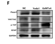
Application: WB    Species: Mouse    Sample: lung tissues

Fig. 3Knockdown of HSP110 inhibits hypoxia-induced autophagy and YAP/TAZ-TEAD4 activity in mice. Relative mRNA level (a) and protein level (b, c) of HSP110 pulmonary arteries in lung tissues in each group (N = 8). d Double immunofluorescence staining of α-SMA (green) and HSP110 (red) in pulmonary arteries (N = 8). White scale bars, 50 μm; Yellow scale bars, 25 μm. White arrows pointed to α-SMA and HSP110 double-positive cells. e Protein levels of LC3II, LC3I, Beclin1, ATG5, ATG7 and p62 in pulmonary arteries (N = 8). f–h Quantitative analysis of relative protein ratio of LC3-II/I and relative protein level of Beclin1, p62, ATG5 and ATG7 (N = 8). i Double immunofluorescence staining of α-SMA (green) and Beclin 1 (red) in pulmonary arteries (N = 8). Scale bars, 50 μm. j Protein levels of p-YAP, YAP, p-TAZ and TAZ in pulmonary arteries (N = 8). k Quantitative analysis of relative protein ratio of p-YAP/t-YAP and p-TAZ/t-TAZ (N = 8). l–n Nuclear protein levels of YAP, TAZ and TEAD4 and quantitative analysis of relative protein level of nuclear YAP, TAZ and TEAD4 (N = 8). o Double immunofluorescence staining of α-SMA (green) and YAP (red) in pulmonary arteries (N = 8). White scale bars, 50 μm; Yellow scale bars, 25 μm. White arrows pointed to α-SMA and YAP double-positive cells. Data are means ± SD from 8 mice per group. *p 

5). Resveratrol Inhibits the Tumorigenesis of Follicular Thyroid Cancer via ST6GAL2-Regulated Activation of the Hippo Signaling Pathway. Molecular Therapy-Oncolytics (PubMed: 32055676) [IF=5.7]

Application: WB    Species: human    Sample: FTC cells

Figure 4.| Upregulation of ST6GAL2 Rescues Tumorigenesis of FTC238 Cells and Resuppresses Hippo Signaling Pathway Activity(A–F) ST6GAL2 knockdown cells were transfected with ST6GAL2 overexpression vectors, and the proliferation,migration, and invasion capacities of FTC cells were enhanced. (G and H) Western blotting was performed to determine the levels of Hippo signaling molecules in FTC cells. *p < 0.05; scale bars, 20 mm.

6). Salvianolic acid B ameliorates atherosclerosis via inhibiting YAP/TAZ/JNK signaling pathway in endothelial cells and pericytes. BIOCHIMICA ET BIOPHYSICA ACTA-MOLECULAR AND CELL BIOLOGY OF LIPIDS (PubMed: 32739616) [IF=4.8]

Application: WB    Species: mouse    Sample: ECs

Fig. 5.| Influence of Sal-B on inflammatory response during YAP/TAZ/JNK signaling pathway in ECs. (A) Expression levels of YAP, TAZ, JNK, NF-κB and TNF-α were monitored by RT-PCR (n = 3). (B–C, F) Proteins (YAP, p-YAP, TAZ, p-TAZ, JNK, Nuclear NF-κB P65, Total P65 and TNF-α) in the pathway were detected by western blot (n = 3).

7). AT1R regulates macrophage polarization through YAP and regulates aortic dissection incidence. Frontiers in Physiology (PubMed: 34305627) [IF=4.0]

Application: WB    Species: Mice    Sample: macrophages

FIGURE 6 Transfecting macrophages with a YAP siRNA further enhanced Ang II–induced macrophage M1 polarization and adhesion. (A) YAP siRNA efficiencies (n = 3, mean and S.D., t-test, &&P < 0.05 compared with siRNA NC;%%P < 0.05 compared with siRNA 860); (B,C) Western Blot of YAP after siRNA transfection *P < 0.05 compared with siRNA NC; (D) Flow cytometry analysis of CD68, CD86, and CD206 expression after 96 h co-culture in both groups. The data in the one-factor histogram represents the cells in the red circle in the upper scatter plot. HAECs + THP-1 + Ang II (1 μM, 24 h) + siRNA NC group: CD68, 41.94%; CD86, 45.73%; CD206, 9.72%. HAECs + THP-1 + Ang II + YAP siRNA group: CD68, 63.13%; CD86, 53.42%; CD206, 7.13%; (E–G) The proportion of CD68 +, CD86 +, and CD206 + cells, respectively (n = 3, mean and S.D., t-test, **P < 0.05 compared with the HAECs + THP-1 + siRNA NC group); (H) Fluorescence staining of adherent macrophages after YAP siRNA transfection. All cells were labeled with DAPI (blue), and the number of adherent macrophages was significantly decreased after transfection with the AT1R siRNA; (I) The ratio between the number of macrophages and the total number of cells (n = 3, mean and S.D., t-test, symbols are the same as in C,D,E).

8). Phosphorylation of YAP Impairs Trophoblast Invasion and Migration: Implications for the Pathogenesis of Fetal Growth Restriction. BIOLOGY OF REPRODUCTION (PubMed: 32582940) [IF=3.6]

Application: WB    Species: Human    Sample: placenta

Fig. 1 Phosphorylation of YAP is upregulated in the trophoblasts of FGR-complicated placentas. (a) IHC staining of YAP and CK7 in first trimester villi of human placenta. CK7 was regarded as a positive control of STB and CTB in trophoblasts; scale bars, 100 μm. (b) IHC staining of YAP and CK7 in Normal and FGR term placenta. CK7 was regarded as a positive control of STB and CTB in trophoblasts; scale bar, 100 μm. (c) IHC staining of YAP and CK7 in E18.5 C57BL/6J mouse placenta. CK7 was regarded as a positive control of trophoblasts. (d) Western blotting of YAP and p-YAP in normal and FGR placentas. **P < 0.01; Mann-Whitney U test. (e) mRNA levels of CTGF, CYR61 and AMOTL2 were measured by qRT-PCR and normalized to β-actin. **P < 0.01, *P < 0.05; Mann-Whitney U test. All data are presented as the mean ± SEM.

Application: IF/ICC    Species: Human    Sample: placenta

Fig. 2 Phosphorylation of YAP in the placenta ascends throughout pregnancy. (a) Images of YAP and p-YAP immunofluorescence staining of gradual gestational age (Week-9, Week-14, Week-17, Week-21, and Week-40) in human placenta. Scale bars, 200 μm. (b) Expression of p-YAP and YAP in human placenta with progressive gestational age determined by Western blotting. (c) Scanning images of YAP and p-YAP immunofluorescence staining of gradual gestational age (E8.5, E9.5, E10.5, E12.5, E13.5, E15.5, E16.5, and E18.5) in mouse placenta. (d) Expression of p-YAP and YAP in mouse placenta with increasing gestational age determined by Western blotting.

9). Resveratrol inhibits proliferation and induces apoptosis via the Hippo/YAP pathway in human colon cancer cells. Biochemical and Biophysical Research Communications (PubMed: 36335870) [IF=3.1]

10). YAP plays a protective role in T-2 toxin-induced inhibition of chondrocyte proliferation and matrix degradation. TOXICON (PubMed: 35697129) [IF=2.8]

加载更多

限制条款

产品的规格、报价、验证数据请以官网为准,官网链接:www.affbiotech.com | www.affbiotech.cn(简体中文)| www.affbiotech.jp(日本語)

产品的数据信息为Affinity所有,未经授权不得收集Affinity官网数据或资料用于商业用途,对抄袭产品数据的行为我们将保留诉诸法律的权利。

产品相关数据会因产品批次、产品检测情况随时调整,如您已订购该产品,请以订购时随货说明书为准,否则请以官网内容为准,官网内容有改动时恕不另行通知。

Affinity保证所销售产品均经过严格质量检测。如您购买的商品在规定时间内出现问题需要售后时,请您在Affinity官方渠道提交售后申请。

产品仅供科学研究使用。不用于诊断和治疗。 

产品未经授权不得转售。

Affinity Biosciences将不会对在使用我们的产品时可能发生的专利侵权或其他侵权行为负责。Affinity Biosciences, Affinity Biosciences标志和所有其他商标所有权归Affinity Biosciences LTD.