产品: Cleaved-Caspase 1 (Asp296), p20 抗体
货号: AF4005
描述: Rabbit polyclonal antibody to Cleaved-Caspase 1 (Asp296), p20
应用: WB IHC
反应: Human, Mouse, Rat
分子量: 20kD(p20), 35~50kD(precursor); 45kD(Calculated).
蛋白号: P29466
RRID: AB_2845463

浏览相似产品>>

   规格 价格 库存
 50ul RMB¥ 1250 现货
 100ul RMB¥ 2300 现货
 200ul RMB¥ 3000 现货

货期: 当天发货

联系销售

产品描述

来源:
Rabbit
应用:
WB 1:500-1:2000, IHC 1:50-1:200
*The optimal dilutions should be determined by the end user.
*Tips:

WB: 适用于变性蛋白样本的免疫印迹检测. IHC: 适用于组织样本的石蜡(IHC-p)或冰冻(IHC-f)切片样本的免疫组化/荧光检测. IF/ICC: 适用于细胞样本的荧光检测. ELISA(peptide): 适用于抗原肽的ELISA检测.

反应:
Human,Mouse,Rat
克隆:
Polyclonal
特异性:
Cleaved-Caspase-1 (Asp296,p20) Antibody detects endogenous levels of fragment of activated Caspase-1 resulting from cleavage adjacent to Asp296.
RRID:
AB_2845463
引用格式: Affinity Biosciences Cat# AF4005, RRID:AB_2845463.
偶联:
Unconjugated.
纯化:
The antiserum was purified by peptide affinity chromatography using SulfoLink™ Coupling Resin (Thermo Fisher Scientific).
保存:
Rabbit IgG in phosphate buffered saline , pH 7.4, 150mM NaCl, 0.02% sodium azide and 50% glycerol. Store at -20 °C. Stable for 12 months from date of receipt.
别名:

展开/折叠

CASP-1; CASP1; CASP1_HUMAN; Caspase 1; Caspase-1 subunit p10; ICE; IL-1 beta-converting enzyme; IL-1BC; IL1 beta converting enzyme; IL1B convertase; Interleukin 1 beta convertase; Interleukin 1B converting enzyme; Interleukin-1 beta convertase; Interleukin-1 beta-converting enzyme; p45;

抗原和靶标

免疫原:
Uniprot:
基因/基因ID:
表达:
P29466 CASP1_HUMAN:

Expressed in larger amounts in spleen and lung. Detected in liver, heart, small intestine, colon, thymus, prostate, skeletal muscle, peripheral blood leukocytes, kidney and testis. No expression in the brain.

序列:
MADKVLKEKRKLFIRSMGEGTINGLLDELLQTRVLNKEEMEKVKRENATVMDKTRALIDSVIPKGAQACQICITYICEEDSYLAGTLGLSADQTSGNYLNMQDSQGVLSSFPAPQAVQDNPAMPTSSGSEGNVKLCSLEEAQRIWKQKSAEIYPIMDKSSRTRLALIICNEEFDSIPRRTGAEVDITGMTMLLQNLGYSVDVKKNLTASDMTTELEAFAHRPEHKTSDSTFLVFMSHGIREGICGKKHSEQVPDILQLNAIFNMLNTKNCPSLKDKPKVIIIQACRGDSPGVVWFKDSVGVSGNLSLPTTEEFEDDAIKKAHIEKDFIAFCSSTPDNVSWRHPTMGSVFIGRLIEHMQEYACSCDVEEIFRKVRFSFEQPDGRAQMPTTERVTLTRCFYLFPGH

翻译修饰 - P29466 作为底物

Site PTM Type Enzyme
S16 Phosphorylation
T21 Phosphorylation
T32 Phosphorylation
K37 Ubiquitination
K44 Ubiquitination
T49 Phosphorylation
K53 Ubiquitination
K134 Ubiquitination
K148 Ubiquitination
S149 Phosphorylation
K158 Ubiquitination
K204 Ubiquitination
T226 Phosphorylation
S227 Phosphorylation
K268 Ubiquitination
K274 Ubiquitination
K278 Ubiquitination
S306 Phosphorylation
K319 Ubiquitination
K320 Ubiquitination
K325 Ubiquitination
S376 Phosphorylation

研究背景

功能:

Thiol protease that cleaves IL-1 beta between an Asp and an Ala, releasing the mature cytokine which is involved in a variety of inflammatory processes. Important for defense against pathogens. Cleaves and activates sterol regulatory element binding proteins (SREBPs). Can also promote apoptosis. Upon inflammasome activation, during DNA virus infection but not RNA virus challenge, controls antiviral immunity through the cleavage of CGAS, rendering it inactive. In apoptotic cells, cleaves SPHK2 which is released from cells and remains enzymatically active extracellularly.

翻译修饰:

The two subunits are derived from the precursor sequence by an autocatalytic mechanism.

细胞定位:

Cytoplasm. Cell membrane.

Extracellular region or secreted Cytosol Plasma membrane Cytoskeleton Lysosome Endosome Peroxisome ER Golgi apparatus Nucleus Mitochondrion Manual annotation Automatic computational assertionSubcellular location
组织特异性:

Expressed in larger amounts in spleen and lung. Detected in liver, heart, small intestine, colon, thymus, prostate, skeletal muscle, peripheral blood leukocytes, kidney and testis. No expression in the brain.

亚基结构:

Heterotetramer that consists of two anti-parallel arranged heterodimers, each one formed by a 20 kDa (p20) and a 10 kDa (p10) subunit. The p20 subunit can also form a heterodimer with the epsilon isoform which then has an inhibitory effect. May be a component of the inflammasome, a protein complex which also includes PYCARD, CARD8 and NALP2 and whose function would be the activation of proinflammatory caspases. Both the p10 and p20 subunits interact with MEFV. Interacts with CARD17/INCA and CARD18. Interacts with SERPINB1; this interaction regulates CASP1 activity.

蛋白家族:

Belongs to the peptidase C14A family.

研究领域

· Cellular Processes > Cell growth and death > Necroptosis.   (View pathway)

· Human Diseases > Neurodegenerative diseases > Amyotrophic lateral sclerosis (ALS).

· Human Diseases > Infectious diseases: Bacterial > Salmonella infection.

· Human Diseases > Infectious diseases: Bacterial > Pertussis.

· Human Diseases > Infectious diseases: Bacterial > Legionellosis.

· Human Diseases > Infectious diseases: Viral > Influenza A.

· Organismal Systems > Immune system > NOD-like receptor signaling pathway.   (View pathway)

· Organismal Systems > Immune system > Cytosolic DNA-sensing pathway.   (View pathway)

文献引用

1). Arsenic induces hepatic insulin resistance via mtROS-NLRP3 inflammasome pathway. JOURNAL OF HAZARDOUS MATERIALS (PubMed: 32544768) [IF=13.6]

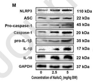
Application: WB    Species: Human    Sample: HepG2 cells

Fig.3 NaAsO2 impairs insulin signaling via activation of NLRP3 inflammasome in HepG2 cells. HepG2 cells were pretreated with 1 μg/ml LPS for 4 hours, 5 μM MCC950, for 4 hours, and were then treated with 4 μM NaAsO2 for 24 hours. Cytosolic fractions were analyzed by Western blot analysis. GAPDH was used as an internal control. The efficiency of MCC950, and its effect on NLRP3, caspase-1 activation, IL-1β and IL- 18 production in NaAsO2-treated HepG2 cells (A-E). HepG2 cells were stimulated with 100 nM insulin for 10 min at the end of treatment. Cytosolic fractions were analyzed by Western blot analysis. GAPDH was used as an internal control. The efficiency of MCC950, and its effect on p-IRS (Ser 307)/IRS1 and p-AKT (Ser473)/AKT1 ratio in NaAsO2-treated HepG2 cells (F-H). Insulin‐ stimulated glucose uptake in HepG2 cells was measured using a glucose assay kit (I). Results are mean ± SEM (n = 3). *P < 0.05 compare with the LPS group. #P<0.05 compare with 4 μM NaAsO2 group.

2). SGLT2 inhibitor canagliflozin alleviates vascular calcification through suppression of NLRP3 inflammasome. Cardiovascular Research (PubMed: 37523743) [IF=10.8]

3). Sodium-glucose cotransporter 2 inhibitor canagliflozin alleviates vascular calcification through suppression of nucleotide-binding domain, leucine-rich-containing family, pyrin domain-containing-3 inflammasome. Cardiovascular research (PubMed: 37523743) [IF=10.8]

4). Hair follicle-MSC-derived small extracellular vesicles as a novel remedy for acute pancreatitis. JOURNAL OF CONTROLLED RELEASE (PubMed: 36402231) [IF=10.8]

5). A unique death pathway keeps RIPK1 D325A mutant mice in check at embryonic day 10.5. PLOS Biology (PubMed: 34437534) [IF=9.8]

6). Activating cGAS–STING axis contributes to neuroinflammation in CVST mouse model and induces inflammasome activation and microglia pyroptosis. Journal of Neuroinflammation (PubMed: 35689216) [IF=9.3]

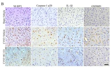
Application: IHC    Species: Mouse    Sample:

Fig. 7Effects of RU.521 on NLRP3 inflammasome and pyroptosis post-CVST. A Western blot assay for NLRP3, caspase-1 p20, pro-IL-1β, cleaved-IL-1β, GSDMD and GSDMD-C expressions from the impaired cortex at 3 days post-CVST. B Typical immunohistochemical staining for NLRP3, caspase-1 p20, IL-1β and GSDMD at diverse groups after CVST. Scale bar = 50 μm. C, D Quantitative analysis of western blot assay. *P < 0.05, **P < 0.01. n = 6. E Representative co-localization staining for IL-1β (green) and microglia (IBA-1, red) in the injured cortex as shown by white arrows, at 3 days after CVST. Scale bar = 50 μm

7). Inhibition of IGF2BP1 attenuates renal injury and inflammation by alleviating m6A modifications and E2F1/MIF pathway. International Journal of Biological Sciences (PubMed: 36632449) [IF=9.2]

8). ELABELA attenuates deoxycorticosterone acetate/salt-induced hypertension and renal injury by inhibition of NADPH oxidase/ROS/NLRP3 inflammasome pathway. Cell Death & Disease (PubMed: 32829380) [IF=9.0]

Application: IF/ICC    Species: mouse    Sample: HK2 cells

Fig. 7 Effects of ELA on the ROS production and NLRP3 imflammasome activation are independent of APJ. APJ knockdown was conducted by the transfection of plasmid pWHAPJg into HK2 cells and confirmed by RT-PCR. Then the HK2 cells with APJ knockdown were pretreated with ELA peptide (1 nM) for 1 h and then treated by Aldosterone (0.1 µM) for 24 h. a Representative immunoblots of APJ in renal cortex and medulla and summarized intensities of blots. b mRNA levels of APJ in HK2 cells with or without APJ knockdown. c ROS production in HK2 cells with APJ knockdown. d, e Representative confocal fluorescent images of co-localization (yellow) between Nlrp3 (green)/Caspase-1 (red) and summarized colocalization coefficient in HK2 cells with APJ knockdown. Scale bar, 10 µm. N = 6 per group. *P < 0.05 versus Ctrl group; #P < 0.05 versus DOCA/ salt group

9). Autophagic-CTSB-inflammasome axis modulates hepatic stellate cells activation in arsenic-induced liver fibrosis. Chemosphere (PubMed: 31669990) [IF=8.8]

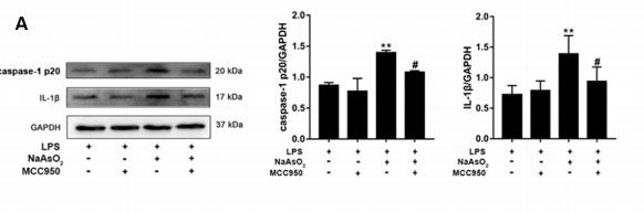
Application: WB    Species: rat    Sample: HSC-t6 cells

Fig.4 |NaAsO2-induced HSCs activation was dependent on the activation of NLRP3 inflammasome. (A, B) HSC-t6 cells were pretreated with or without MCC950 for 2h and then treated with NaAsO2 for 24 h. The expression level of caspase-1 p20, IL-1β, Collagen-, a-SMA, MMP-1, and TIMP-1 were determined by Western blot.

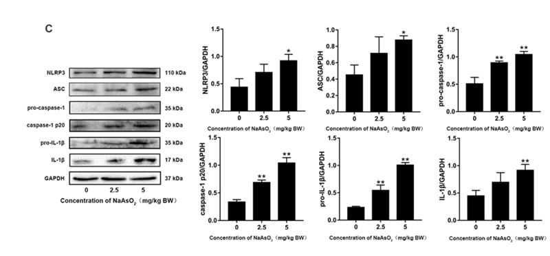
Application: WB    Species: rat    Sample: livers

Fig.2 NaAsO2 induced autophagy, activation of NLRP3 inflammasome and 662 hepatic stellate cell in vivo. (A) The expression level of LC3 and p62 in rat livers 663 following exposure to NaAsO2. The protein fraction was analyzed by Western blot. (B) 664 The expression level of cytoplasmic CTSB in rat livers following exposure to NaAsO2. 665 The protein fraction was analyzed by Western blot. (C) The expression level of NLRP3 inflammasome-associated proteins in rat livers following exposure to NaAsO2. 667 The protein fraction was analyzed by Western blot. (D) The expression level of 668 Collagen- , α-SMA, MMP-1, and TIMP-1in rat livers following exposure to NaAsO2. 669 The protein fraction was analyzed by Western blot. (E) α-SMA was detected by IF. 670 White arrows indicate positive staining. Values are mean ± SD, and n = 5. *p < 0.05, 671 **p < 0.01 compared with the control group; Scale bar = 100 µm

10). Mesenchymal stem cells protect against TBI-induced pyroptosis in vivo and in vitro through TSG-6. Cell Communication and Signaling (PubMed: 35982465) [IF=8.4]

加载更多

限制条款

产品的规格、报价、验证数据请以官网为准,官网链接:www.affbiotech.com | www.affbiotech.cn(简体中文)| www.affbiotech.jp(日本語)

产品的数据信息为Affinity所有,未经授权不得收集Affinity官网数据或资料用于商业用途,对抄袭产品数据的行为我们将保留诉诸法律的权利。

产品相关数据会因产品批次、产品检测情况随时调整,如您已订购该产品,请以订购时随货说明书为准,否则请以官网内容为准,官网内容有改动时恕不另行通知。

Affinity保证所销售产品均经过严格质量检测。如您购买的商品在规定时间内出现问题需要售后时,请您在Affinity官方渠道提交售后申请。

产品仅供科学研究使用。不用于诊断和治疗。 

产品未经授权不得转售。

Affinity Biosciences将不会对在使用我们的产品时可能发生的专利侵权或其他侵权行为负责。Affinity Biosciences, Affinity Biosciences标志和所有其他商标所有权归Affinity Biosciences LTD.