产品: Caspase 9 抗体
货号: AF6348
描述: Rabbit polyclonal antibody to Caspase 9
应用: WB IHC IF/ICC
反应: Human, Mouse, Rat
预测: Pig, Bovine, Horse, Sheep
分子量: 46kDa; 46kD(Calculated).
蛋白号: P55211
RRID: AB_2835042

浏览相似产品>>

   规格 价格 库存
 50ul RMB¥ 1250 现货
 100ul RMB¥ 2300 现货
 200ul RMB¥ 3000 现货

货期: 当天发货

联系销售

产品描述

来源:
Rabbit
应用:
WB 1:500-1:2000, IHC 1:50-1:200, IF/ICC 1:100-1:500
*The optimal dilutions should be determined by the end user.
*Tips:

WB: 适用于变性蛋白样本的免疫印迹检测. IHC: 适用于组织样本的石蜡(IHC-p)或冰冻(IHC-f)切片样本的免疫组化/荧光检测. IF/ICC: 适用于细胞样本的荧光检测. ELISA(peptide): 适用于抗原肽的ELISA检测.

反应:
Human,Mouse,Rat
预测:
Pig(88%), Bovine(82%), Horse(91%), Sheep(90%)
克隆:
Polyclonal
特异性:
Caspase 9 Antibody detects endogenous levels of total Caspase 9.
RRID:
AB_2835042
引用格式: Affinity Biosciences Cat# AF6348, RRID:AB_2835042.
偶联:
Unconjugated.
纯化:
The antiserum was purified by peptide affinity chromatography using SulfoLink™ Coupling Resin (Thermo Fisher Scientific).
保存:
Rabbit IgG in phosphate buffered saline , pH 7.4, 150mM NaCl, 0.02% sodium azide and 50% glycerol. Store at -20 °C. Stable for 12 months from date of receipt.
别名:

展开/折叠

APAF-3; APAF3; Apoptosis related cysteine peptidase; Apoptotic protease Mch-6; Apoptotic protease-activating factor 3; CASP-9; CASP9; CASP9_HUMAN; Caspase 9 apoptosis related cysteine peptidase; Caspase 9 Dominant Negative; Caspase 9c; Caspase-9; Caspase-9 subunit p10; ICE LAP6; ICE like apoptotic protease 6; ICE-LAP6; ICE-like apoptotic protease 6; MCH6; PPP1R56; protein phosphatase 1, regulatory subunit 56; RNCASP9;

抗原和靶标

免疫原:
Uniprot:
基因/基因ID:
表达:
P55211 CASP9_HUMAN:

Ubiquitous, with highest expression in the heart, moderate expression in liver, skeletal muscle, and pancreas. Low levels in all other tissues. Within the heart, specifically expressed in myocytes.

描述:
This gene encodes a protein which is a member of the cysteine-aspartic acid protease (caspase) family. Sequential activation of caspases plays a central role in the execution-phase of cell apoptosis. Caspases exist as inactive proenzymes which undergo proteolytic processing at conserved aspartic residues to produce 2 subunits, large and small, that dimerize to form the active enzyme.
序列:
MDEADRRLLRRCRLRLVEELQVDQLWDALLSRELFRPHMIEDIQRAGSGSRRDQARQLIIDLETRGSQALPLFISCLEDTGQDMLASFLRTNRQAAKLSKPTLENLTPVVLRPEIRKPEVLRPETPRPVDIGSGGFGDVGALESLRGNADLAYILSMEPCGHCLIINNVNFCRESGLRTRTGSNIDCEKLRRRFSSLHFMVEVKGDLTAKKMVLALLELAQQDHGALDCCVVVILSHGCQASHLQFPGAVYGTDGCPVSVEKIVNIFNGTSCPSLGGKPKLFFIQACGGEQKDHGFEVASTSPEDESPGSNPEPDATPFQEGLRTFDQLDAISSLPTPSDIFVSYSTFPGFVSWRDPKSGSWYVETLDDIFEQWAHSEDLQSLLLRVANAVSVKGIYKQMPGCFNFLRKKLFFKTS

种属预测

种属预测:

score>80的预测可信度较高,可尝试用于WB检测。*预测模型主要基于免疫原序列比对,结果仅作参考,不作为质保凭据。

Species
Results
Score
Horse
91
Sheep
90
Pig
88
Bovine
82
Rabbit
78
Xenopus
71
Zebrafish
63
Dog
60
Chicken
0
Model Confidence:
High(score>80) Medium(80>score>50) Low(score<50) No confidence

翻译修饰 - P55211 作为底物

Site PTM Type Enzyme
K97 Ubiquitination
S99 Phosphorylation P17612 (PRKACA)
K100 Ubiquitination
T107 Phosphorylation
K117 Ubiquitination
T125 Phosphorylation P27361 (MAPK3) , P28482 (MAPK1) , Q13627 (DYRK1A) , P06493 (CDK1)
S133 Phosphorylation
S144 Phosphorylation Q05513 (PRKCZ)
Y153 Phosphorylation P00519 (ABL1) , A0A173G4P4 (Abl fusion)
S175 Phosphorylation
S183 Phosphorylation P17612 (PRKACA)
K189 Ubiquitination
S195 Phosphorylation P17612 (PRKACA)
S196 Phosphorylation Q9Y243 (AKT3) , P31751 (AKT2) , P31749 (AKT1) , Q13237 (PRKG2)
K204 Ubiquitination
T208 Phosphorylation
K210 Ubiquitination
K211 Ubiquitination
Y251 Phosphorylation
K278 Ubiquitination
T301 Phosphorylation
S302 Phosphorylation
S307 Phosphorylation
S310 Phosphorylation
K394 Ubiquitination
Y397 Phosphorylation

研究背景

功能:

Involved in the activation cascade of caspases responsible for apoptosis execution. Binding of caspase-9 to Apaf-1 leads to activation of the protease which then cleaves and activates caspase-3. Promotes DNA damage-induced apoptosis in a ABL1/c-Abl-dependent manner. Proteolytically cleaves poly(ADP-ribose) polymerase (PARP).

Isoform 2 lacks activity is an dominant-negative inhibitor of caspase-9.

翻译修饰:

Cleavages at Asp-315 by granzyme B and at Asp-330 by caspase-3 generate the two active subunits. Caspase-8 and -10 can also be involved in these processing events.

Phosphorylated at Thr-125 by MAPK1/ERK2. Phosphorylation at Thr-125 is sufficient to block caspase-9 processing and subsequent caspase-3 activation. Phosphorylation on Tyr-153 by ABL1/c-Abl; occurs in the response of cells to DNA damage.

组织特异性:

Ubiquitous, with highest expression in the heart, moderate expression in liver, skeletal muscle, and pancreas. Low levels in all other tissues. Within the heart, specifically expressed in myocytes.

亚基结构:

Heterotetramer that consists of two anti-parallel arranged heterodimers, each one formed by a 35 kDa (p35) and a 10 kDa (p10) subunit. Caspase-9 and APAF1 bind to each other via their respective NH2-terminal CED-3 homologous domains in the presence of cytochrome C and ATP. Interacts (inactive form) with EFHD2. Interacts with HAX1. Interacts with BIRC2/c-IAP1, XIAP/BIRC4, BIRC5/survivin, BIRC6/bruce and BIRC7/livin. Interacts with ABL1 (via SH3 domain); the interaction is direct and increases in the response of cells to genotoxic stress and ABL1/c-Abl activation. Interacts with NleF from pathogenic E.coli.

蛋白家族:

Belongs to the peptidase C14A family.

研究领域

· Cellular Processes > Cell growth and death > p53 signaling pathway.   (View pathway)

· Cellular Processes > Cell growth and death > Apoptosis.   (View pathway)

· Cellular Processes > Cell growth and death > Apoptosis - multiple species.   (View pathway)

· Environmental Information Processing > Signal transduction > PI3K-Akt signaling pathway.   (View pathway)

· Human Diseases > Drug resistance: Antineoplastic > Platinum drug resistance.

· Human Diseases > Neurodegenerative diseases > Alzheimer's disease.

· Human Diseases > Neurodegenerative diseases > Parkinson's disease.

· Human Diseases > Neurodegenerative diseases > Amyotrophic lateral sclerosis (ALS).

· Human Diseases > Neurodegenerative diseases > Huntington's disease.

· Human Diseases > Infectious diseases: Bacterial > Legionellosis.

· Human Diseases > Infectious diseases: Parasitic > Toxoplasmosis.

· Human Diseases > Infectious diseases: Bacterial > Tuberculosis.

· Human Diseases > Infectious diseases: Viral > Hepatitis B.

· Human Diseases > Infectious diseases: Viral > Influenza A.

· Human Diseases > Cancers: Overview > Pathways in cancer.   (View pathway)

· Human Diseases > Cancers: Specific types > Colorectal cancer.   (View pathway)

· Human Diseases > Cancers: Specific types > Pancreatic cancer.   (View pathway)

· Human Diseases > Cancers: Specific types > Endometrial cancer.   (View pathway)

· Human Diseases > Cancers: Specific types > Prostate cancer.   (View pathway)

· Human Diseases > Cancers: Specific types > Small cell lung cancer.   (View pathway)

· Human Diseases > Cancers: Specific types > Non-small cell lung cancer.   (View pathway)

· Human Diseases > Cardiovascular diseases > Viral myocarditis.

· Organismal Systems > Endocrine system > Thyroid hormone signaling pathway.   (View pathway)

文献引用

1). Upregulation of BCL-2 by acridone derivative through gene promoter i-motif for alleviating liver damage of NAFLD/NASH. NUCLEIC ACIDS RESEARCH (PubMed: 32710621) [IF=14.9]

Application: WB    Species: human    Sample: HepG2

Figure 4. Effect of A22 on anti-apoptosis in 0.5 mM palmitic acid oil (PA) induced cell model. (A) Effect of A22 on cell viability for anti-apoptotic protective effect. (B) Effect of A22 on transcription of BCL-2 and BAX with measurement of mRNA levels. (C) Effect of A22 on protein expressions related with apoptosis (left), which were quantitatively analyzed (right). All the experiments were repeated for three times.

2). Formation of di-cysteine acrolein adduct decreases cytotoxicity of acrolein by ROS alleviation and apoptosis intervention. Journal of Hazardous Materials (PubMed: 31780296) [IF=13.6]

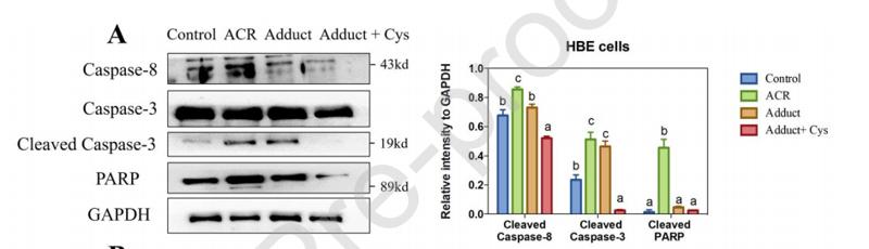
Application: WB    Species: Human    Sample: HBE (A) and Caco-2 (B) cells

Fig. 8 Effect of acrolein (ACR) or its adduct ACR-di-Cys with/without addition of 1mM cysteine on the cleavage of caspase-3, caspase-8 and PARP in HBE (A) and Caco-2 (B) cells. Left: western blot. Right: The data were expressed as relative intensity to GAPDH; Error bars represent standard deviation (n=3); Different letters indicate significant differences (p < 0.05) between treatments. 42 J

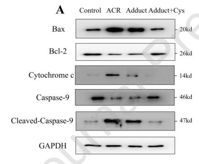
Application: WB    Species: Human    Sample: HBE and Caco-2 cells

Fig. 9 Effect of acrolein (ACR) or its adduct ACR-di-Cys with/without addition of 1mM cysteine on the expression of Bax, Bcl-2, cytochrome c and the cleavage of caspase-9 in HBE (A) and Caco-2 (B) cells. Left: western blot. Right: The data were expressed as relative intensity to GAPDH; Error bars represent standard deviation (n=3); Different letters indicate significant differences (p < 0.05) between treatments.

3). Echinatin inhibits the growth and metastasis of human osteosarcoma cells through Wnt/β-catenin and p38 signaling pathways. Pharmacological Research (PubMed: 37023991) [IF=9.3]

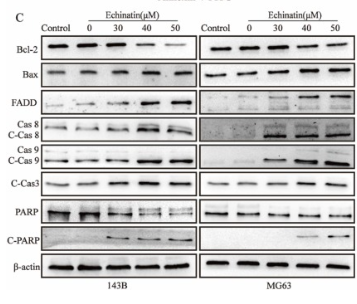
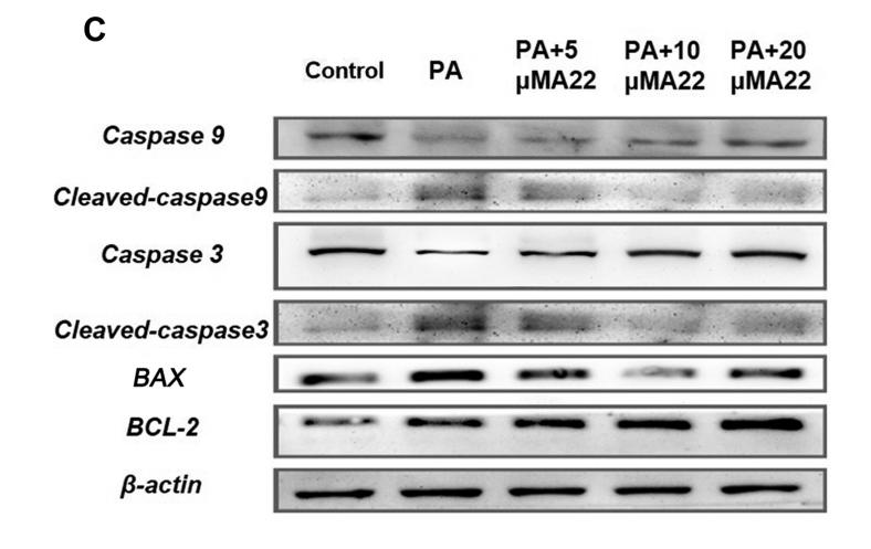
Application: WB    Species: Human    Sample: OS cells

Fig. 4. Ecn inhibits the migration and invasion of OS cells. (A) The effect of Ecn on the migration of OS cells (Wound healing assay, 100 ×). (B) The effect of Ecn on the migration of OS cells (Transwell assay, 100 ×). (C) The effect of Ecn on the invasion of OS cells (Matrigel-coated Transwell assay, 100 ×). (D) The effect of Ecn on the protein level of MMP2, MMP7, MMP9, Snail, Vimentin, N-Cadherin and E-Cadherin in OS cells (Western blot). ##P 

4). Increased autophagy in EOC re-ascites cells can inhibit cell death and promote drug resistance. Cell Death & Disease (PubMed: 29549251) [IF=9.0]

Application: IHC    Species: human    Sample: EOC ascites cells

Fig. 2| Autophagy is increased and apoptosis is reduced in the EOC re-ascites group. a IHC analysis of LC-3, Belin-1, CASP-9, and c-CASP-3 expression in EOC ascites cells (scale bar =30µm). b, c Western blot analysis of autophagy/apoptosis-related proteins expressed in the chemosensitive group and re-ascites group (***p<0.001, **p<0.01, *p<0.05). d Immunofluorescence analysis of the chemosensitive group and re-ascites group with LC3 (green) and LAMP2 (red) staining. Nuclei were counterstained with DAPI (blue). e qRT-PCR analysis of the average relative mRNA expression levels of autophagy/apoptosis-related genes in the chemosensitive group and re-ascites group (**p<0.01, *p<0.05)

Application: IF/ICC    Species: human    Sample: ascites cells

Fig. 5 |Apoptosis is increased in the EOC chemosensitive group. a, b IHC and IF analysis of apoptotic protein expression in ovarian cancer samples. c, d Western blot analysis of apoptotic proteins expressed in the no-chemotherapy group and chemosensitive group (***p<0.001,**p<0.01, *p<0.05). e qRT-PCR analysis of the average relative mRNA expression levels of apoptosis-related genes in the no-chemotherapy group and chemosensitive group (*p<0.05)

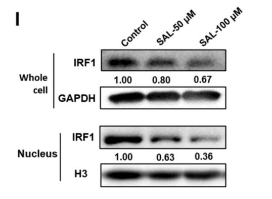
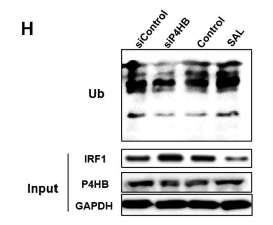
Application: WB    Species: human    Sample: ascites cells

Fig. 5 |Apoptosis is increased in the EOC chemosensitive group. a, b IHC and IF analysis of apoptotic protein expression in ovarian cancer samples. c, d Western blot analysis of apoptotic proteins expressed in the no-chemotherapy group and chemosensitive group (***p<0.001,**p<0.01, *p<0.05). e qRT-PCR analysis of the average relative mRNA expression levels of apoptosis-related genes in the no-chemotherapy group and chemosensitive group (*p<0.05)

5). LNC473 regulating APAF1 IRES-dependent translation via competitive sponging miR574 and miR15b: Implications in colorectal cancer. Molecular Therapy-Nucleic Acids (PubMed: 32784109) [IF=8.8]

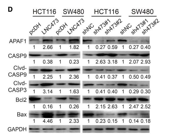
Application: WB    Species: Human    Sample: HCT116 and SW480 cells

Figure 6. LNC473-miR574/miR15b-APAF1 Signaling Axis in HCT116 and SW480 Cells (A) IF and ISH combination assay revealing the co-localization of APAF1 protein with included ncRNAs in HCT116 cells. Scale bars, 5 mm. (B and C) The levels of APAF1 mRNA (B) and protein (C) were determined after interfering LNC473 expression in HCT116 and SW480 cells by qPCR and IF assays. Scale bars, 20 mm. (D) The expression of apoptosis-related proteins including APAF1 was detected after interfering LNC473 expression in HCT116 and SW480 cells by western blot assay. (E and F) Rescue experiments showing the APAF1 levels in HCT116 and SW480 cells with exposure to the co-transfection of pcDH-LNC473 vector and miR574-5p or miR15b-5p mimic by qPCR (E) and western blot (F) assays. (G) Pattern diagram of APAF1-CDS and APAF1-IRES-CDS vectors. (H and I) APAF1 protein expression was determined in HCT116 and SW480 cells treated with APAF1-IRES-CDS vector, or pcDH-LNC473 and APAF1-IRES-CD co-transfection by western blot (H) and IF assays (I). Scale bars, 5 mm. (J) The percentage (%) of cell apoptosis in cells upon co-overexpressing APAF1-CDS or APAF1-IRE-CDS and LNC473 as assayed by flow cytometry. All tests were performed at least three times. Data were expressed as mean ± SD. ns (nonsignificant), p > 0.05; *p < 0.05, **p < 0.01, ***p < 0.001, ****p < 0.0001.

6). Cytotoxicity of adducts formed between quercetin and methylglyoxal in PC-12 cells. Food Chemistry (PubMed: 33706136) [IF=8.8]

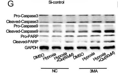
Application: WB    Species: Rat    Sample: PC-12 cells

Fig. 5. Effect of treatments of MGO, Que-mono-MGO, and Que-di-MGO on the expression levels of apoptotic markers and components of AKT and Nrf2-HO-1/NQO-1 signaling pathways. Significant differences (p < 0.05) between samples of different treatments are marked with different letters on each column.

7). Long noncoding RNAs regulated spermatogenesis in varicocele‐induced spermatogenic dysfunction. CELL PROLIFERATION (PubMed: 35297519) [IF=8.5]

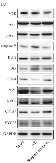
Application: WB    Species: Human    Sample: spermatogenic cell

FIGURE 13Validation of regulated signalling pathways, spermatogenic cell apoptosis and proliferation, and meiotic spermatocytes by Western blot. Representative Western blot images of PI3K, Akt, p-Akt, caspase-9, Bcl-2, Bax, PCNA, PLZF, REC8, STRA8, and SYCP3. (B) Statistical analysis of band intensity by Student's t-test. *p < 0.05; **p < 0.01; ***p < 0.001; ****p < 0.0001. NS, not significant; Sham, sham group; Surgical treatment, surgical treatment group; VC, varicocele group

8). Synthesis, characterization, and cytotoxicity analysis of selenium nanoparticles stabilized by Morchella sextelata polysaccharide. International Journal of Biological Macromolecules (PubMed: 37247714) [IF=8.2]

9). Development of anisamide-targeted PEGylated gold nanorods to deliver epirubicin for chemo-photothermal therapy in tumor-bearing mice. International Journal of Nanomedicine (PubMed: 30880982) [IF=8.0]

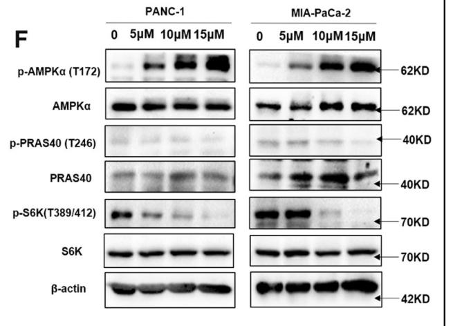
Application: WB    Species:    Sample: PC-3 cells

Figure 6 |The expression of caspase 3, cleaved caspase 3 (phosphorylated form), caspase 9, cleaved caspase 9 (phosphorylated form), Bcl-2, Bax, and β-actin was determined using Western blotting.

10). Quercetin induces autophagy via FOXO1-dependent pathways and autophagy suppression enhances quercetin-induced apoptosis in PASMCs in hypoxia. FREE RADICAL BIOLOGY AND MEDICINE (PubMed: 27979659) [IF=7.4]

Application: WB    Species: rat    Sample:


加载更多

限制条款

产品的规格、报价、验证数据请以官网为准,官网链接:www.affbiotech.com | www.affbiotech.cn(简体中文)| www.affbiotech.jp(日本語)

产品的数据信息为Affinity所有,未经授权不得收集Affinity官网数据或资料用于商业用途,对抄袭产品数据的行为我们将保留诉诸法律的权利。

产品相关数据会因产品批次、产品检测情况随时调整,如您已订购该产品,请以订购时随货说明书为准,否则请以官网内容为准,官网内容有改动时恕不另行通知。

Affinity保证所销售产品均经过严格质量检测。如您购买的商品在规定时间内出现问题需要售后时,请您在Affinity官方渠道提交售后申请。

产品仅供科学研究使用。不用于诊断和治疗。 

产品未经授权不得转售。

Affinity Biosciences将不会对在使用我们的产品时可能发生的专利侵权或其他侵权行为负责。Affinity Biosciences, Affinity Biosciences标志和所有其他商标所有权归Affinity Biosciences LTD.