产品: AFP 抗体
货号: AF5134
描述: Rabbit polyclonal antibody to AFP
应用: WB IHC
反应: Human, Mouse, Rat
预测: Pig, Bovine, Horse, Sheep, Rabbit, Dog
分子量: 62-65 kDa; 69kD(Calculated).
蛋白号: P02771
RRID: AB_2837620

浏览相似产品>>

   规格 价格 库存
 50ul RMB¥ 1250 现货
 100ul RMB¥ 2300 现货
 200ul RMB¥ 3000 现货

货期: 当天发货

联系销售

产品描述

来源:
Rabbit
应用:
WB 1:500-1:2000, IHC 1:50-1:200
*The optimal dilutions should be determined by the end user.
*Tips:

WB: 适用于变性蛋白样本的免疫印迹检测. IHC: 适用于组织样本的石蜡(IHC-p)或冰冻(IHC-f)切片样本的免疫组化/荧光检测. IF/ICC: 适用于细胞样本的荧光检测. ELISA(peptide): 适用于抗原肽的ELISA检测.

反应:
Human,Mouse,Rat
预测:
Pig(100%), Bovine(92%), Horse(100%), Sheep(92%), Rabbit(92%), Dog(100%)
克隆:
Polyclonal
特异性:
AFP Antibody detects endogenous levels of total AFP.
RRID:
AB_2837620
引用格式: Affinity Biosciences Cat# AF5134, RRID:AB_2837620.
偶联:
Unconjugated.
纯化:
The antiserum was purified by peptide affinity chromatography using SulfoLink™ Coupling Resin (Thermo Fisher Scientific).
保存:
Rabbit IgG in phosphate buffered saline , pH 7.4, 150mM NaCl, 0.02% sodium azide and 50% glycerol. Store at -20 °C. Stable for 12 months from date of receipt.
别名:

展开/折叠

Afp; AFPD; Alpha fetoglobulin; Alpha fetoprotein; Alpha fetoprotein precursor; Alpha-1-fetoprotein; Alpha-fetoglobulin; Alpha-fetoprotein; alpha-fetoprotein, Hereditary persistence of, included; FETA; FETA_HUMAN; Hereditary persistence of alpha fetoprotein; HPAFP;

抗原和靶标

免疫原:
Uniprot:
基因/基因ID:
表达:
P02771 FETA_HUMAN:

Plasma. Synthesized by the fetal liver and yolk sac.

描述:
Binds copper, nickel, and fatty acids as well as, and bilirubin less well than, serum albumin. Only a small percentage (less than 2%) of the human AFP shows estrogen-binding properties.
序列:
MKWVESIFLIFLLNFTESRTLHRNEYGIASILDSYQCTAEISLADLATIFFAQFVQEATYKEVSKMVKDALTAIEKPTGDEQSSGCLENQLPAFLEELCHEKEILEKYGHSDCCSQSEEGRHNCFLAHKKPTPASIPLFQVPEPVTSCEAYEEDRETFMNKFIYEIARRHPFLYAPTILLWAARYDKIIPSCCKAENAVECFQTKAATVTKELRESSLLNQHACAVMKNFGTRTFQAITVTKLSQKFTKVNFTEIQKLVLDVAHVHEHCCRGDVLDCLQDGEKIMSYICSQQDTLSNKITECCKLTTLERGQCIIHAENDEKPEGLSPNLNRFLGDRDFNQFSSGEKNIFLASFVHEYSRRHPQLAVSVILRVAKGYQELLEKCFQTENPLECQDKGEEELQKYIQESQALAKRSCGLFQKLGEYYLQNAFLVAYTKKAPQLTSSELMAITRKMAATAATCCQLSEDKLLACGEGAADIIIGHLCIRHEMTPVNPGVGQCCTSSYANRRPCFSSLVVDETYVPPAFSDDKFIFHKDLCQAQGVALQTMKQEFLINLVKQKPQITEEQLEAVIADFSGLLEKCCQGQEQEVCFAEEGQKLISKTRAALGV

种属预测

种属预测:

score>80的预测可信度较高,可尝试用于WB检测。*预测模型主要基于免疫原序列比对,结果仅作参考,不作为质保凭据。

Species
Results
Score
Pig
100
Horse
100
Dog
100
Bovine
92
Sheep
92
Rabbit
92
Chicken
55
Xenopus
0
Zebrafish
0
Model Confidence:
High(score>80) Medium(80>score>50) Low(score<50) No confidence

翻译修饰 - P02771 作为底物

Site PTM Type Enzyme
S111 Phosphorylation
S115 Phosphorylation
S117 Phosphorylation
T248 Phosphorylation
S344 Phosphorylation
S368 Phosphorylation
K413 Acetylation
S415 Phosphorylation
S444 Phosphorylation
S445 Phosphorylation

研究背景

功能:

Binds copper, nickel, and fatty acids as well as, and bilirubin less well than, serum albumin. Only a small percentage (less than 2%) of the human AFP shows estrogen-binding properties.

翻译修饰:

Independent studies suggest heterogeneity of the N-terminal sequence of the mature protein and of the cleavage site of the signal sequence.

Sulfated.

细胞定位:

Secreted.

Extracellular region or secreted Cytosol Plasma membrane Cytoskeleton Lysosome Endosome Peroxisome ER Golgi apparatus Nucleus Mitochondrion Manual annotation Automatic computational assertionSubcellular location
组织特异性:

Plasma. Synthesized by the fetal liver and yolk sac.

亚基结构:

Dimeric and trimeric forms have been found in addition to the monomeric form.

蛋白家族:

Belongs to the ALB/AFP/VDB family.

研究领域

· Environmental Information Processing > Signal transduction > Hippo signaling pathway.   (View pathway)

文献引用

1). Human umbilical cord-derived mesenchymal stem cells improve the function of liver in rats with acute-on-chronic liver failure via downregulating Notch and Stat1/Stat3 signaling. Stem Cell Research & Therapy (PubMed: 34256837) [IF=7.5]

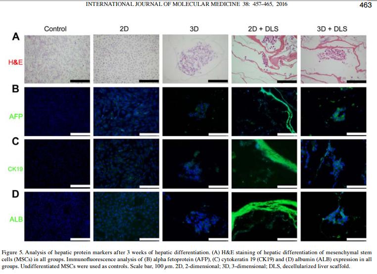
Application: IHC    Species: Rat    Sample: liver tissue

Fig. 7 The effects of hUC-MSC transplantation on hepatocyte regeneration in ACLF rats. Liver sections from ACLF rats 12 and 24 h post-hUC-MSC transplantation or 0.9% sodium chloride injection as a control were used for immunohistochemical staining of AFP, CK18, HGF, and PCNA and microscopic examination (n = 4/group). Representative photographs are shown (a). Positive staining was quantified and is presented as the mean ± SD (b, c). d Serum levels of HGF were detected by ELISA (4h, n = 3/group; 12h and 24h, n = 4/group). Data are presented as the mean ± SD. *P < 0.05, **P < 0.01, ***P < 0.001

2). Enhanced hepatic differentiation of rat bone marrow-derived mesenchymal stem cells in spheroidal aggregate culture on a decellularized liver scaffold. INTERNATIONAL JOURNAL OF MOLECULAR MEDICINE (PubMed: 27314916) [IF=5.4]

Application: IF/ICC    Species: rat    Sample:

Figure 5. Analysis of hepatic protein markers after 3 weeks of hepatic differentiation. (A) H&E staining of hepatic differentiation of mesenchymal stem cells (MSCs) in all groups. Immunofluorescence analysis of (B) alpha fetoprotein (AFP), (C) cytokeratin 19 (CK19) and (D) albumin (ALB) expression in all groups. Undifferentiated MSCs were used as controls. Scale bar, 100 µm. 2D, 2-dimensional; 3D, 3-dimensional; DLS, decellularized liver scaffold.

3). Glycyrrhizic Acid-Induced Differentiation Repressed Stemness in Hepatocellular Carcinoma by Targeting c-Jun N-Terminal Kinase 1. Frontiers in Oncology (PubMed: 31998631) [IF=4.7]

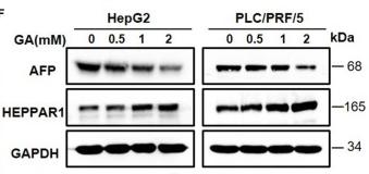
Application: WB    Species: Mice    Sample: hepatic cancer cells

Figure 1 GA reduces stemness and induces differentiation of hepatic cancer cells. (A) Survival of HepG2 and PLC/PRF/5 cells incubated with the indicated amounts of GA for 48 h. (B) Cell proliferation was assessed through a colony formation assay. The number of colonies was compared (one-way ANOVA; *P < 0.05, **P < 0.01, ***P < 0.001). (C) Images of sphere formation assay of HepG2 and PLC/PRF/5. The number of spheres was compared (one-way ANOVA; *P < 0.05, **P < 0.01, ***P < 0.001). (D) Expression of representative CSC markers (SOX2 and OCT4) was analyzed by Western blot assay (one-way ANOVA; *P < 0.05, **P < 0.01, ***P < 0.001). (E) Images of HepG2 and PLC/PRF/5 cells were taken after incubation with different GA concentrations. (F) Expressions of representative differentiation markers (AFP and HEPPAR1) were analyzed by Western blot assay (one-way ANOVA; *P < 0.05, **P < 0.01). Scale bars in C and E are at 200 and 20 μm, respectively.

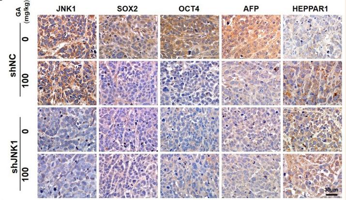
Application: IHC    Species: Mice    Sample: tumors tissue

Figure 6 GA suppresses tumor growth and enhances the antitumor effect of sorafenib. Images of tumors (A), tumor volumes (B), and body weight (C) of PLC/PRF/5 transfected with shNC or shJNK1 in BALB/c nu/nu mice treated with 100 mg/kg GA or saline as control. (D) IHC staining indicates the expression of CSC markers (SOX2 and OCT4) and differentiation markers (AFP and HEPPAR1) in tumors. (E) IHC analysis of SOX2, OCT4, AFP, and HEPPAR1 in tumors (one-way ANOVA; ***P < 0.001). Scale bars in (D), 30 μm. Images of tumors (F), tumor volumes (G) and body weight (H) of PLC/PRF/5-bearing BALB/c nu/nu mice. GA or sorafenib treatment obviously inhibited tumor growth. Furthermore, GA could enhance the anti-tumor effect of sorafenib.

4). Transient Inhibition of mTORC1 Signaling Ameliorates Irradiation-Induced Liver Damage. Frontiers in Physiology (PubMed: 30984007) [IF=4.0]

Application: WB    Species: mouse    Sample: Liver

FIGURE 1 | Irradiation-induced liver damage in mice. C57BL/6J mice were exposed to 8.0 Gy of total body irradiation (TBI). Liver tissues were harvested at days 1, 3, and 7 post-exposure (n = 6). (A) Representative pictures by HE staining are shown. Scale bar = 50 μm. Arrows indicate edema hepatocytes. (B and C) ALB and AFP expression in liver tissues (n = 3). Expression of ALB (B) and AFP (C) was detected by western blotting.

5). Hepatic differentiation of mouse bone marrow‑derived mesenchymal stem cells using a novel 3D culture system. Molecular Medicine Reports (PubMed: 29152658) [IF=3.4]

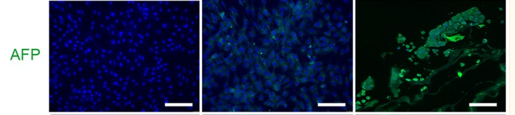
Application: IF/ICC    Species: Mouse    Sample:

Figure 4. Analysis of hepatic protein markers in mouse MSCs following 23 days of hepatic differentiation. Green immunofluorescence staining of ALB, AFP and CK19 in undifferentiated MSC control, 2D and 3D groups. Nuclei were stained blue with DAPI. Scale bars, 100 µm. MSCs, mesenchymal stem cells; ALB, albumin; AFP, α-fetoprotein; CK19, cytokeratin-19.

6). Clinicopathologic Features and Prognostic Factors in Alpha-Fetoprotein-Producing Colorectal Cancer: Analysis of 78 Cases. CELLULAR PHYSIOLOGY AND BIOCHEMISTRY (PubMed: 30522102)

限制条款

产品的规格、报价、验证数据请以官网为准,官网链接:www.affbiotech.com | www.affbiotech.cn(简体中文)| www.affbiotech.jp(日本語)

产品的数据信息为Affinity所有,未经授权不得收集Affinity官网数据或资料用于商业用途,对抄袭产品数据的行为我们将保留诉诸法律的权利。

产品相关数据会因产品批次、产品检测情况随时调整,如您已订购该产品,请以订购时随货说明书为准,否则请以官网内容为准,官网内容有改动时恕不另行通知。

Affinity保证所销售产品均经过严格质量检测。如您购买的商品在规定时间内出现问题需要售后时,请您在Affinity官方渠道提交售后申请。

产品仅供科学研究使用。不用于诊断和治疗。 

产品未经授权不得转售。

Affinity Biosciences将不会对在使用我们的产品时可能发生的专利侵权或其他侵权行为负责。Affinity Biosciences, Affinity Biosciences标志和所有其他商标所有权归Affinity Biosciences LTD.