产品: Claudin 5 抗体
货号: AF5216
描述: Rabbit polyclonal antibody to Claudin 5
应用: WB IHC IF/ICC
反应: Human, Mouse, Rat, Pig, Bovine, Monkey
预测: Pig, Bovine, Rabbit, Chicken, Xenopus
分子量: 23 kDa; 23kD(Calculated).
蛋白号: O00501
RRID: AB_2837702

浏览相似产品>>

   规格 价格 库存
 50ul RMB¥ 1250 现货
 100ul RMB¥ 2300 现货
 200ul RMB¥ 3000 现货

货期: 当天发货

联系销售

产品描述

来源:
Rabbit
应用:
WB 1:500-1:2000, IHC 1:50-1:200, IF/ICC 1:100-1:500
*The optimal dilutions should be determined by the end user.
*Tips:

WB: 适用于变性蛋白样本的免疫印迹检测. IHC: 适用于组织样本的石蜡(IHC-p)或冰冻(IHC-f)切片样本的免疫组化/荧光检测. IF/ICC: 适用于细胞样本的荧光检测. ELISA(peptide): 适用于抗原肽的ELISA检测.

反应:
Human,Mouse,Rat,Pig,Bovine,Monkey
预测:
Rabbit(93%), Chicken(80%), Xenopus(80%)
克隆:
Polyclonal
特异性:
Claudin 5 Antibody detects endogenous levels of total Claudin 5.
RRID:
AB_2837702
引用格式: Affinity Biosciences Cat# AF5216, RRID:AB_2837702.
偶联:
Unconjugated.
纯化:
The antiserum was purified by peptide affinity chromatography using SulfoLink™ Coupling Resin (Thermo Fisher Scientific).
保存:
Rabbit IgG in phosphate buffered saline , pH 7.4, 150mM NaCl, 0.02% sodium azide and 50% glycerol. Store at -20 °C. Stable for 12 months from date of receipt.
别名:

展开/折叠

Androgen withdrawal and apoptosis induced protein RVP1 like; AWAL; BEC 1; BEC1; Claudin 5 (transmembrane protein deleted in velocardiofacial syndrome); Claudin-5; Claudin5; CLD5_HUMAN; CLDN 5; Cldn5; CPETR L1; CPETRL 1; CPETRL1; TMDVCF; TMVCF; Transmembrane protein deleted in VCFS; Transmembrane protein deleted in velocardiofacial syndrome;

抗原和靶标

免疫原:
Uniprot:
基因/基因ID:
描述:
Plays a major role in tight junction-specific obliteration of the intercellular space.
序列:
MGSAALEILGLVLCLVGWGGLILACGLPMWQVTAFLDHNIVTAQTTWKGLWMSCVVQSTGHMQCKVYDSVLALSTEVQAARALTVSAVLLAFVALFVTLAGAQCTTCVAPGPAKARVALTGGVLYLFCGLLALVPLCWFANIVVREFYDPSVPVSQKYELGAALYIGWAATALLMVGGCLLCCGAWVCTGRPDLSFPVKYSAPRRPTATGDYDKKNYV

种属预测

种属预测:

score>80的预测可信度较高,可尝试用于WB检测。*预测模型主要基于免疫原序列比对,结果仅作参考,不作为质保凭据。

Species
Results
Score
Pig
100
Bovine
93
Rabbit
93
Xenopus
80
Chicken
80
Horse
0
Sheep
0
Dog
0
Zebrafish
0
Model Confidence:
High(score>80) Medium(80>score>50) Low(score<50) No confidence

翻译修饰 - O00501 作为底物

Site PTM Type Enzyme
S86 Phosphorylation
Y148 Phosphorylation
S155 Phosphorylation
T207 Phosphorylation
T209 Phosphorylation
Y212 Phosphorylation
Y217 Phosphorylation

研究背景

功能:

Plays a major role in tight junction-specific obliteration of the intercellular space.

细胞定位:

Cell junction>Tight junction. Cell membrane>Multi-pass membrane protein.

Extracellular region or secreted Cytosol Plasma membrane Cytoskeleton Lysosome Endosome Peroxisome ER Golgi apparatus Nucleus Mitochondrion Manual annotation Automatic computational assertionSubcellular location
亚基结构:

Directly interacts with TJP1/ZO-1, TJP2/ZO-2 and TJP3/ZO-3. Interacts with MPDZ (By similarity).

蛋白家族:

Belongs to the claudin family.

研究领域

· Cellular Processes > Cellular community - eukaryotes > Tight junction.   (View pathway)

· Environmental Information Processing > Signaling molecules and interaction > Cell adhesion molecules (CAMs).   (View pathway)

· Human Diseases > Infectious diseases: Viral > Hepatitis C.

· Organismal Systems > Immune system > Leukocyte transendothelial migration.   (View pathway)

文献引用

1). Activation of Wnt/β-catenin pathway mitigates blood–brain barrier dysfunction in Alzheimer's disease. BRAIN (PubMed: 35788280) [IF=14.5]

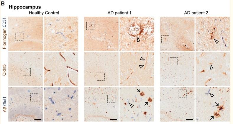
Application: IHC    Species: Human    Sample: hippocampus

Figure 1 BBB dysfunction and BEC disruption in post-mortem Alzheimer’s disease brains. (A) Fibrinogen/CD31, Cldn5 and Aβ/Glut1 staining in the cortex and (B) hippocampus of post-mortem health control and patients with Alzheimer’s disease. (C) Quantification of Cldn5 and (D) Glut1 intensity, and (E) Aβ plaques in the cortex and hippocampus of healthy controls and patients with Alzheimer’s disease. Black frames in the low-magnification images indicate the location of the high-magnification images to the right. Arrows indicate Aβ plaques, and arrowheads indicate the fibrinogen leakage and decreased Cldn5 and Glut1 in patients with Alzheimer’s disease. Scale bar = 200 µm in A and B. Data are presented as mean ± SEM, n = 5, *P < 0.05, **P < 0.01.

2). Protein palmitoylation-mediated palmitic acid sensing causes blood-testis barrier damage via inducing ER stress. Redox Biology (PubMed: 35803125) [IF=11.4]

Application: WB    Species: Mouse    Sample: TM4 Sertoli cells

Fig. 4. Inhibition of protein palmitoylation ameliorates PA induced Sertoli cell dysfunction. (A) Analysis of the palmitoylation levels of proteins extracted from the testes of mice administered or not administered the PA injection, with or without gavage of 2-BP (n = 6 for Control, n = 7 for PA and 2-BP + PA). (B) Analysis of the palmitoylation levels of proteins extracted from primary Sertoli cells, Leydig cells and germ cells, which were treated with or without 0.4 mM PA (n = 3). (C) Inhibition of palmitoylation by 2-BP suppressed PA-induced ER stress in Sertoli cells. Translational expression levels of ER stress-related genes were analyzed using Western blotting (n = 3). (D) ROS detection using DCFH-DA staining. The fluorescence densities were calculated using ImageJ (n = 3). Scale bar: 50 μm. (E) TER detection of primary Sertoli cell barriers. The cells were incubated with PA (PA), with PA combined with 2-BP (2-BP + PA), or with the vehicle (Control) for 3 days after barriers were formed on day 4 (n = 5). (F) FITC-dextran permeability assessment of primary Sertoli cell barriers. The cells were treated PA (PA), with PA combined with 2-BP (2-BP + PA), or with the vehicle (Control) for 24 h after cell barriers were formed (n = 5). (G)Tight junction protein levels were examined by western blotting in TM4 Sertoli cells (n = 3). The relative intensities of bands in western blotting results were quantified by ImageJ and normalized to β-actin levels. Data are presented as mean ± SD. n. s., no significant difference vs. Control group. *P < 0.05, **P < 0.01 and ***P < 0.001 vs. Control group. #P < 0.05, ##P < 0.01 and ###P < 0.001 vs. PA group.

3). A novel IL-1RA-PEP fusion protein alleviates blood-brain barrier disruption after ischemia-reperfusion in male rats. Journal of Neuroinflammation (PubMed: 29334965) [IF=9.3]

4). Robo4 inhibits gamma radiation-induced permeability of a murine microvascular endothelial cell by regulating the junctions. Cellular & Molecular Biology Letters (PubMed: 36647012) [IF=8.3]

5). Icariside II attenuates cerebral ischemia/reperfusion-induced blood–brain barrier dysfunction in rats via regulating the balance of MMP9/TIMP1. Acta Pharmacologica Sinica (PubMed: 32488170) [IF=8.2]

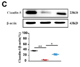
Application: WB    Species: Rat    Sample: Cerebrum

Fig. 6 ICS II improved BBB integrity after cerebral I/R by upregulating the expression of tight junction-related proteins. a Representative images of immunoblotting staining of claudin 5, occludin and ZO 1 in the penumbra. b Quantitative analysis of occludin. c Quantitative analysis of claudin 5. d Quantitative analysis of ZO 1. *P < 0.05, **P < 0.01 vs sham; #P < 0.05, ##P < 0.01 vs MCAO; n = 6 per group

Application: WB    Species: rat    Sample:

Fig. 6| ICS II improved BBB integrity after cerebral I/R by upregulating the expression of tight junction-related proteins. a Representative images of immunoblotting staining of claudin 5, occludin and ZO 1 in the penumbra.

6). Tetrastigma hemsleyanum Diels et Gilg ameliorates lipopolysaccharide induced sepsis via repairing the intestinal mucosal barrier. Biomedicine & Pharmacotherapy (PubMed: 35217279) [IF=7.5]

7). Human menstrual blood-derived stem cell transplantation suppresses liver injury in DDC-induced chronic cholestasis. Stem Cell Research & Therapy (PubMed: 35123555) [IF=7.5]

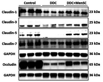
Application: WB    Species: Mice    Sample: liver tissue

Fig. 5 MenSCs promoted TJ- and bile transport function-related protein expression and reduced fibrosis-related protein expression. A Western blot analysis protein levels of Claudin-1, Claudin-3, Claudin-5, Claudin-7 and Occludin in liver tissue of different groups (n = 3 for each group). B BSEP, OATP2 and NTCP1 expression in liver tissues of different groups (n = 3 for each group). C, D Liver COL1A1, α-SMA, TGF-β1 and β-catenin expression among different groups (n = 3 for each group). *p < 0.05, **p < 0.01, ***p < 0.001, ****p < 0.0001

8). P-Glycoprotein Aggravates Blood Brain Barrier Dysfunction in Experimental Ischemic Stroke by Inhibiting Endothelial Autophagy. Aging and Disease (PubMed: 36186136) [IF=7.4]

Application: WB    Species: Mouse    Sample:

Figure 2. P-glycoprotein silence attenuates adhesion molecule expression, myeloid cell accumulation and blood-brain barrier damage in experimental ischemic stroke. Mice were intracerebroventricularly injected with P-glycoprotein (P-gp) or negative control (NC) siRNA (1.5 μL/10 g body weight) 48 h prior to MCAO/R surgery. Twenty-four hours after the surgery, brains were harvested for immunohistochemical and Western-blotting assays. (A) Immunohistochemical localization and quantification of ICAM-1 and VCAM-1 expression (n = 6). (B) Representative immunostaining images and quantification of neutrophiles (MPO) and macrophages (F4/80) (n = 6). (C-E) Representative Western-blotting images and quantification of tight junction proteins (Claudin-5, Occludin, and ZO-1) (n = 3). Scale bars: 40 μm. One-way ANOVA followed by the post hoc Tukey test. All data are mean ± SD, *P

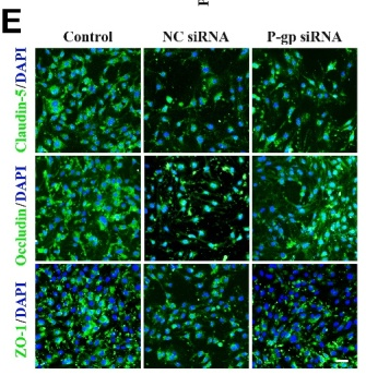
Application: IF/ICC    Species: Mouse    Sample:

Figure 5. P-glycoprotein silence alleviates endothelial dysfunction following oxygen glucose deprivation/reoxygenation. Endothelial cells (bEnd.3) were transfected with P-glycoprotein (P-gp) or negative control (NC) siRNA, or un-transfected, and then subjected to either oxygen glucose deprivation/reoxygenation (OGD/R) treatment or normal culture conditions. Twenty-four hours thereafter, cells were harvested for immunofluorescence staining, adhesion and transendothelial migration, real-time PCR gene expression and Western-blotting analyses. (A) Representative Western-blotting images and quantification of P-gp levels (n = 3). (B) Quantification of mRNA levels for TNF-α, IL-1β, MMP-2, and MMP-9 via quantitative real-time PCR assay (n = 4). (C) Representative immunofluorescence staining images and quantification of ICAM-1 and VCAM-1 expression (n = 3). (D) Representative images and quantification for fluorochrome-labeled leukocyte adhesion to endothelial cells and transendothelial migration (n = 3). (E) Representative immunofluorescence staining images for tight junction proteins (Claudin-5, Occludin, and ZO-1). (F) Representative immunoblots and quantification for the expressions of Claudin-5, Occludin, and ZO-1 (n = 3). Scale bars, 40 μm. One-way ANOVA followed by the post hoc Tukey test for A, E, and F. Mann-Whitney test for B, C, and D. All data are mean ± SD, *P

9). Stealthy nanoparticles protect endothelial barrier from leakiness by resisting the absorption of VE-cadherin. Nanoscale (PubMed: 34259298) [IF=6.7]

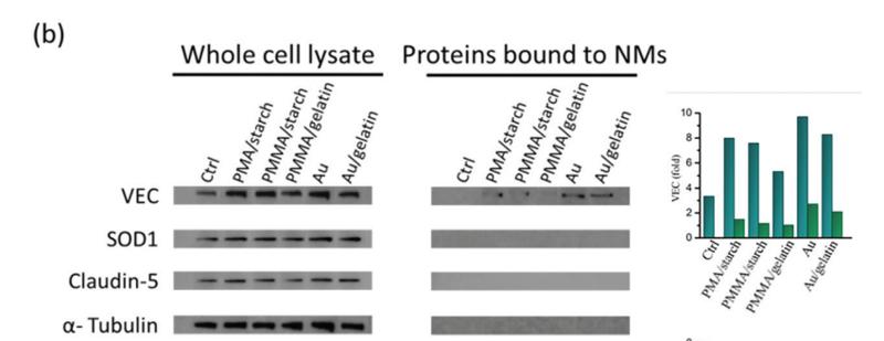
Application: WB    Species: Human    Sample: HMVEC cells

Fig. 4 (a) Schematic showing the protein pull down experimental setup. Immunoblotting (left panel) and its semi-quantitative analysis (right panel) of (b) VEC, SOD1, claudin-5 and α-tublin from whole cell lysate and pulled down by different NPs. (c) Y658 (p-VEC(Y658)), Y731 (p-VEC(Y731), VEC and α-tublin from the whole cell lysate with/without the Src kinase inhibitor, PP1.

10). Asiatic acid attenuates diabetic retinopathy through TLR4/MyD88/NF-κB p65 mediated modulation of microglia polarization. Life Sciences (PubMed: 33965378) [IF=6.1]

加载更多

限制条款

产品的规格、报价、验证数据请以官网为准,官网链接:www.affbiotech.com | www.affbiotech.cn(简体中文)| www.affbiotech.jp(日本語)

产品的数据信息为Affinity所有,未经授权不得收集Affinity官网数据或资料用于商业用途,对抄袭产品数据的行为我们将保留诉诸法律的权利。

产品相关数据会因产品批次、产品检测情况随时调整,如您已订购该产品,请以订购时随货说明书为准,否则请以官网内容为准,官网内容有改动时恕不另行通知。

Affinity保证所销售产品均经过严格质量检测。如您购买的商品在规定时间内出现问题需要售后时,请您在Affinity官方渠道提交售后申请。

产品仅供科学研究使用。不用于诊断和治疗。 

产品未经授权不得转售。

Affinity Biosciences将不会对在使用我们的产品时可能发生的专利侵权或其他侵权行为负责。Affinity Biosciences, Affinity Biosciences标志和所有其他商标所有权归Affinity Biosciences LTD.