
产品描述
*The optimal dilutions should be determined by the end user.
*Tips:
WB: 适用于变性蛋白样本的免疫印迹检测. IHC: 适用于组织样本的石蜡(IHC-p)或冰冻(IHC-f)切片样本的免疫组化/荧光检测. IF/ICC: 适用于细胞样本的荧光检测. ELISA(peptide): 适用于抗原肽的ELISA检测.
引用格式: Affinity Biosciences Cat# AF5304, RRID:AB_2837789.
展开/折叠
DKFZp781H1925; E2AK3_HUMAN; EC 2.7.11.1; Eif2ak3; Eukaryotic translation initiation factor 2 alpha kinase 3; Eukaryotic translation initiation factor 2-alpha kinase 3; Heme regulated EIF2 alpha kinase; HRI; HsPEK; Pancreatic eIF2 alpha kinase; Pancreatic eIF2-alpha kinase; PEK; PRKR like endoplasmic reticulum kinase; PRKR-like endoplasmic reticulum kinase; WRS;
抗原和靶标
- Q9NZJ5 E2AK3_HUMAN:
- Protein BLAST With
- NCBI/
- ExPASy/
- Uniprot
MERAISPGLLVRALLLLLLLLGLAARTVAAGRARGLPAPTAEAAFGLGAAAAPTSATRVPAAGAVAAAEVTVEDAEALPAAAGEQEPRGPEPDDETELRPRGRSLVIISTLDGRIAALDPENHGKKQWDLDVGSGSLVSSSLSKPEVFGNKMIIPSLDGALFQWDQDRESMETVPFTVESLLESSYKFGDDVVLVGGKSLTTYGLSAYSGKVRYICSALGCRQWDSDEMEQEEDILLLQRTQKTVRAVGPRSGNEKWNFSVGHFELRYIPDMETRAGFIESTFKPNENTEESKIISDVEEQEAAIMDIVIKVSVADWKVMAFSKKGGHLEWEYQFCTPIASAWLLKDGKVIPISLFDDTSYTSNDDVLEDEEDIVEAARGATENSVYLGMYRGQLYLQSSVRISEKFPSSPKALESVTNENAIIPLPTIKWKPLIHSPSRTPVLVGSDEFDKCLSNDKFSHEEYSNGALSILQYPYDNGYYLPYYKRERNKRSTQITVRFLDNPHYNKNIRKKDPVLLLHWWKEIVATILFCIIATTFIVRRLFHPHPHRQRKESETQCQTENKYDSVSGEANDSSWNDIKNSGYISRYLTDFEPIQCLGRGGFGVVFEAKNKVDDCNYAIKRIRLPNRELAREKVMREVKALAKLEHPGIVRYFNAWLEAPPEKWQEKMDEIWLKDESTDWPLSSPSPMDAPSVKIRRMDPFATKEHIEIIAPSPQRSRSFSVGISCDQTSSSESQFSPLEFSGMDHEDISESVDAAYNLQDSCLTDCDVEDGTMDGNDEGHSFELCPSEASPYVRSRERTSSSIVFEDSGCDNASSKEEPKTNRLHIGNHCANKLTAFKPTSSKSSSEATLSISPPRPTTLSLDLTKNTTEKLQPSSPKVYLYIQMQLCRKENLKDWMNGRCTIEERERSVCLHIFLQIAEAVEFLHSKGLMHRDLKPSNIFFTMDDVVKVGDFGLVTAMDQDEEEQTVLTPMPAYARHTGQVGTKLYMSPEQIHGNSYSHKVDIFSLGLILFELLYPFSTQMERVRTLTDVRNLKFPPLFTQKYPCEYVMVQDMLSPSPMERPEAINIIENAVFEDLDFPGKTVLRQRSRSLSSSGTKHSRQSNNSHSPLPSN
种属预测
score>80的预测可信度较高,可尝试用于WB检测。*预测模型主要基于免疫原序列比对,结果仅作参考,不作为质保凭据。
High(score>80) Medium(80>score>50) Low(score<50) No confidence
翻译修饰 - Q9NZJ5 作为底物
| Site | PTM Type | Enzyme | Source |
|---|---|---|---|
| T177 | Phosphorylation | Uniprot | |
| Y268 | Phosphorylation | Uniprot | |
| T441 | Phosphorylation | Uniprot | |
| S447 | Phosphorylation | Uniprot | |
| K452 | Ubiquitination | Uniprot | |
| S455 | Phosphorylation | Uniprot | |
| Y464 | Phosphorylation | Uniprot | |
| Y474 | Phosphorylation | Uniprot | |
| Y480 | Phosphorylation | Uniprot | |
| Y481 | Phosphorylation | Uniprot | |
| Y484 | Phosphorylation | Uniprot | |
| Y485 | Phosphorylation | Uniprot | |
| S555 | Phosphorylation | Uniprot | |
| T557 | Phosphorylation | Uniprot | |
| T561 | Phosphorylation | Uniprot | |
| S567 | Phosphorylation | Uniprot | |
| K581 | Ubiquitination | Uniprot | |
| Y585 | Phosphorylation | Uniprot | |
| Y619 | Phosphorylation | Q9NZJ5 (EIF2AK3) | Uniprot |
| K622 | Ubiquitination | Uniprot | |
| K669 | Ubiquitination | Uniprot | |
| S688 | Phosphorylation | Uniprot | |
| S694 | Phosphorylation | Uniprot | |
| T705 | Phosphorylation | Uniprot | |
| S715 | Phosphorylation | Uniprot | |
| S719 | Phosphorylation | Uniprot | |
| T802 | Phosphorylation | P31751 (AKT2) , P31749 (AKT1) | Uniprot |
| S803 | Phosphorylation | Uniprot | |
| S804 | Phosphorylation | Uniprot | |
| S811 | Phosphorylation | Uniprot | |
| S844 | Phosphorylation | Uniprot | |
| S845 | Phosphorylation | Uniprot | |
| S854 | Phosphorylation | Uniprot | |
| S856 | Phosphorylation | Uniprot | |
| T861 | Phosphorylation | Uniprot | |
| T862 | Phosphorylation | Uniprot | |
| T982 | Phosphorylation | Uniprot | |
| S1094 | Phosphorylation | Uniprot | |
| S1096 | Phosphorylation | Uniprot | |
| S1109 | Phosphorylation | Uniprot | |
| S1111 | Phosphorylation | Uniprot |
翻译修饰 - Q9NZJ5 作为激酶
研究背景
Metabolic-stress sensing protein kinase that phosphorylates the alpha subunit of eukaryotic translation initiation factor 2 (eIF-2-alpha/EIF2S1) on 'Ser-52' during the unfolded protein response (UPR) and in response to low amino acid availability. Converts phosphorylated eIF-2-alpha/EIF2S1 either in a global protein synthesis inhibitor, leading to a reduced overall utilization of amino acids, or to a translation initiation activator of specific mRNAs, such as the transcriptional activator ATF4, and hence allowing ATF4-mediated reprogramming of amino acid biosynthetic gene expression to alleviate nutrient depletion. Serves as a critical effector of unfolded protein response (UPR)-induced G1 growth arrest due to the loss of cyclin-D1 (CCND1). Involved in control of mitochondrial morphology and function.
Oligomerization of the N-terminal ER luminal domain by ER stress promotes PERK trans-autophosphorylation of the C-terminal cytoplasmic kinase domain at multiple residues including Thr-982 on the kinase activation loop (By similarity). Autophosphorylated. Phosphorylated at Tyr-619 following endoplasmic reticulum stress, leading to activate its tyrosine-protein kinase activity. Dephosphorylated by PTPN1/TP1B, leading to inactivate its enzyme activity.
N-glycosylated.
ADP-ribosylated by PARP16 upon ER stress, which increases kinase activity.
Endoplasmic reticulum membrane>Single-pass type I membrane protein.
Ubiquitous. A high level expression is seen in secretory tissues.
Forms dimers with HSPA5/BIP in resting cells (By similarity). Oligomerizes in ER-stressed cells (By similarity). Interacts with DNAJC3 and MFN2 (By similarity). Interacts with TMEM33. Interacts with PDIA6.
The lumenal domain senses perturbations in protein folding in the ER, probably through reversible interaction with HSPA5/BIP.
Belongs to the protein kinase superfamily. Ser/Thr protein kinase family. GCN2 subfamily.
研究领域
· Cellular Processes > Transport and catabolism > Autophagy - animal. (View pathway)
· Cellular Processes > Cell growth and death > Apoptosis. (View pathway)
· Genetic Information Processing > Folding, sorting and degradation > Protein processing in endoplasmic reticulum. (View pathway)
· Human Diseases > Endocrine and metabolic diseases > Non-alcoholic fatty liver disease (NAFLD).
· Human Diseases > Neurodegenerative diseases > Alzheimer's disease.
· Human Diseases > Infectious diseases: Viral > Hepatitis C.
· Human Diseases > Infectious diseases: Viral > Measles.
· Human Diseases > Infectious diseases: Viral > Influenza A.
· Human Diseases > Infectious diseases: Viral > Herpes simplex infection.
· Human Diseases > Infectious diseases: Viral > Epstein-Barr virus infection.
文献引用
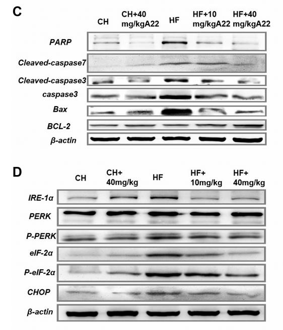
Application: WB Species: mouse Sample: liver
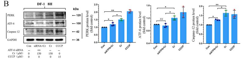
Application: WB Species: chicken Sample: DF-1 cells
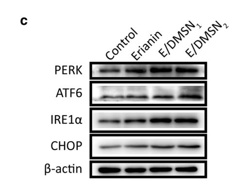
Application: WB Species: Human Sample: HaCaT cells
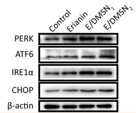
Application: WB Species: Human Sample: HaCaT cells
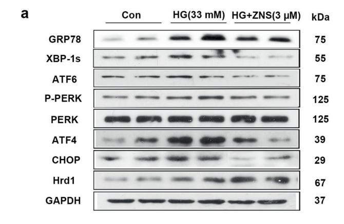
Application: WB Species: mice Sample: NRCMs
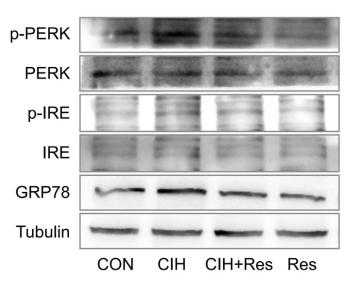
Application: WB Species: Rat Sample: heart tissue

Application: WB Species: human Sample: MDA-MB-231 cells

Application: WB Species: rat Sample: liver
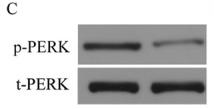
Application: WB Species: Human Sample:
限制条款
产品的规格、报价、验证数据请以官网为准,官网链接:www.affbiotech.com | www.affbiotech.cn(简体中文)| www.affbiotech.jp(日本語)产品的数据信息为Affinity所有,未经授权不得收集Affinity官网数据或资料用于商业用途,对抄袭产品数据的行为我们将保留诉诸法律的权利。
产品相关数据会因产品批次、产品检测情况随时调整,如您已订购该产品,请以订购时随货说明书为准,否则请以官网内容为准,官网内容有改动时恕不另行通知。
Affinity保证所销售产品均经过严格质量检测。如您购买的商品在规定时间内出现问题需要售后时,请您在Affinity官方渠道提交售后申请。产品仅供科学研究使用。不用于诊断和治疗。
产品未经授权不得转售。
Affinity Biosciences将不会对在使用我们的产品时可能发生的专利侵权或其他侵权行为负责。Affinity Biosciences, Affinity Biosciences标志和所有其他商标所有权归Affinity Biosciences LTD.