DRP1 Antibody - #DF7037

产品描述
*The optimal dilutions should be determined by the end user.
*Tips:
WB: 适用于变性蛋白样本的免疫印迹检测. IHC: 适用于组织样本的石蜡(IHC-p)或冰冻(IHC-f)切片样本的免疫组化/荧光检测. IF/ICC: 适用于细胞样本的荧光检测. ELISA(peptide): 适用于抗原肽的ELISA检测.
引用格式: Affinity Biosciences Cat# DF7037, RRID:AB_2838993.
展开/折叠
DLP1; dnm1l; DNM1L_HUMAN; Dnm1p/Vps1p-like protein; dnml1; DRP1; DVLP; Dymple; Dynamin 1 like; Dynamin family member proline-rich carboxyl-terminal domain less; Dynamin like protein; Dynamin related protein 1; Dynamin-1-like protein; Dynamin-like protein 4; Dynamin-like protein; Dynamin-like protein IV; Dynamin-related protein 1; DYNIV 11; EMPF; EMPF1; FLJ41912; HdynIV; VPS1;
抗原和靶标
Ubiquitously expressed with highest levels found in skeletal muscles, heart, kidney and brain. Isoform 1 is brain-specific. Isoform 2 and isoform 3 are predominantly expressed in testis and skeletal muscles respectively. Isoform 4 is weakly expressed in brain, heart and kidney. Isoform 5 is dominantly expressed in liver, heart and kidney. Isoform 6 is expressed in neurons.
- O00429 DNM1L_HUMAN:
- Protein BLAST With
- NCBI/
- ExPASy/
- Uniprot
MEALIPVINKLQDVFNTVGADIIQLPQIVVVGTQSSGKSSVLESLVGRDLLPRGTGIVTRRPLILQLVHVSQEDKRKTTGEENGVEAEEWGKFLHTKNKLYTDFDEIRQEIENETERISGNNKGVSPEPIHLKIFSPNVVNLTLVDLPGMTKVPVGDQPKDIELQIRELILRFISNPNSIILAVTAANTDMATSEALKISREVDPDGRRTLAVITKLDLMDAGTDAMDVLMGRVIPVKLGIIGVVNRSQLDINNKKSVTDSIRDEYAFLQKKYPSLANRNGTKYLARTLNRLLMHHIRDCLPELKTRINVLAAQYQSLLNSYGEPVDDKSATLLQLITKFATEYCNTIEGTAKYIETSELCGGARICYIFHETFGRTLESVDPLGGLNTIDILTAIRNATGPRPALFVPEVSFELLVKRQIKRLEEPSLRCVELVHEEMQRIIQHCSNYSTQELLRFPKLHDAIVEVVTCLLRKRLPVTNEMVHNLVAIELAYINTKHPDFADACGLMNNNIEEQRRNRLARELPSAVSRDKSSKVPSALAPASQEPSPAASAEADGKLIQDSRRETKNVASGGGGVGDGVQEPTTGNWRGMLKTSKAEELLAEEKSKPIPIMPASPQKGHAVNLLDVPVPVARKLSAREQRDCEVIERLIKSYFLIVRKNIQDSVPKAVMHFLVNHVKDTLQSELVGQLYKSSLLDDLLTESEDMAQRRKEAADMLKALQGASQIIAEIRETHLW
种属预测
score>80的预测可信度较高,可尝试用于WB检测。*预测模型主要基于免疫原序列比对,结果仅作参考,不作为质保凭据。
High(score>80) Medium(80>score>50) Low(score<50) No confidence
翻译修饰 - O00429 作为底物
| Site | PTM Type | Enzyme | Source |
|---|---|---|---|
| M1 | Acetylation | Uniprot | |
| S40 | Phosphorylation | P49841 (GSK3B) | Uniprot |
| S44 | Phosphorylation | P49841 (GSK3B) | Uniprot |
| T78 | Phosphorylation | Uniprot | |
| T79 | Phosphorylation | Uniprot | |
| K92 | Ubiquitination | Uniprot | |
| K97 | Ubiquitination | Uniprot | |
| K99 | Ubiquitination | Uniprot | |
| T102 | Phosphorylation | Uniprot | |
| R108 | Methylation | Uniprot | |
| K123 | Ubiquitination | Uniprot | |
| S126 | Phosphorylation | Uniprot | |
| K133 | Ubiquitination | Uniprot | |
| S136 | Phosphorylation | Uniprot | |
| K160 | Ubiquitination | Uniprot | |
| S179 | Phosphorylation | Uniprot | |
| T193 | Phosphorylation | Uniprot | |
| S200 | Phosphorylation | Uniprot | |
| K238 | Ubiquitination | Uniprot | |
| K255 | Ubiquitination | Uniprot | |
| K256 | Ubiquitination | Uniprot | |
| Y266 | Phosphorylation | Uniprot | |
| K271 | Ubiquitination | Uniprot | |
| K272 | Ubiquitination | Uniprot | |
| K283 | Acetylation | Uniprot | |
| S330 | Phosphorylation | Uniprot | |
| C361 | S-Nitrosylation | Uniprot | |
| C367 | S-Nitrosylation | Uniprot | |
| Y368 | Phosphorylation | Uniprot | |
| T394 | Phosphorylation | Uniprot | |
| T400 | Phosphorylation | Uniprot | |
| S412 | Phosphorylation | Uniprot | |
| Y449 | Phosphorylation | Uniprot | |
| S529 | Phosphorylation | Uniprot | |
| K532 | Sumoylation | Uniprot | |
| K535 | Sumoylation | Uniprot | |
| K535 | Ubiquitination | Uniprot | |
| S544 | Phosphorylation | Uniprot | |
| S548 | Phosphorylation | Uniprot | |
| S552 | Phosphorylation | Uniprot | |
| K558 | Sumoylation | Uniprot | |
| K568 | Sumoylation | Uniprot | |
| K568 | Ubiquitination | Uniprot | |
| S572 | Phosphorylation | Uniprot | |
| T586 | Phosphorylation | Uniprot | |
| K594 | Sumoylation | Uniprot | |
| T595 | Phosphorylation | Uniprot | |
| K597 | Sumoylation | Uniprot | |
| K597 | Ubiquitination | Uniprot | |
| K606 | Sumoylation | Uniprot | |
| S607 | Phosphorylation | Uniprot | |
| K608 | Sumoylation | Uniprot | |
| S616 | Phosphorylation | P06493 (CDK1) , P28482 (MAPK1) , P27361 (MAPK3) , Q05655 (PRKCD) , Q00535 (CDK5) , P24941 (CDK2) | Uniprot |
| S637 | Phosphorylation | P17612 (PRKACA) , Q13464 (ROCK1) , Q13131 (PRKAA1) | Uniprot |
| C644 | S-Nitrosylation | Uniprot | |
| S693 | Phosphorylation | P49841 (GSK3B) | Uniprot |
| T701 | Phosphorylation | Uniprot | |
| S724 | Phosphorylation | Uniprot |
研究背景
Functions in mitochondrial and peroxisomal division. Mediates membrane fission through oligomerization into membrane-associated tubular structures that wrap around the scission site to constrict and sever the mitochondrial membrane through a GTP hydrolysis-dependent mechanism. The specific recruitment at scission sites is mediated by membrane receptors like MFF, MIEF1 and MIEF2 for mitochondrial membranes. While the recruitment by the membrane receptors is GTP-dependent, the following hydrolysis of GTP induces the dissociation from the receptors and allows DNM1L filaments to curl into closed rings that are probably sufficient to sever a double membrane. Through its function in mitochondrial division, ensures the survival of at least some types of postmitotic neurons, including Purkinje cells, by suppressing oxidative damage. Required for normal brain development, including that of cerebellum. Facilitates developmentally regulated apoptosis during neural tube formation. Required for a normal rate of cytochrome c release and caspase activation during apoptosis; this requirement may depend upon the cell type and the physiological apoptotic cues. Plays an important role in mitochondrial fission during mitosis. Required for formation of endocytic vesicles. Proposed to regulate synaptic vesicle membrane dynamics through association with BCL2L1 isoform Bcl-X(L) which stimulates its GTPase activity in synaptic vesicles; the function may require its recruitment by MFF to clathrin-containing vesicles. Required for programmed necrosis execution. Rhythmic control of its activity following phosphorylation at Ser-637 is essential for the circadian control of mitochondrial ATP production.
Inhibits peroxisomal division when overexpressed.
Inhibits peroxisomal division when overexpressed.
Phosphorylation/dephosphorylation events on two sites near the GED domain regulate mitochondrial fission. Phosphorylation on Ser-637 inhibits the GTPase activity, leading to a defect in mitochondrial fission promoting mitochondrial elongation. Dephosphorylated on this site by PPP3CA which promotes mitochondrial fission. Phosphorylation on Ser-616 activates the GTPase activity and promotes mitochondrial fission. Phosphorylated in a circadian manner at Ser-637.
Sumoylated on various lysine residues within the B domain, probably by MUL1. Sumoylation positively regulates mitochondrial fission. Desumoylated by SENP5 during G2/M transition of mitosis. Appears to be linked to its catalytic activity.
S-nitrosylation increases DNM1L dimerization, mitochondrial fission and causes neuronal damage.
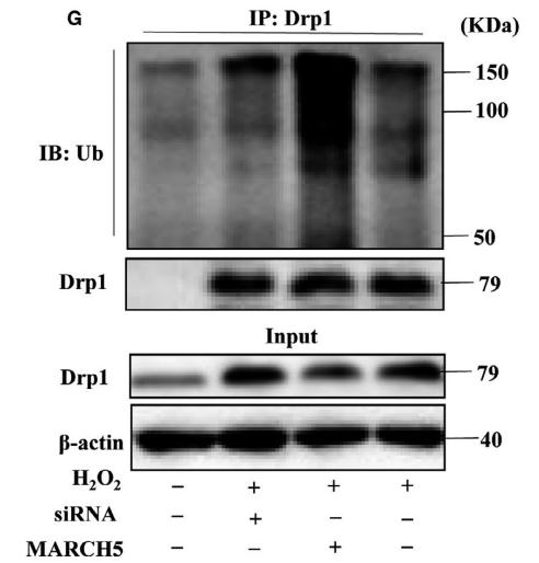
Ubiquitination by MARCHF5 affects mitochondrial morphology.
O-GlcNAcylation augments the level of the GTP-bound active form of DRP1 and induces translocation from the cytoplasm to mitochondria in cardiomyocytes. It also decreases phosphorylation at Ser-637 (By similarity).
Cytoplasm>Cytosol. Golgi apparatus. Endomembrane system>Peripheral membrane protein. Mitochondrion outer membrane>Peripheral membrane protein. Peroxisome. Membrane>Clathrin-coated pit. Cytoplasmic vesicle>Secretory vesicle>Synaptic vesicle membrane.
Note: Mainly cytosolic. Translocated to the mitochondrial membrane through O-GlcNAcylation and interaction with FIS1. Recruited to the mitochondrial outer membrane by interaction with MIEF1. Colocalized with MARCHF5 at mitochondrial membrane. Localizes to mitochondria at sites of division. Localizes to mitochondria following necrosis induction. Associated with peroxisomal membranes, partly recruited there by PEX11B. May also be associated with endoplasmic reticulum tubules and cytoplasmic vesicles and found to be perinuclear. In some cell types, localizes to the Golgi complex. Binds to phospholipid membranes.
Ubiquitously expressed with highest levels found in skeletal muscles, heart, kidney and brain. Isoform 1 is brain-specific. Isoform 2 and isoform 3 are predominantly expressed in testis and skeletal muscles respectively. Isoform 4 is weakly expressed in brain, heart and kidney. Isoform 5 is dominantly expressed in liver, heart and kidney. Isoform 6 is expressed in neurons.
Homotetramer; dimerizes through the N-terminal GTP-middle region of one molecule binding to the GED domain of another DNM1L molecule. Oligomerizes in a GTP-dependent manner to form membrane-associated tubules with a spiral pattern. Interacts with GSK3B and MARCHF5. Interacts (via the GTPase and B domains) with UBE2I; the interaction promotes sumoylation of DNM1L, mainly in its B domain. Interacts with PPP3CA; the interaction dephosphorylates DNM1L and regulates its transition to mitochondria. Interacts with BCL2L1 isoform BCL-X(L) and CLTA; DNM1L and BCL2L1 isoform BCL-X(L) may form a complex in synaptic vesicles that also contains clathrin and MFF. Interacts with FIS1. Interacts with MIEF2 and MIEF1; GTP-dependent this regulates GTP hydrolysis and DNM1L oligomerization. Interacts with PGAM5; this interaction leads to dephosphorylation at Ser-656 and activation of GTPase activity and eventually to mitochondria fragmentation.
The GED domain folds back to interact, in cis, with the GTP-binding domain and middle domain, and interacts, in trans, with the GED domains of other DNM1L molecules, and is thus critical for activating GTPase activity and for DNM1L dimerization.
Belongs to the TRAFAC class dynamin-like GTPase superfamily. Dynamin/Fzo/YdjA family.
研究领域
· Cellular Processes > Cell growth and death > Necroptosis. (View pathway)
· Environmental Information Processing > Signal transduction > TNF signaling pathway. (View pathway)
· Organismal Systems > Immune system > NOD-like receptor signaling pathway. (View pathway)
文献引用
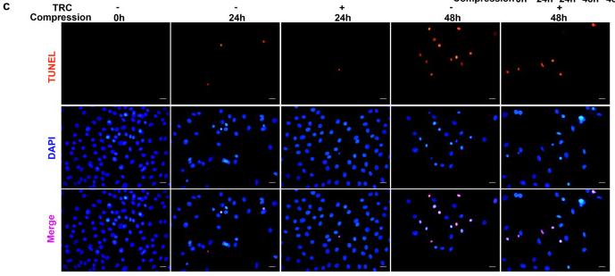
Application: IF/ICC Species: Rat Sample: NP cells
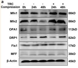
Application: WB Species: Rat Sample: NP cells
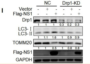
Application: WB Species: Human Sample:
Application: WB Species: Human Sample:
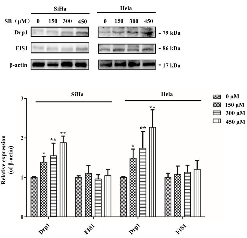
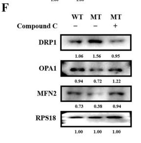
Application: WB Species: human Sample: cervical cancer cells
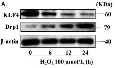
Application: WB Species: mouse Sample: H9C2 cells
Application: WB Species: mouse Sample: H9C2 cells
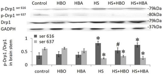
Application: WB Species: rat Sample: brain stem
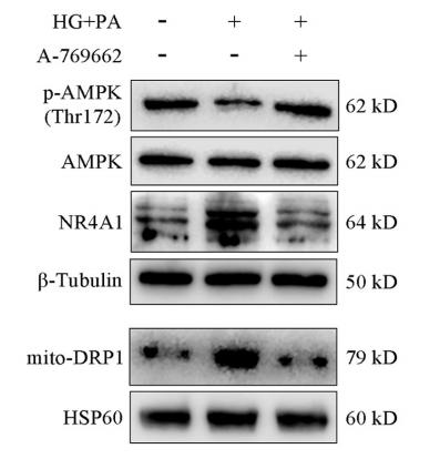
Application: WB Species: rat Sample: H9c2 cells
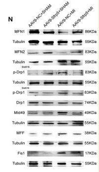
Application: WB Species: Mouse Sample: Heart tissue
限制条款
产品的规格、报价、验证数据请以官网为准,官网链接:www.affbiotech.com | www.affbiotech.cn(简体中文)| www.affbiotech.jp(日本語)产品的数据信息为Affinity所有,未经授权不得收集Affinity官网数据或资料用于商业用途,对抄袭产品数据的行为我们将保留诉诸法律的权利。
产品相关数据会因产品批次、产品检测情况随时调整,如您已订购该产品,请以订购时随货说明书为准,否则请以官网内容为准,官网内容有改动时恕不另行通知。
Affinity保证所销售产品均经过严格质量检测。如您购买的商品在规定时间内出现问题需要售后时,请您在Affinity官方渠道提交售后申请。产品仅供科学研究使用。不用于诊断和治疗。
产品未经授权不得转售。
Affinity Biosciences将不会对在使用我们的产品时可能发生的专利侵权或其他侵权行为负责。Affinity Biosciences, Affinity Biosciences标志和所有其他商标所有权归Affinity Biosciences LTD.