
产品描述
*The optimal dilutions should be determined by the end user.
*Tips:
WB: 适用于变性蛋白样本的免疫印迹检测. IHC: 适用于组织样本的石蜡(IHC-p)或冰冻(IHC-f)切片样本的免疫组化/荧光检测. IF/ICC: 适用于细胞样本的荧光检测. ELISA(peptide): 适用于抗原肽的ELISA检测.
引用格式: Affinity Biosciences Cat# AF0146, RRID:AB_2833328.
展开/折叠
CYC; CYC_HUMAN; CYCS; Cytochrome c; Cytochrome c somatic; HCS; THC4;
抗原和靶标
- P99999 CYC_HUMAN:
- Protein BLAST With
- NCBI/
- ExPASy/
- Uniprot
MGDVEKGKKIFIMKCSQCHTVEKGGKHKTGPNLHGLFGRKTGQAPGYSYTAANKNKGIIWGEDTLMEYLENPKKYIPGTKMIFVGIKKKEERADLIAYLKKATNE
种属预测
score>80的预测可信度较高,可尝试用于WB检测。*预测模型主要基于免疫原序列比对,结果仅作参考,不作为质保凭据。
High(score>80) Medium(80>score>50) Low(score<50) No confidence
翻译修饰 - P99999 作为底物
| Site | PTM Type | Enzyme | Source |
|---|---|---|---|
| G2 | Acetylation | Uniprot | |
| K9 | Acetylation | Uniprot | |
| K28 | Sumoylation | Uniprot | |
| K28 | Ubiquitination | Uniprot | |
| T29 | Phosphorylation | Uniprot | |
| K40 | Ubiquitination | Uniprot | |
| T41 | Phosphorylation | Uniprot | |
| Y47 | Phosphorylation | Uniprot | |
| S48 | Phosphorylation | Uniprot | |
| Y49 | Phosphorylation | Uniprot | |
| T50 | Phosphorylation | Uniprot | |
| K54 | Ubiquitination | Uniprot | |
| Y68 | Phosphorylation | Uniprot | |
| K73 | Acetylation | Uniprot | |
| K73 | Ubiquitination | Uniprot | |
| K74 | Acetylation | Uniprot | |
| K74 | Ubiquitination | Uniprot | |
| Y75 | Phosphorylation | Uniprot | |
| T79 | Phosphorylation | Uniprot | |
| K80 | Ubiquitination | Uniprot | |
| K87 | Acetylation | Uniprot | |
| K87 | Ubiquitination | Uniprot | |
| K88 | Acetylation | Uniprot | |
| K89 | Acetylation | Uniprot | |
| Y98 | Phosphorylation | Uniprot | |
| K100 | Acetylation | Uniprot | |
| K100 | Methylation | Uniprot | |
| K100 | Ubiquitination | Uniprot | |
| K101 | Methylation | Uniprot |
研究背景
Electron carrier protein. The oxidized form of the cytochrome c heme group can accept an electron from the heme group of the cytochrome c1 subunit of cytochrome reductase. Cytochrome c then transfers this electron to the cytochrome oxidase complex, the final protein carrier in the mitochondrial electron-transport chain.
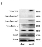
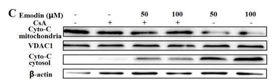
Plays a role in apoptosis. Suppression of the anti-apoptotic members or activation of the pro-apoptotic members of the Bcl-2 family leads to altered mitochondrial membrane permeability resulting in release of cytochrome c into the cytosol. Binding of cytochrome c to Apaf-1 triggers the activation of caspase-9, which then accelerates apoptosis by activating other caspases.
Binds 1 heme group per subunit.
Phosphorylation at Tyr-49 and Tyr-98 both reduce by half the turnover in the reaction with cytochrome c oxidase, down-regulating mitochondrial respiration.
Mitochondrion intermembrane space.
Note: Loosely associated with the inner membrane.
Belongs to the cytochrome c family.
研究领域
· Cellular Processes > Cell growth and death > p53 signaling pathway. (View pathway)
· Cellular Processes > Cell growth and death > Apoptosis. (View pathway)
· Cellular Processes > Cell growth and death > Apoptosis - multiple species. (View pathway)
· Human Diseases > Drug resistance: Antineoplastic > Platinum drug resistance.
· Human Diseases > Endocrine and metabolic diseases > Non-alcoholic fatty liver disease (NAFLD).
· Human Diseases > Neurodegenerative diseases > Alzheimer's disease.
· Human Diseases > Neurodegenerative diseases > Parkinson's disease.
· Human Diseases > Neurodegenerative diseases > Amyotrophic lateral sclerosis (ALS).
· Human Diseases > Neurodegenerative diseases > Huntington's disease.
· Human Diseases > Infectious diseases: Bacterial > Legionellosis.
· Human Diseases > Infectious diseases: Parasitic > Toxoplasmosis.
· Human Diseases > Infectious diseases: Bacterial > Tuberculosis.
· Human Diseases > Infectious diseases: Viral > Hepatitis B.
· Human Diseases > Infectious diseases: Viral > Influenza A.
· Human Diseases > Infectious diseases: Viral > Herpes simplex infection.
· Human Diseases > Cancers: Overview > Pathways in cancer. (View pathway)
· Human Diseases > Cancers: Specific types > Colorectal cancer. (View pathway)
· Human Diseases > Cancers: Specific types > Small cell lung cancer. (View pathway)
· Human Diseases > Cardiovascular diseases > Viral myocarditis.
· Metabolism > Energy metabolism > Sulfur metabolism.
· Metabolism > Global and overview maps > Metabolic pathways.
文献引用
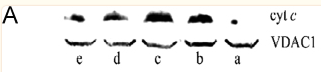
Application: WB Species: Human Sample:
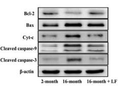
Application: WB Species: Mouse Sample: B cells
Application: WB Species: human Sample:
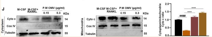
Application: WB Species: mouse Sample: Osteoclasts
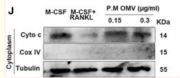
Application: WB Species: Mouse Sample: osteoclasts
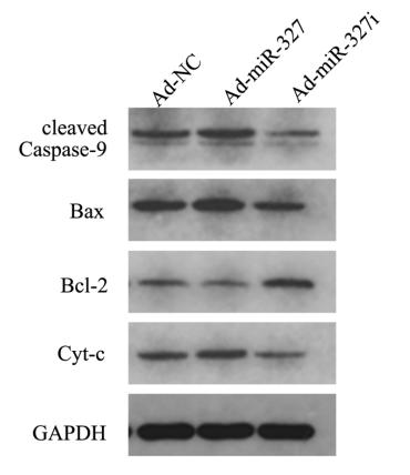
Application: WB Species: Mice Sample:
Application: WB Species: rat Sample: heart

Application: WB Species: Human Sample: MGC-803 cells
限制条款
产品的规格、报价、验证数据请以官网为准,官网链接:www.affbiotech.com | www.affbiotech.cn(简体中文)| www.affbiotech.jp(日本語)产品的数据信息为Affinity所有,未经授权不得收集Affinity官网数据或资料用于商业用途,对抄袭产品数据的行为我们将保留诉诸法律的权利。
产品相关数据会因产品批次、产品检测情况随时调整,如您已订购该产品,请以订购时随货说明书为准,否则请以官网内容为准,官网内容有改动时恕不另行通知。
Affinity保证所销售产品均经过严格质量检测。如您购买的商品在规定时间内出现问题需要售后时,请您在Affinity官方渠道提交售后申请。产品仅供科学研究使用。不用于诊断和治疗。
产品未经授权不得转售。
Affinity Biosciences将不会对在使用我们的产品时可能发生的专利侵权或其他侵权行为负责。Affinity Biosciences, Affinity Biosciences标志和所有其他商标所有权归Affinity Biosciences LTD.