
产品描述
*The optimal dilutions should be determined by the end user.
*Tips:
WB: 适用于变性蛋白样本的免疫印迹检测. IHC: 适用于组织样本的石蜡(IHC-p)或冰冻(IHC-f)切片样本的免疫组化/荧光检测. IF/ICC: 适用于细胞样本的荧光检测. ELISA(peptide): 适用于抗原肽的ELISA检测.
引用格式: Affinity Biosciences Cat# DF2989, RRID:AB_2840968.
展开/折叠
Beta catenin; Beta-catenin; Cadherin associated protein; Catenin (cadherin associated protein), beta 1, 88kDa; Catenin beta 1; Catenin beta-1; CATNB; CHBCAT; CTNB1_HUMAN; CTNNB; CTNNB1; DKFZp686D02253; FLJ25606; FLJ37923; OTTHUMP00000162082; OTTHUMP00000165222; OTTHUMP00000165223; OTTHUMP00000209288; OTTHUMP00000209289;
抗原和靶标
Expressed in several hair follicle cell types: basal and peripheral matrix cells, and cells of the outer and inner root sheaths. Expressed in colon. Present in cortical neurons (at protein level). Expressed in breast cancer tissues (at protein level) (PubMed:29367600).
- P35222 CTNB1_HUMAN:
- Protein BLAST With
- NCBI/
- ExPASy/
- Uniprot
MATQADLMELDMAMEPDRKAAVSHWQQQSYLDSGIHSGATTTAPSLSGKGNPEEEDVDTSQVLYEWEQGFSQSFTQEQVADIDGQYAMTRAQRVRAAMFPETLDEGMQIPSTQFDAAHPTNVQRLAEPSQMLKHAVVNLINYQDDAELATRAIPELTKLLNDEDQVVVNKAAVMVHQLSKKEASRHAIMRSPQMVSAIVRTMQNTNDVETARCTAGTLHNLSHHREGLLAIFKSGGIPALVKMLGSPVDSVLFYAITTLHNLLLHQEGAKMAVRLAGGLQKMVALLNKTNVKFLAITTDCLQILAYGNQESKLIILASGGPQALVNIMRTYTYEKLLWTTSRVLKVLSVCSSNKPAIVEAGGMQALGLHLTDPSQRLVQNCLWTLRNLSDAATKQEGMEGLLGTLVQLLGSDDINVVTCAAGILSNLTCNNYKNKMMVCQVGGIEALVRTVLRAGDREDITEPAICALRHLTSRHQEAEMAQNAVRLHYGLPVVVKLLHPPSHWPLIKATVGLIRNLALCPANHAPLREQGAIPRLVQLLVRAHQDTQRRTSMGGTQQQFVEGVRMEEIVEGCTGALHILARDVHNRIVIRGLNTIPLFVQLLYSPIENIQRVAAGVLCELAQDKEAAEAIEAEGATAPLTELLHSRNEGVATYAAAVLFRMSEDKPQDYKKRLSVELTSSLFRTEPMAWNETADLGLDIGAQGEPLGYRQDDPSYRSFHSGGYGQDALGMDPMMEHEMGGHHPGADYPVDGLPDLGHAQDLMDGLPPGDSNQLAWFDTDL
翻译修饰 - P35222 作为底物
| Site | PTM Type | Enzyme | Source |
|---|---|---|---|
| A2 | Acetylation | Uniprot | |
| K19 | Acetylation | Uniprot | |
| K19 | Ubiquitination | Uniprot | |
| S23 | O-Glycosylation | Uniprot | |
| S23 | Phosphorylation | Uniprot | |
| S29 | Phosphorylation | P68400 (CSNK2A1) , P67870 (CSNK2B) , P19784 (CSNK2A2) | Uniprot |
| Y30 | Phosphorylation | Uniprot | |
| S33 | Phosphorylation | O15111 (CHUK) , P49841 (GSK3B) , P45983 (MAPK8) , Q9H2X6 (HIPK2) , P49840 (GSK3A) | Uniprot |
| S37 | Phosphorylation | P49840 (GSK3A) , P45983 (MAPK8) , P49841 (GSK3B) , O15111 (CHUK) , Q9H2X6 (HIPK2) | Uniprot |
| T41 | Phosphorylation | P49840 (GSK3A) , P45983 (MAPK8) , O15111 (CHUK) , P49841 (GSK3B) | Uniprot |
| S45 | Phosphorylation | O15111 (CHUK) , P48730 (CSNK1D) , P17612 (PRKACA) , P48729 (CSNK1A1) , P49674 (CSNK1E) , Q8N752 (CSNK1A1L) , Q00534 (CDK6) , Q05513 (PRKCZ) | Uniprot |
| S47 | Phosphorylation | Uniprot | |
| K49 | Acetylation | Uniprot | |
| K49 | Methylation | Uniprot | |
| K49 | Ubiquitination | Uniprot | |
| Y64 | Phosphorylation | Q13882 (PTK6) | Uniprot |
| S73 | Phosphorylation | Uniprot | |
| Y86 | Phosphorylation | P00519 (ABL1) , P12931 (SRC) | Uniprot |
| T102 | Phosphorylation | P68400 (CSNK2A1) , P19784 (CSNK2A2) , P67870 (CSNK2B) | Uniprot |
| T112 | Phosphorylation | Q15139 (PRKD1) , P67870 (CSNK2B) , P19784 (CSNK2A2) , P68400 (CSNK2A1) | Uniprot |
| T120 | Phosphorylation | Q15139 (PRKD1) | Uniprot |
| K133 | Ubiquitination | Uniprot | |
| Y142 | Phosphorylation | P04629 (NTRK1) , Q13882 (PTK6) , P21802 (FGFR2) , P22607 (FGFR3) , P06241 (FYN) , P16591 (FER) , P00533 (EGFR) | Uniprot |
| K158 | Ubiquitination | Uniprot | |
| K170 | Ubiquitination | Uniprot | |
| S179 | Phosphorylation | Uniprot | |
| K180 | Ubiquitination | Uniprot | |
| S191 | Phosphorylation | P45984 (MAPK9) , Q00535 (CDK5) | Uniprot |
| S196 | Phosphorylation | Uniprot | |
| S222 | Phosphorylation | Uniprot | |
| K233 | Ubiquitination | Uniprot | |
| S246 | Phosphorylation | Q00535 (CDK5) | Uniprot |
| K288 | Ubiquitination | Uniprot | |
| Y331 | Phosphorylation | Q13882 (PTK6) | Uniprot |
| T332 | Phosphorylation | Q9UQM7 (CAMK2A) , Q13554 (CAMK2B) | Uniprot |
| Y333 | Phosphorylation | P12931 (SRC) , Q13882 (PTK6) | Uniprot |
| K335 | Ubiquitination | Uniprot | |
| K345 | Acetylation | Uniprot | |
| K345 | Ubiquitination | Uniprot | |
| K354 | Acetylation | Uniprot | |
| K354 | Ubiquitination | Uniprot | |
| T393 | Phosphorylation | P68400 (CSNK2A1) | Uniprot |
| K394 | Ubiquitination | Uniprot | |
| K435 | Acetylation | Uniprot | |
| K435 | Ubiquitination | Uniprot | |
| T461 | Phosphorylation | Uniprot | |
| T472 | Phosphorylation | Q13554 (CAMK2B) , Q9UQM7 (CAMK2A) | Uniprot |
| S473 | Phosphorylation | Uniprot | |
| Y489 | Phosphorylation | P00519 (ABL1) | Uniprot |
| K496 | Ubiquitination | Uniprot | |
| K508 | Ubiquitination | Uniprot | |
| T510 | Phosphorylation | Uniprot | |
| C520 | S-Nitrosylation | Uniprot | |
| T547 | Phosphorylation | Uniprot | |
| T551 | Phosphorylation | Uniprot | |
| S552 | Phosphorylation | Q9UQM7 (CAMK2A) , P17612 (PRKACA) , P54646 (PRKAA2) , P31751 (AKT2) , Q13554 (CAMK2B) , P31749 (AKT1) | Uniprot |
| T556 | Phosphorylation | Uniprot | |
| T574 | Phosphorylation | Uniprot | |
| S605 | Phosphorylation | P53778 (MAPK12) , P45984 (MAPK9) | Uniprot |
| K625 | Ubiquitination | Uniprot | |
| S646 | Phosphorylation | Uniprot | |
| T653 | Phosphorylation | Uniprot | |
| Y654 | Phosphorylation | P07949 (RET) , P04626 (ERBB2) , P00533 (EGFR) , P00519 (ABL1) , P36888 (FLT3) , P12931 (SRC) | Uniprot |
| S663 | Phosphorylation | Uniprot | |
| Y670 | Phosphorylation | Uniprot | |
| K671 | Ubiquitination | Uniprot | |
| S675 | Phosphorylation | O96013 (PAK4) , Q13153 (PAK1) , P17612 (PRKACA) | Uniprot |
| T679 | Phosphorylation | Uniprot | |
| S680 | Phosphorylation | Uniprot | |
| S715 | Phosphorylation | Q05655 (PRKCD) | Uniprot |
| Y716 | Phosphorylation | Uniprot | |
| S718 | Phosphorylation | P53350 (PLK1) | Uniprot |
| S721 | Phosphorylation | Uniprot | |
| Y724 | Phosphorylation | Uniprot | |
| Y748 | Phosphorylation | Uniprot |
研究背景
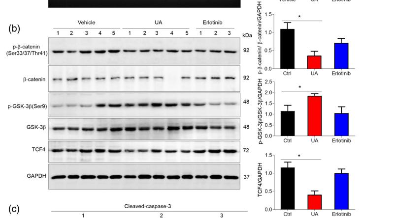
Key downstream component of the canonical Wnt signaling pathway. In the absence of Wnt, forms a complex with AXIN1, AXIN2, APC, CSNK1A1 and GSK3B that promotes phosphorylation on N-terminal Ser and Thr residues and ubiquitination of CTNNB1 via BTRC and its subsequent degradation by the proteasome. In the presence of Wnt ligand, CTNNB1 is not ubiquitinated and accumulates in the nucleus, where it acts as a coactivator for transcription factors of the TCF/LEF family, leading to activate Wnt responsive genes. Involved in the regulation of cell adhesion, as component of an E-cadherin:catenin adhesion complex. Acts as a negative regulator of centrosome cohesion. Involved in the CDK2/PTPN6/CTNNB1/CEACAM1 pathway of insulin internalization. Blocks anoikis of malignant kidney and intestinal epithelial cells and promotes their anchorage-independent growth by down-regulating DAPK2. Disrupts PML function and PML-NB formation by inhibiting RANBP2-mediated sumoylation of PML. Promotes neurogenesis by maintaining sympathetic neuroblasts within the cell cycle (By similarity).
Phosphorylation at Ser-552 by AMPK promotes stabilizion of the protein, enhancing TCF/LEF-mediated transcription (By similarity). Phosphorylation by GSK3B requires prior phosphorylation of Ser-45 by another kinase. Phosphorylation proceeds then from Thr-41 to Ser-37 and Ser-33. Phosphorylated by NEK2. EGF stimulates tyrosine phosphorylation. Phosphorylation on Tyr-654 decreases CDH1 binding and enhances TBP binding. Phosphorylated on Ser-33 and Ser-37 by HIPK2 and GSK3B, this phosphorylation triggers proteasomal degradation. Phosphorylation on Ser-191 and Ser-246 by CDK5. Phosphorylation by CDK2 regulates insulin internalization. Phosphorylation by PTK6 at Tyr-64, Tyr-142, Tyr-331 and/or Tyr-333 with the predominant site at Tyr-64 is not essential for inhibition of transcriptional activity.
Ubiquitinated by the SCF(BTRC) E3 ligase complex when phosphorylated by GSK3B, leading to its degradation. Ubiquitinated by a E3 ubiquitin ligase complex containing UBE2D1, SIAH1, CACYBP/SIP, SKP1, APC and TBL1X, leading to its subsequent proteasomal degradation (By similarity).
S-nitrosylation at Cys-619 within adherens junctions promotes VEGF-induced, NO-dependent endothelial cell permeability by disrupting interaction with E-cadherin, thus mediating disassembly adherens junctions.
O-glycosylation at Ser-23 decreases nuclear localization and transcriptional activity, and increases localization to the plasma membrane and interaction with E-cadherin CDH1.
Deacetylated at Lys-49 by SIRT1.
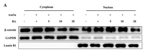
Cytoplasm. Nucleus. Cytoplasm>Cytoskeleton. Cell junction>Adherens junction. Cell junction. Cell membrane. Cytoplasm>Cytoskeleton>Microtubule organizing center>Centrosome. Cytoplasm>Cytoskeleton>Spindle pole. Cell junction>Synapse. Cytoplasm>Cytoskeleton>Cilium basal body.
Note: Colocalized with RAPGEF2 and TJP1 at cell-cell contacts (By similarity). Cytoplasmic when it is unstabilized (high level of phosphorylation) or bound to CDH1. Translocates to the nucleus when it is stabilized (low level of phosphorylation). Interaction with GLIS2 and MUC1 promotes nuclear translocation. Interaction with EMD inhibits nuclear localization. The majority of beta-catenin is localized to the cell membrane. In interphase, colocalizes with CROCC between CEP250 puncta at the proximal end of centrioles, and this localization is dependent on CROCC and CEP250. In mitosis, when NEK2 activity increases, it localizes to centrosomes at spindle poles independent of CROCC. Colocalizes with CDK5 in the cell-cell contacts and plasma membrane of undifferentiated and differentiated neuroblastoma cells. Interaction with FAM53B promotes translocation to the nucleus (PubMed:25183871).
Expressed in several hair follicle cell types: basal and peripheral matrix cells, and cells of the outer and inner root sheaths. Expressed in colon. Present in cortical neurons (at protein level). Expressed in breast cancer tissues (at protein level).
Two separate complex-associated pools are found in the cytoplasm. The majority is present as component of an E-cadherin:catenin adhesion complex composed of at least E-cadherin/CDH1 and beta-catenin/CTNNB1, and possibly alpha-catenin/CTNNA1; the complex is located to adherens junctions. The stable association of CTNNA1 is controversial as CTNNA1 was shown not to bind to F-actin when assembled in the complex. Alternatively, the CTNNA1-containing complex may be linked to F-actin by other proteins such as LIMA1. Another cytoplasmic pool is part of a large complex containing AXIN1, AXIN2, APC, CSNK1A1 and GSK3B that promotes phosphorylation on N-terminal Ser and Thr residues and ubiquitination of CTNNB1 via BTRC and its subsequent degradation by the proteasome. Wnt-dependent activation of DVL antagonizes the action of GSK3B. When GSK3B activity is inhibited the complex dissociates, CTNNB1 is dephosphorylated and is no longer targeted for destruction. The stabilized protein translocates to the nucleus, where it binds TCF/LEF-1 family members, TBP, BCL9, BCL9L and possibly also RUVBL1 and CHD8. Binds CTNNBIP and EP300. CTNNB1 forms a ternary complex with LEF1 and EP300 that is disrupted by CTNNBIP1 binding. Interacts with TAX1BP3 (via the PDZ domain); this interaction inhibits the transcriptional activity of CTNNB1. Interacts with AJAP1, BAIAP1, CARM1, CTNNA3, CXADR and PCDH11Y. Binds SLC9A3R1. Interacts with GLIS2 and MUC1. Interacts with SLC30A9. Interacts with XIRP1. Interacts directly with AXIN1; the interaction is regulated by CDK2 phosphorylation of AXIN1. Interacts with SCRIB. Interacts with RAPGEF2. Interacts with PTPRU (via the cytoplasmic juxtamembrane domain). Interacts with EMD. Interacts with TNIK and TCF7L2. Interacts with SESTD1 and TRPC4. Interacts with CAV1. Interacts with TRPV4. The TRPV4 and CTNNB1 complex can interact with CDH1. Interacts with VCL. Interacts with PTPRJ. Interacts with PKT7 and CDK2. Interacts with FAT1 (via the cytoplasmic domain). Interacts with NANOS1 and NDRG2. Interacts with isoform 1 of NEK2. Interacts with both isoform 1 and isoform 2 of CDK5. Interacts with PTK6. Interacts with SOX7; this interaction may lead to proteasomal degradation of active CTNNB1 and thus inhibition of Wnt/beta-catenin-stimulated transcription. Identified in a complex with HINT1 and MITF. Interacts with FHIT. The CTNNB1 and TCF7L2/TCF4 complex interacts with PML (isoform PML-4). Interacts with FERMT2. Identified in a complex with TCF7L2/TCF4 and FERMT2. Interacts with RORA. May interact with P-cadherin/CDH3. Interacts with RNF220. Interacts with CTNND2. Interacts (via the C-terminal region) with CBY1. The complex composed, at least, of APC, CTNNB1 and GSK3B interacts with JPT1; the interaction requires the inactive form of GSK3B (phosphorylated at 'Ser-9'). Interacts with DLG5 (By similarity). Interacts with FAM53B; promoting translocation to the nucleus. Interacts with TMEM170B. Interacts with AHI1. Interacts with GID8. Component of an cadherin:catenin adhesion complex composed of at least of CDH26, beta-catenin/CTNNB1, alpha-catenin/CTNNA1 and p120 catenin/CTNND1. Forms a complex comprising APPL1, RUVBL2, APPL2, HDAC1 and HDAC2. Interacts with IRF2BPL; mediates the ubiquitination and degradation of CTNNB1. Interacts with AMFR (By similarity). Interacts with LMBR1L. Interacts with SOX30; prevents interaction of CTNNB1 with TCF7L2/TCF4 and leads to inhibition of Wnt signaling.
(Microbial infection) Interacts with herpes virus 8 protein vPK; this interaction inhibits the Wnt signaling pathway.
Belongs to the beta-catenin family.
研究领域
· Cellular Processes > Cellular community - eukaryotes > Focal adhesion. (View pathway)
· Cellular Processes > Cellular community - eukaryotes > Adherens junction. (View pathway)
· Cellular Processes > Cellular community - eukaryotes > Signaling pathways regulating pluripotency of stem cells. (View pathway)
· Environmental Information Processing > Signal transduction > Rap1 signaling pathway. (View pathway)
· Environmental Information Processing > Signal transduction > Wnt signaling pathway. (View pathway)
· Environmental Information Processing > Signal transduction > Hippo signaling pathway. (View pathway)
· Human Diseases > Infectious diseases: Bacterial > Bacterial invasion of epithelial cells.
· Human Diseases > Infectious diseases: Bacterial > Pathogenic Escherichia coli infection.
· Human Diseases > Infectious diseases: Viral > Human papillomavirus infection.
· Human Diseases > Infectious diseases: Viral > HTLV-I infection.
· Human Diseases > Cancers: Overview > Pathways in cancer. (View pathway)
· Human Diseases > Cancers: Overview > Proteoglycans in cancer.
· Human Diseases > Cancers: Specific types > Colorectal cancer. (View pathway)
· Human Diseases > Cancers: Specific types > Endometrial cancer. (View pathway)
· Human Diseases > Cancers: Specific types > Prostate cancer. (View pathway)
· Human Diseases > Cancers: Specific types > Thyroid cancer. (View pathway)
· Human Diseases > Cancers: Specific types > Basal cell carcinoma. (View pathway)
· Human Diseases > Cancers: Specific types > Breast cancer. (View pathway)
· Human Diseases > Cancers: Specific types > Hepatocellular carcinoma. (View pathway)
· Human Diseases > Cancers: Specific types > Gastric cancer. (View pathway)
· Human Diseases > Cardiovascular diseases > Arrhythmogenic right ventricular cardiomyopathy (ARVC).
· Organismal Systems > Immune system > Leukocyte transendothelial migration. (View pathway)
· Organismal Systems > Endocrine system > Melanogenesis.
· Organismal Systems > Endocrine system > Thyroid hormone signaling pathway. (View pathway)
文献引用
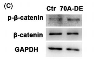
Application: WB Species: mice Sample: Oocytes
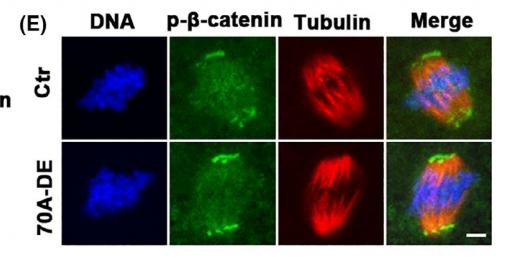
Application: IF/ICC Species: mice Sample: Oocytes
Application: WB Species: Mice Sample: Mlg and PPF cells

Application: IF/ICC Species: Mice Sample: Mlg and PPF cells
Application: WB Species: mouse Sample: tumour
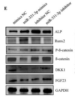
Application: WB Species: Mouse Sample: bone tissues
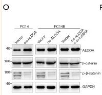
Application: WB Species: Human Sample: LAD cells
限制条款
产品的规格、报价、验证数据请以官网为准,官网链接:www.affbiotech.com | www.affbiotech.cn(简体中文)| www.affbiotech.jp(日本語)产品的数据信息为Affinity所有,未经授权不得收集Affinity官网数据或资料用于商业用途,对抄袭产品数据的行为我们将保留诉诸法律的权利。
产品相关数据会因产品批次、产品检测情况随时调整,如您已订购该产品,请以订购时随货说明书为准,否则请以官网内容为准,官网内容有改动时恕不另行通知。
Affinity保证所销售产品均经过严格质量检测。如您购买的商品在规定时间内出现问题需要售后时,请您在Affinity官方渠道提交售后申请。产品仅供科学研究使用。不用于诊断和治疗。
产品未经授权不得转售。
Affinity Biosciences将不会对在使用我们的产品时可能发生的专利侵权或其他侵权行为负责。Affinity Biosciences, Affinity Biosciences标志和所有其他商标所有权归Affinity Biosciences LTD.