BOK Antibody - #DF3829

产品描述
*The optimal dilutions should be determined by the end user.
*Tips:
WB: 适用于变性蛋白样本的免疫印迹检测. IHC: 适用于组织样本的石蜡(IHC-p)或冰冻(IHC-f)切片样本的免疫组化/荧光检测. IF/ICC: 适用于细胞样本的荧光检测. ELISA(peptide): 适用于抗原肽的ELISA检测.
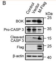
引用格式: Affinity Biosciences Cat# DF3829, RRID:AB_2836186.
展开/折叠
Bcl 2 related ovarian killer protein; Bcl-2-like protein 9; Bcl-2-related ovarian killer protein; BCL2 related ovarian killer; Bcl2-L-9; BCL2L9; BOK; BOK_HUMAN; BOKL; Hbok; MGC4631;
抗原和靶标
Expressed mainly in oocytes; weak expression in granulosa cells of the developing follicles. In adult human ovaries, expressed in granulosa cells at all follicular stages, but expression in primordial/primary follicles granulosa cell is stronger than in secondary and antral follicles.
- Q9UMX3 BOK_HUMAN:
- Protein BLAST With
- NCBI/
- ExPASy/
- Uniprot
MEVLRRSSVFAAEIMDAFDRSPTDKELVAQAKALGREYVHARLLRAGLSWSAPERAAPVPGRLAEVCAVLLRLGDELEMIRPSVYRNVARQLHISLQSEPVVTDAFLAVAGHIFSAGITWGKVVSLYAVAAGLAVDCVRQAQPAMVHALVDCLGEFVRKTLATWLRRRGGWTDVLKCVVSTDPGLRSHWLVAALCSFGRFLKAAFFVLLPER
种属预测
score>80的预测可信度较高,可尝试用于WB检测。*预测模型主要基于免疫原序列比对,结果仅作参考,不作为质保凭据。
High(score>80) Medium(80>score>50) Low(score<50) No confidence
翻译修饰 - Q9UMX3 作为底物
| Site | PTM Type | Enzyme | Source |
|---|---|---|---|
| S7 | Phosphorylation | Uniprot | |
| S8 | Phosphorylation | Uniprot | |
| K25 | Ubiquitination | Uniprot | |
| S196 | Phosphorylation | Uniprot |
研究背景
Apoptosis regulator that functions through different apoptotic signaling pathways. Plays a roles as pro-apoptotic protein that positively regulates intrinsic apoptotic process in a BAX- and BAK1-dependent manner or in a BAX- and BAK1-independent manner. In response to endoplasmic reticulum stress promotes mitochondrial apoptosis through downstream BAX/BAK1 activation and positive regulation of PERK-mediated unfolded protein response (By similarity). Activates apoptosis independently of heterodimerization with survival-promoting BCL2 and BCL2L1 through induction of mitochondrial outer membrane permeabilization, in a BAX- and BAK1-independent manner, in response to inhibition of ERAD-proteasome degradation system, resulting in cytochrome c release. In response to DNA damage, mediates intrinsic apoptotic process in a TP53-dependent manner. Plays a role in granulosa cell apoptosis by CASP3 activation. Plays a roles as anti-apoptotic protein during neuronal apoptotic process, by negatively regulating poly ADP-ribose polymerase-dependent cell death through regulation of neuronal calcium homeostasis and mitochondrial bioenergetics in response to NMDA excitation (By similarity). In addition to its role in apoptosis, may regulate trophoblast cell proliferation during the early stages of placental development, by acting on G1/S transition through regulation of CCNE1 expression. May also play a role as an inducer of autophagy by disrupting interaction between MCL1 and BECN1.
Pro-apoptotic molecule exerting its function through the mitochondrial pathway.
Ubiquitinated by AMFR/gp78 E3 ubiquitin ligase complex; mediates degradation by ubiquitin-proteasome pathway in a VCP/p97-dependent manner; prevents from pro-apoptotic activity; promotes degradation of newly synthesized proteins that are not ITPR1 associated.
Mitochondrion membrane>Single-pass membrane protein. Endoplasmic reticulum membrane>Single-pass membrane protein. Mitochondrion inner membrane. Cytoplasm. Nucleus. Mitochondrion. Endoplasmic reticulum. Mitochondrion outer membrane. Early endosome membrane. Recycling endosome membrane. Nucleus outer membrane. Golgi apparatus>cis-Golgi network membrane. Golgi apparatus>trans-Golgi network membrane. Membrane.
Note: Nuclear and cytoplasmic compartments in the early stages of apoptosis and during apoptosis it associates with mitochondria (PubMed:19942931). In healthy cells, associates loosely with the membrane in a hit-and-run mode. The insertion and accumulation on membranes is enhanced through the activity of death signals, resulting in the integration of the membrane-bound protein into the membrane (PubMed:15868100). The transmembrane domain controls subcellular localization; constitutes a tail-anchor. Localizes in early and late endosome upon blocking of apoptosis. Must localize to the mitochondria to induce mitochondrial outer membrane permeabilization and apoptosis (By similarity).
Membrane. Cytoplasm.
Expressed mainly in oocytes; weak expression in granulosa cells of the developing follicles. In adult human ovaries, expressed in granulosa cells at all follicular stages, but expression in primordial/primary follicles granulosa cell is stronger than in secondary and antral follicles.
Monomer; positively regulates apoptotic process. Homodimer (By similarity). Heterodimer (By similarity). Oligomer; promoted by apoptotic stimuli and BH3-only proteins; mediates constitutive activation. Interacts (via BH4 domain) with ITPR1; enhances BOK expression and stabilization; limits apoptosis and prevents ubiquitination and then degradation; protects ITPR1 from proteolysis by CASP3 during apoptosis. Interacts with ITPR2 AND ITPR3; binds most strongly to ITPR2, and barely to ITPR3; regulates their expression (By similarity). Interacts with XPO1; translocates to the cytoplasm. Interacts with BNIP3; promotes oligomerization.
BH4 domain mediates interaction with ITPR1.
Belongs to the Bcl-2 family.
研究领域
· Cellular Processes > Cell growth and death > Apoptosis - multiple species. (View pathway)
文献引用
Application: WB Species: Mouse Sample: lung tissue
限制条款
产品的规格、报价、验证数据请以官网为准,官网链接:www.affbiotech.com | www.affbiotech.cn(简体中文)| www.affbiotech.jp(日本語)产品的数据信息为Affinity所有,未经授权不得收集Affinity官网数据或资料用于商业用途,对抄袭产品数据的行为我们将保留诉诸法律的权利。
产品相关数据会因产品批次、产品检测情况随时调整,如您已订购该产品,请以订购时随货说明书为准,否则请以官网内容为准,官网内容有改动时恕不另行通知。
Affinity保证所销售产品均经过严格质量检测。如您购买的商品在规定时间内出现问题需要售后时,请您在Affinity官方渠道提交售后申请。产品仅供科学研究使用。不用于诊断和治疗。
产品未经授权不得转售。
Affinity Biosciences将不会对在使用我们的产品时可能发生的专利侵权或其他侵权行为负责。Affinity Biosciences, Affinity Biosciences标志和所有其他商标所有权归Affinity Biosciences LTD.