产品: CD8 抗体
货号: AF5126
描述: Rabbit polyclonal antibody to CD8
应用: WB IHC
反应: Human, Mouse, Rat
分子量: 25 kDa; 26kD(Calculated).
蛋白号: P01732
RRID: AB_2837612

浏览相似产品>>

   规格 价格 库存
 50ul RMB¥ 1250 现货
 100ul RMB¥ 2300 现货
 200ul RMB¥ 3000 现货

货期: 当天发货

联系销售

产品描述

来源:
Rabbit
应用:
WB 1:500-1:2000, IHC 1:50-1:200
*The optimal dilutions should be determined by the end user.
*Tips:

WB: 适用于变性蛋白样本的免疫印迹检测. IHC: 适用于组织样本的石蜡(IHC-p)或冰冻(IHC-f)切片样本的免疫组化/荧光检测. IF/ICC: 适用于细胞样本的荧光检测. ELISA(peptide): 适用于抗原肽的ELISA检测.

反应:
Human,Mouse,Rat
克隆:
Polyclonal
特异性:
CD8 Antibody detects endogenous levels of total CD8.
RRID:
AB_2837612
引用格式: Affinity Biosciences Cat# AF5126, RRID:AB_2837612.
偶联:
Unconjugated.
纯化:
The antiserum was purified by peptide affinity chromatography using SulfoLink™ Coupling Resin (Thermo Fisher Scientific).
保存:
Rabbit IgG in phosphate buffered saline , pH 7.4, 150mM NaCl, 0.02% sodium azide and 50% glycerol. Store at -20 °C. Stable for 12 months from date of receipt.
别名:

展开/折叠

alpha polypeptide (p32); CD8; CD8 antigen alpha polypeptide; CD8 antigen alpha polypeptide (p32); CD8a; CD8A antigen; CD8A molecule; CD8A_HUMAN; Leu2; Leu2 T lymphocyte antigen; Ly3; LYT3; MAL; OKT8 T cell antigen; OTTHUMP00000160760; OTTHUMP00000160764; OTTHUMP00000203528; OTTHUMP00000203721; p32; T cell antigen Leu2; T cell co receptor; T-cell surface glycoprotein CD8 alpha chain; T-lymphocyte differentiation antigen T8/Leu-2; T8 T cell antigen; T8/Leu-2 T-lymphocyte differentiation antigen;

抗原和靶标

免疫原:
Uniprot:
基因/基因ID:
表达:
P01732 CD8A_HUMAN:

CD8 on thymus-derived T-cells usually consists of a disulfide-linked alpha/CD8A and a beta/CD8B chain. Less frequently, CD8 can be expressed as a CD8A homodimer. A subset of natural killer cells, memory T-cells, intraepithelial lymphocytes, monocytes and dendritic cells expresses CD8A homodimers. Expressed at the cell surface of plasmacytoid dendritic cells upon herpes simplex virus-1 stimulation.

描述:
Identifies cytotoxic/suppressor T-cells that interact with MHC class I bearing targets. CD8 is thought to play a role in the process of T-cell mediated killing. CD8 alpha chains binds to class I MHC molecules alpha-3 domains.
序列:
MALPVTALLLPLALLLHAARPSQFRVSPLDRTWNLGETVELKCQVLLSNPTSGCSWLFQPRGAAASPTFLLYLSQNKPKAAEGLDTQRFSGKRLGDTFVLTLSDFRRENEGYYFCSALSNSIMYFSHFVPVFLPAKPTTTPAPRPPTPAPTIASQPLSLRPEACRPAAGGAVHTRGLDFACDIYIWAPLAGTCGVLLLSLVITLYCNHRNRRRVCKCPRPVVKSGDKPSLSARYV

翻译修饰 - P01732 作为底物

Site PTM Type Enzyme
S224 Phosphorylation
S229 Phosphorylation
S231 Phosphorylation

研究背景

功能:

Integral membrane glycoprotein that plays an essential role in the immune response and serves multiple functions in responses against both external and internal offenses. In T-cells, functions primarily as a coreceptor for MHC class I molecule:peptide complex. The antigens presented by class I peptides are derived from cytosolic proteins while class II derived from extracellular proteins. Interacts simultaneously with the T-cell receptor (TCR) and the MHC class I proteins presented by antigen presenting cells (APCs). In turn, recruits the Src kinase LCK to the vicinity of the TCR-CD3 complex. LCK then initiates different intracellular signaling pathways by phosphorylating various substrates ultimately leading to lymphokine production, motility, adhesion and activation of cytotoxic T-lymphocytes (CTLs). This mechanism enables CTLs to recognize and eliminate infected cells and tumor cells. In NK-cells, the presence of CD8A homodimers at the cell surface provides a survival mechanism allowing conjugation and lysis of multiple target cells. CD8A homodimer molecules also promote the survival and differentiation of activated lymphocytes into memory CD8 T-cells.

翻译修饰:

Palmitoylated, but association with CD8B seems to be more important for the enrichment of CD8A in lipid rafts.

O-glycosylated.

Phosphorylated in cytotoxic T-lymphocytes (CTLs) following activation.

细胞定位:

Cell membrane>Single-pass type I membrane protein.
Note: CD8A localizes to lipid rafts only when associated with its partner CD8B.

Secreted.

Extracellular region or secreted Cytosol Plasma membrane Cytoskeleton Lysosome Endosome Peroxisome ER Golgi apparatus Nucleus Mitochondrion Manual annotation Automatic computational assertionSubcellular location
组织特异性:

CD8 on thymus-derived T-cells usually consists of a disulfide-linked alpha/CD8A and a beta/CD8B chain. Less frequently, CD8 can be expressed as a CD8A homodimer. A subset of natural killer cells, memory T-cells, intraepithelial lymphocytes, monocytes and dendritic cells expresses CD8A homodimers. Expressed at the cell surface of plasmacytoid dendritic cells upon herpes simplex virus-1 stimulation.

亚基结构:

Forms disulfide-linked heterodimers with CD8B at the cell surface. Forms also homodimers in several cell types including NK-cells or peripheral blood T-lymphocytes. Interacts with the MHC class I HLA-A/B2M dimer. One HLA-A molecule (mainly via nonpolymorphic alpha-3 domain) interacts with one CD8A homodimer (via CDR-like loop). Interacts with LCK in a zinc-dependent manner. Interacts with HLA-G; this interaction is direct and might down-regulate T cell receptor signaling.

研究领域

· Environmental Information Processing > Signaling molecules and interaction > Cell adhesion molecules (CAMs).   (View pathway)

· Human Diseases > Immune diseases > Primary immunodeficiency.

· Organismal Systems > Immune system > Antigen processing and presentation.   (View pathway)

· Organismal Systems > Immune system > Hematopoietic cell lineage.   (View pathway)

· Organismal Systems > Immune system > T cell receptor signaling pathway.   (View pathway)

文献引用

1). Alantolactone-loaded chitosan/hyaluronic acid nanoparticles suppress psoriasis by deactivating STAT3 pathway and restricting immune cell recruitment. Asian Journal of Pharmaceutical Sciences (PubMed: 35582636) [IF=10.2]

Application: IF/ICC    Species: mouse    Sample: skin

Fig. 8| CHALT attenuates splenomegaly and immune cell recruitment in IMQ-induced skin. On Day 7, (A) the spleens were collected and photographed, and (B) the weight ratio of spleen/body was calculated. Immunfluorescence staining of (C) CD 4 and (E) CD 8 in the skin, and the relative amount of (D) CD 4 and (F) CD 8 was quantified (the normal group as the control).

2). SIRPα antibody combined with oncolytic virus OH2 protects against tumours by activating innate immunity and reprogramming the tumour immune microenvironment. BMC Medicine (PubMed: 36310169) [IF=9.3]

Application: IHC    Species: Mouse    Sample:

Fig. 5M-IHC and IHC staining of the anti-SIRPα antibody treatment model. A Representative M-IHC results of the OH2 combined anti-SIRPα antibody group, OH2 combined isotype, OH2 group, and control group with larger tumour volume sat the initial treatment. CD8, green, CD16, sky blue, F4/80, purple, CD86, orange, and CD206, red. Scale bar, 100μm. B Quantitative results of NK cell (CD16) staining in the OH2 combined anti-SIRPα antibody group, OH2 combined isotype, OH2 group, and control group. n=3 samples/group. C Quantitative result of M2 macrophage (F4/80+CD206+) staining in the OH2 combined anti-SIRPα antibody group, OH2 combined isotype, OH2 group, and control group. n=3 samples/group. D Quantitative results of M1 macrophage (F4/80+CD86+) staining in the OH2 combined anti-SIRPα antibody group, OH2 combined isotype, OH2 group, and control group. n=3 samples/group. E Quantitative results of CD8 T cell staining in the OH2 combined anti-SIRPα antibody group, OH2 combined Isotype, OH2 group, and control group. n=3 samples/group. F Representative IHC staining for CD8, CD16, F4/80, CD86, and CD206 of the OH2 combined anti-SIRPα antibody group, OH2 combined isotype group, OH2 group, and control group with larger tumour volumes at the initial treatment. Original magnification, ×200. n=5 samples/group. G Quantitative results of CD8, CD16, F4/80, CD86 and CD206 staining in the OH2 combined anti-SIRPα antibody group, OH2 combined isotype, OH2 group and control group. n=5 samples/group. Statistical analysis was performed using ANOVA with multiple comparisons. ns, no significant differences, *, p<0.05, **, p<0.01, ***, p<0.001, ****, p<0.0001 (proportion represented the percentage of the cell to the total number of cells)

3). Icariside II attenuates cerebral ischemia/reperfusion-induced blood–brain barrier dysfunction in rats via regulating the balance of MMP9/TIMP1. Acta Pharmacologica Sinica (PubMed: 32488170) [IF=8.2]

4). Machine-Learning Algorithm-Based Prediction of Diagnostic Gene Biomarkers Related to Immune Infiltration in Patients With Chronic Obstructive Pulmonary Disease. Frontiers in Immunology (PubMed: 35350787) [IF=7.3]

Application: IHC    Species: mouse    Sample: lung

FIGURE 8 | Immune cell infiltration in COPD mouse models.(J) Representative immunohistochemistry images of CD8a and CD57 (400x).

5). PD-1-Positive Tumor-Associated Macrophages Define Poor Clinical Outcomes in Patients With Muscle Invasive Bladder Cancer Through Potential CD68/PD-1 Complex Interactions. Frontiers in Oncology (PubMed: 34079767) [IF=4.7]

6). ASPM Is a Prognostic Biomarker and Correlates With Immune Infiltration in Kidney Renal Clear Cell Carcinoma and Liver Hepatocellular Carcinoma. Frontiers in Oncology (PubMed: 35515103) [IF=4.7]

Application: IHC    Species: Human    Sample: KIRC and LIHC tissues

Figure 7 Expression analysis of ASPM in KIRC and LIHC tissues. (A) Relative level of ASPM mRNA using quantitative RT-PCR; **p < 0.01. (B) The expression of ASPM was analyzed by Western-blot analysis using a compound samples. (C) Tumor infiltration of B cells, CD8+ T cells, and M2 macrophages in KIRC and LIHC using immunohistochemistry. 20 diagnosed cases of KIRC and 20 diagnosed cases of LIHC samples for immunohistochemistry. We validate the relationship between ASPM expression and B cells (marker: CD19), CD8+ T cells (marker: CD8A), and M2 macrophages (marker: CD163), we performed immunohistochemistry to assess ASPM, CD19, CD8A, and CD163. Muscle and lymph nodes as control samples. (a-d, f-i) Tumor infiltration of B cells, CD8+ T cells, and M2 macrophages in KIRC. (k-n, p-s) Tumor infiltration of B cells, CD8+ T cells, and M2 macrophages in LIHC. (a-d) High expression of ASPM (+++), CD8A (++), CD19 (++), and CD163 (++) in KIRC. (f-i) Low expression of ASPM (+), CD8A (+), CD19 (+), and CD163 (+) in KIRC. (k-n) High expression of ASPM (+++), CD8A (++), CD19 (++), and CD163 (++) in LIHC. (p-s) Low expression of ASPM (+), CD8A (+), CD19 (+), and CD163 (+) in LIHC. (e, o) lymph nodes was used as a positive control in KIRC (+++) and LIHC (+++). (j, t) muscle was used as a negative control in KIRC (-) and LIHC (-). The expression density of ASPM, CD8A, CD19, and CD163 in KIRC and LIHC tissues were quantitated by scoring staining intensity, including negative (–) and weak (+) staining, moderate (++) and strong (+ + +) staining, respectively.

7). CircPAN3 contributes to drug resistance in acute myeloid leukemia through regulation of autophagy. LEUKEMIA RESEARCH (PubMed: 31401408) [IF=2.7]

8). Depression promotes lung carcinoma progression by regulating the tumor microenvironment in tumor-bearing models of C57BL/6J mice. NEUROSCIENCE LETTERS (PubMed: 33781910) [IF=2.5]

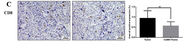
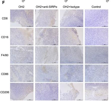
Application: IHC    Species: Mice    Sample: tumor tissue

Fig. 5. Relative protein levels of CD8, MMP2, MMP9, PD-L1 and VEGF in tumor tissue specimens. (A)- (E) The protein expression in the tumor tissues by immunohistochemical staining. Statistical analyses of single comparison were conducted through the Student’s t-test. All data are presented as mean ± SD, *p < 0.05, **p < 0.01, n = 3 per group. Bar: 50 μm (low magnification); 20 μm (high magnification).

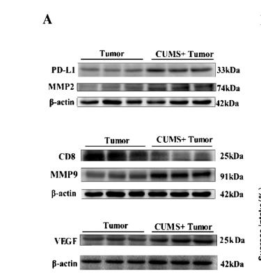
Application: WB    Species: Mice    Sample: tumor tissues

Fig. 6. WB of protein expression in tumor tissues and correlation analysis between the depression-like behaviors and protein expression. (A) The bands of protein expression. (B)-(F) Correlation analysis between the depression-like behaviors and protein expression. Statistical analyses of single comparison were conducted through the Student’s t-test. All data are presented as mean ± SD, *p < 0.05, **p < 0.01, n = 3 per group.

9). Gemcitabine and Celecoxib Synergistically Promote Long-Lasting Antitumor Efficacy of Pd-1 Antibody by Triggering Immunogenic Cell Death.

10). GZMA as a Potential Therapeutic Target Involved in Immune Infiltration in Breast Cancer.

限制条款

产品的规格、报价、验证数据请以官网为准,官网链接:www.affbiotech.com | www.affbiotech.cn(简体中文)| www.affbiotech.jp(日本語)

产品的数据信息为Affinity所有,未经授权不得收集Affinity官网数据或资料用于商业用途,对抄袭产品数据的行为我们将保留诉诸法律的权利。

产品相关数据会因产品批次、产品检测情况随时调整,如您已订购该产品,请以订购时随货说明书为准,否则请以官网内容为准,官网内容有改动时恕不另行通知。

Affinity保证所销售产品均经过严格质量检测。如您购买的商品在规定时间内出现问题需要售后时,请您在Affinity官方渠道提交售后申请。

产品仅供科学研究使用。不用于诊断和治疗。 

产品未经授权不得转售。

Affinity Biosciences将不会对在使用我们的产品时可能发生的专利侵权或其他侵权行为负责。Affinity Biosciences, Affinity Biosciences标志和所有其他商标所有权归Affinity Biosciences LTD.