
产品描述
*The optimal dilutions should be determined by the end user.
*Tips:
WB: 适用于变性蛋白样本的免疫印迹检测. IHC: 适用于组织样本的石蜡(IHC-p)或冰冻(IHC-f)切片样本的免疫组化/荧光检测. IF/ICC: 适用于细胞样本的荧光检测. ELISA(peptide): 适用于抗原肽的ELISA检测.
引用格式: Affinity Biosciences Cat# AF5128, RRID:AB_2837614.
展开/折叠
APG6; ATG 6; ATG6; ATG6 autophagy related 6 homolog; Bcl-2-interacting protein beclin; Beclin 1 (coiled coil moesin like BCL2 interacting protein); Beclin 1 autophagy related; Beclin-1; Beclin1; BECN 1; Becn1; BECN1_HUMAN; Coiled coil myosin like BCL2 interacting protein; Coiled-coil myosin-like BCL2-interacting protein; GT197; Protein GT197; VPS 30; VPS30;
抗原和靶标
- Q14457 BECN1_HUMAN:
- Protein BLAST With
- NCBI/
- ExPASy/
- Uniprot
MEGSKTSNNSTMQVSFVCQRCSQPLKLDTSFKILDRVTIQELTAPLLTTAQAKPGETQEEETNSGEEPFIETPRQDGVSRRFIPPARMMSTESANSFTLIGEASDGGTMENLSRRLKVTGDLFDIMSGQTDVDHPLCEECTDTLLDQLDTQLNVTENECQNYKRCLEILEQMNEDDSEQLQMELKELALEEERLIQELEDVEKNRKIVAENLEKVQAEAERLDQEEAQYQREYSEFKRQQLELDDELKSVENQMRYAQTQLDKLKKTNVFNATFHIWHSGQFGTINNFRLGRLPSVPVEWNEINAAWGQTVLLLHALANKMGLKFQRYRLVPYGNHSYLESLTDKSKELPLYCSGGLRFFWDNKFDHAMVAFLDCVQQFKEEVEKGETRFCLPYRMDVEKGKIEDTGGSGGSYSIKTQFNSEEQWTKALKFMLTNLKWGLAWVSSQFYNK
种属预测
score>80的预测可信度较高,可尝试用于WB检测。*预测模型主要基于免疫原序列比对,结果仅作参考,不作为质保凭据。
High(score>80) Medium(80>score>50) Low(score<50) No confidence
翻译修饰 - Q14457 作为底物
| Site | PTM Type | Enzyme | Source |
|---|---|---|---|
| Ubiquitination | Uniprot | ||
| M1 | Acetylation | Uniprot | |
| S15 | Phosphorylation | Uniprot | |
| K26 | Ubiquitination | Uniprot | |
| S30 | Phosphorylation | Uniprot | |
| K32 | Ubiquitination | Uniprot | |
| T38 | Phosphorylation | Uniprot | |
| K53 | Ubiquitination | Uniprot | |
| T57 | Phosphorylation | Uniprot | |
| T62 | Phosphorylation | Uniprot | |
| S64 | Phosphorylation | Uniprot | |
| S90 | Phosphorylation | P49137 (MAPKAPK2) , Q16644 (MAPKAPK3) | Uniprot |
| T91 | Phosphorylation | Uniprot | |
| S93 | Phosphorylation | Uniprot | |
| S96 | Phosphorylation | Uniprot | |
| T108 | Phosphorylation | Q13043 (STK4) | Uniprot |
| K117 | Ubiquitination | Uniprot | |
| T119 | Phosphorylation | P53355 (DAPK1) | Uniprot |
| T141 | Phosphorylation | Uniprot | |
| K203 | Ubiquitination | Uniprot | |
| K206 | Acetylation | Uniprot | |
| K214 | Ubiquitination | Uniprot | |
| Y229 | Phosphorylation | P00533 (EGFR) | Uniprot |
| Y233 | Phosphorylation | P00533 (EGFR) | Uniprot |
| S234 | Phosphorylation | Uniprot | |
| K237 | Ubiquitination | Uniprot | |
| K248 | Ubiquitination | Uniprot | |
| K263 | Ubiquitination | Uniprot | |
| S279 | Phosphorylation | Uniprot | |
| S295 | Phosphorylation | P31749 (AKT1) | Uniprot |
| K324 | Ubiquitination | Uniprot | |
| S337 | Phosphorylation | Uniprot | |
| Y352 | Phosphorylation | P00533 (EGFR) | Uniprot |
| T388 | Phosphorylation | Uniprot | |
| K402 | Ubiquitination | Uniprot | |
| T406 | Phosphorylation | P78368 (CSNK1G2) | Uniprot |
| S409 | Phosphorylation | P78368 (CSNK1G2) | Uniprot |
| K427 | Ubiquitination | Uniprot | |
| K430 | Acetylation | Uniprot | |
| K437 | Acetylation | Uniprot | |
| K437 | Ubiquitination | Uniprot |
研究背景
Plays a central role in autophagy. Acts as core subunit of the PI3K complex that mediates formation of phosphatidylinositol 3-phosphate; different complex forms are believed to play a role in multiple membrane trafficking pathways: PI3KC3-C1 is involved in initiation of autophagosomes and PI3KC3-C2 in maturation of autophagosomes and endocytosis. Involved in regulation of degradative endocytic trafficking and required for the abcission step in cytokinesis, probably in the context of PI3KC3-C2. Essential for the formation of PI3KC3-C2 but not PI3KC3-C1 PI3K complex forms. Involved in endocytosis. Protects against infection by a neurovirulent strain of Sindbis virus. May play a role in antiviral host defense.
Beclin-1-C 35 kDa localized to mitochondria can promote apoptosis; it induces the mitochondrial translocation of BAX and the release of proapoptotic factors.
Phosphorylation at Thr-119 by DAPK1 reduces its interaction with BCL2 and BCL2L1 and promotes induction of autophagy. In response to autophagic stimuli, phosphorylated at serine residues by AMPK in an ATG14-dependent manner, and this phosphorylation is critical for maximally efficient autophagy.
Polyubiquitinated by NEDD4, both with 'Lys-11'- and 'Lys-63'-linkages. 'Lys-11'-linked polyubiquitination leads to degradation and is enhanced when the stabilizing interaction partner VPS34 is depleted. Deubiquitinated by USP10 and USP13, leading to stabilize the PIK3C3/VPS34-containing complexes. Polyubiquitinated at Lys-402 with 'Lys-48'-linkages. 'Lys-48'-linked polyubiquitination of Lys-402 leads to degradation. Deubiquitinated by ATXN3, leading to stabilization.
Proteolytically processed by caspases including CASP8 and CASP3; the C-terminal fragments lack autophagy-inducing capacity and are proposed to induce apoptosis. Thus the cleavage is proposed to be an determinant to switch from autophagy to apoptosis pathways affecting cellular homeostasis including viral infections and survival of tumor cells.
Cytoplasm. Golgi apparatus>trans-Golgi network membrane>Peripheral membrane protein. Endosome membrane>Peripheral membrane protein. Endoplasmic reticulum membrane>Peripheral membrane protein. Mitochondrion membrane>Peripheral membrane protein. Endosome. Cytoplasmic vesicle>Autophagosome.
Note: Interaction with ATG14 promotes translocation to autophagosomes. Expressed in dendrites and cell bodies of cerebellar Purkinje cells (By similarity).
Mitochondrion. Nucleus. Cytoplasm.
Mitochondrion.
Ubiquitous.
A homodimeric form is proposed to exist; this metastable form readily transits to ATG14- or UVRAG-containing complexes with BECN1:UVRAG being more stable than BECN1:ATG14 (By similarity). Component of the PI3K (PI3KC3/PI3K-III/class III phosphatidylinositol 3-kinase) complex the core of which is composed of the catalytic subunit PIK3C3, the regulatory subunit PIK3R4 and BECN1 associating with additional regulatory/auxilliary subunits to form alternative complex forms. Alternative complex forms containing a forth regulatory subunit in a mutually exclusive manner are PI3K complex I (PI3KC3-C1) containing ATG14, and PI3K complex II (PI3KC3-C2) containing UVRAG. PI3KC3-C1 displays a V-shaped architecture with PIK3R4 serving as a bridge between PIK3C3 and the ATG14:BECN1 subcomplex. Both, PI3KC3-C1 and PI3KC3-C2, can associate with further regulatory subunits, such as RUBCN, SH3GLB1/Bif-1 and AMBRA1. PI3KC3-C1 probably associates with PIK3CB (By similarity). Interacts with AMBRA1, GOPC, GRID2 (By similarity). Interacts with BCL2 and BCL2L1 isoform Bcl-X(L); the interaction inhibits BECN1 function in promoting autophagy by interfering with the formation of the PI3K complex. Interacts with cytosolic HMGB1; inhibits the interaction of BECN1 and BCL2 leading to promotion of autophagy. Interacts with USP10, USP13, VMP1, DAPK1, RAB39A. Interacts with the poly-Gln domain of ATXN3; the interaction causes deubiquitination at Lys-402 and stabilizes BECN1. Interacts with SLAMF1. Interacts with TRIM5; the interaction causes activation of BECN1 by causing its dissociation from its inhibitors BCL2 and TAB2. Interacts with active ULK1 (phosphorylated on 'Ser-317') and MEFV simultaneously. Interacts with WDR81 and WDR91; negatively regulates the PI3 kinase/PI3K activity associated with endosomal membranes. Interacts with LAPTM4B; competes with EGFR for LAPTM4B binding; regulates EGFR activity. Interacts with TRIM50. Interacts with TRIM16.
(Microbial infection) Interacts with human cytomegalovirus/HHV-5 protein TRS1.
(Microbial infection) Interacts with murine gammaherpesvirus 68 M11.
(Microbial infection) Interacts with herpes simplex virus 1 (HHV-1) protein ICP34.5; this interaction antagonizes the host autophagy response.
The coiled coil domain can form antiparallel homodimers and mediates dimerization with the coiled coil domains of ATG14 or UVRAG involved in the formation of PI3K complexes.
The C-terminal evolutionary conserved domain (ECD) contains poly-Gln-binding domains such as the ATXN3 poly-Gln motif, consistent with structural docking models revealing two highly scored poly-Gln-binding pockets in the ECD (PubMed:28445460). As some binding is observed with BECN1 lacking the ECD, other domains of BECN1 may also interact with ATXN3 (PubMed:28445460).
Belongs to the beclin family.
研究领域
· Cellular Processes > Transport and catabolism > Autophagy - other. (View pathway)
· Cellular Processes > Transport and catabolism > Autophagy - animal. (View pathway)
· Cellular Processes > Cell growth and death > Apoptosis - multiple species. (View pathway)
· Environmental Information Processing > Signal transduction > Apelin signaling pathway. (View pathway)
文献引用
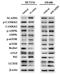
Application: WB Species: Human Sample: CRC cells

Application: WB Species: human Sample: MDA-MB-231 cells

Application: WB Species: Human Sample: MDA-MB-231 cells

Application: WB Species: mouse Sample: MSCs
Application: WB Species: Rat Sample: MC3T3-E1 cells

Application: IHC Species: Rat Sample: MC3T3-E1 cells
Application: WB Species: Rat Sample:
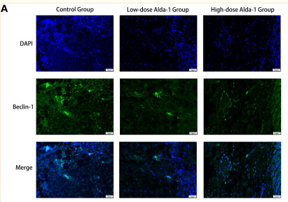
Application: IF/ICC Species: Mouse Sample:
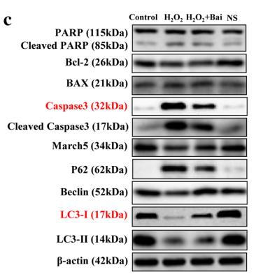
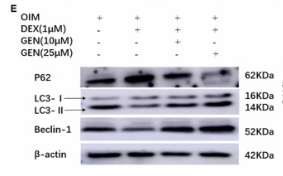
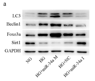
Application: WB Species: human Sample: L02 cells
限制条款
产品的规格、报价、验证数据请以官网为准,官网链接:www.affbiotech.com | www.affbiotech.cn(简体中文)| www.affbiotech.jp(日本語)产品的数据信息为Affinity所有,未经授权不得收集Affinity官网数据或资料用于商业用途,对抄袭产品数据的行为我们将保留诉诸法律的权利。
产品相关数据会因产品批次、产品检测情况随时调整,如您已订购该产品,请以订购时随货说明书为准,否则请以官网内容为准,官网内容有改动时恕不另行通知。
Affinity保证所销售产品均经过严格质量检测。如您购买的商品在规定时间内出现问题需要售后时,请您在Affinity官方渠道提交售后申请。产品仅供科学研究使用。不用于诊断和治疗。
产品未经授权不得转售。
Affinity Biosciences将不会对在使用我们的产品时可能发生的专利侵权或其他侵权行为负责。Affinity Biosciences, Affinity Biosciences标志和所有其他商标所有权归Affinity Biosciences LTD.