VDAC1 Antibody - #DF6140
产品描述
*The optimal dilutions should be determined by the end user.
*Tips:
WB: 适用于变性蛋白样本的免疫印迹检测. IHC: 适用于组织样本的石蜡(IHC-p)或冰冻(IHC-f)切片样本的免疫组化/荧光检测. IF/ICC: 适用于细胞样本的荧光检测. ELISA(peptide): 适用于抗原肽的ELISA检测.
引用格式: Affinity Biosciences Cat# DF6140, RRID:AB_2838107.
展开/折叠
N2441; OMP2; POR1; hVDAC1; MGC111064; Mitochondrial Porin; Outer mitochondrial membrane protein porin 1; Plasmalemmal porin; Porin 31HL; Porin 31HM; VDAC; VDAC-1; Vdac1; VDAC1_HUMAN; Voltage dependent anion channel 1; Voltage dependent anion selective channel protein 1; Voltage-dependent anion-selective channel protein 1; YNL055C; YNL2441C;
抗原和靶标
- P21796 VDAC1_HUMAN:
- Protein BLAST With
- NCBI/
- ExPASy/
- Uniprot
MAVPPTYADLGKSARDVFTKGYGFGLIKLDLKTKSENGLEFTSSGSANTETTKVTGSLETKYRWTEYGLTFTEKWNTDNTLGTEITVEDQLARGLKLTFDSSFSPNTGKKNAKIKTGYKREHINLGCDMDFDIAGPSIRGALVLGYEGWLAGYQMNFETAKSRVTQSNFAVGYKTDEFQLHTNVNDGTEFGGSIYQKVNKKLETAVNLAWTAGNSNTRFGIAAKYQIDPDACFSAKVNNSSLIGLGYTQTLKPGIKLTLSALLDGKNVNAGGHKLGLGLEFQA
种属预测
score>80的预测可信度较高,可尝试用于WB检测。*预测模型主要基于免疫原序列比对,结果仅作参考,不作为质保凭据。
High(score>80) Medium(80>score>50) Low(score<50) No confidence
翻译修饰 - P21796 作为底物
| Site | PTM Type | Enzyme | Source |
|---|---|---|---|
| A2 | Acetylation | Uniprot | |
| K12 | Acetylation | Uniprot | |
| K12 | Ubiquitination | Uniprot | |
| S13 | Phosphorylation | Uniprot | |
| T19 | Phosphorylation | Uniprot | |
| K20 | Acetylation | Uniprot | |
| K20 | Ubiquitination | Uniprot | |
| Y22 | Phosphorylation | Uniprot | |
| K28 | Acetylation | Uniprot | |
| K28 | Ubiquitination | Uniprot | |
| K34 | Acetylation | Uniprot | |
| K34 | Ubiquitination | Uniprot | |
| S43 | Phosphorylation | Uniprot | |
| S44 | Phosphorylation | Uniprot | |
| S46 | Phosphorylation | Uniprot | |
| K53 | Ubiquitination | Uniprot | |
| S57 | Phosphorylation | Uniprot | |
| K61 | Acetylation | Uniprot | |
| K61 | Ubiquitination | Uniprot | |
| Y62 | Phosphorylation | Uniprot | |
| T65 | Phosphorylation | Uniprot | |
| Y67 | Phosphorylation | Uniprot | |
| T98 | Phosphorylation | Uniprot | |
| S101 | Phosphorylation | Uniprot | |
| S102 | Phosphorylation | Uniprot | |
| S104 | Phosphorylation | Uniprot | |
| T107 | Phosphorylation | Uniprot | |
| K109 | Acetylation | Uniprot | |
| K109 | Sumoylation | Uniprot | |
| K109 | Ubiquitination | Uniprot | |
| K110 | Ubiquitination | Uniprot | |
| K119 | Acetylation | Uniprot | |
| K161 | Ubiquitination | Uniprot | |
| R163 | Methylation | Uniprot | |
| T165 | Phosphorylation | Uniprot | |
| S167 | Phosphorylation | Uniprot | |
| Y173 | Phosphorylation | Uniprot | |
| K174 | Acetylation | Uniprot | |
| K174 | Methylation | Uniprot | |
| S193 | Phosphorylation | Q96PY6 (NEK1) | Uniprot |
| Y195 | Phosphorylation | Uniprot | |
| K197 | Ubiquitination | Uniprot | |
| T204 | Phosphorylation | Uniprot | |
| S215 | Phosphorylation | Uniprot | |
| K224 | Acetylation | Uniprot | |
| K224 | Ubiquitination | Uniprot | |
| Y225 | Phosphorylation | Uniprot | |
| C232 | S-Nitrosylation | Uniprot | |
| S234 | Phosphorylation | Uniprot | |
| K236 | Acetylation | Uniprot | |
| S240 | Phosphorylation | Uniprot | |
| S241 | Phosphorylation | Uniprot | |
| Y247 | Phosphorylation | Uniprot | |
| T250 | Phosphorylation | Uniprot | |
| K252 | Acetylation | Uniprot | |
| K252 | Ubiquitination | Uniprot | |
| S260 | Phosphorylation | Uniprot | |
| K266 | Acetylation | Uniprot | |
| K266 | Ubiquitination | Uniprot | |
| K274 | Acetylation | Uniprot | |
| K274 | Sumoylation | Uniprot | |
| K274 | Ubiquitination | Uniprot |
研究背景
Forms a channel through the mitochondrial outer membrane and also the plasma membrane. The channel at the outer mitochondrial membrane allows diffusion of small hydrophilic molecules; in the plasma membrane it is involved in cell volume regulation and apoptosis. It adopts an open conformation at low or zero membrane potential and a closed conformation at potentials above 30-40 mV. The open state has a weak anion selectivity whereas the closed state is cation-selective. May participate in the formation of the permeability transition pore complex (PTPC) responsible for the release of mitochondrial products that triggers apoptosis.
Phosphorylation at Ser-193 by NEK1 promotes the open conformational state preventing excessive mitochondrial membrane permeability and subsequent apoptotic cell death after injury. Phosphorylation by the AKT-GSK3B axis stabilizes the protein probably by preventing ubiquitin-mediated proteasomal degradation.
Ubiquitinated by PRKN during mitophagy, leading to its degradation and enhancement of mitophagy. Deubiquitinated by USP30.
Mitochondrion outer membrane>Multi-pass membrane protein. Cell membrane>Multi-pass membrane protein. Membrane raft>Multi-pass membrane protein.
Heart, liver and skeletal muscle.
Interacts with hexokinases including HK1. The HK1-VDAC1 complex interacts with ATF2. Interacts with BCL2L1. Interacts with BAK1. Interacts with RTL10/BOP (via BH3 domain). Interacts with amyloid-beta and APP; induces VDAC1 dephosphorylation. Component of the mitochondrial permeability transition pore complex (mPTPC), at least composed of SPG7, VDAC1 and PPIF. Interacts with SPG7, NIPSNAP2 and SLC25A30.
(Microbial infection) Interacts with influenza A virus PB1-F2 protein.
Consists mainly of a membrane-spanning beta-barrel formed by 19 beta-strands. The helical N-terminus folds back into the pore opening and plays a role in voltage-gated channel activity.
Belongs to the eukaryotic mitochondrial porin family.
研究领域
· Cellular Processes > Cell growth and death > Necroptosis. (View pathway)
· Cellular Processes > Cell growth and death > Cellular senescence. (View pathway)
· Environmental Information Processing > Signal transduction > Calcium signaling pathway. (View pathway)
· Environmental Information Processing > Signal transduction > cGMP-PKG signaling pathway. (View pathway)
· Human Diseases > Neurodegenerative diseases > Parkinson's disease.
· Human Diseases > Neurodegenerative diseases > Huntington's disease.
· Human Diseases > Infectious diseases: Viral > Influenza A.
· Human Diseases > Infectious diseases: Viral > HTLV-I infection.
· Organismal Systems > Immune system > NOD-like receptor signaling pathway. (View pathway)
· Organismal Systems > Digestive system > Cholesterol metabolism.
文献引用
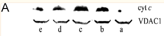
Application: WB Species: Human Sample:
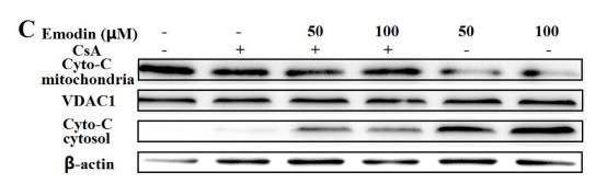
Application: WB Species: human Sample:

Application: WB Species: Mice Sample: cholangiocarcinoma cell

Application: WB Species: mouse Sample: L02 cells
限制条款
产品的规格、报价、验证数据请以官网为准,官网链接:www.affbiotech.com | www.affbiotech.cn(简体中文)| www.affbiotech.jp(日本語)产品的数据信息为Affinity所有,未经授权不得收集Affinity官网数据或资料用于商业用途,对抄袭产品数据的行为我们将保留诉诸法律的权利。
产品相关数据会因产品批次、产品检测情况随时调整,如您已订购该产品,请以订购时随货说明书为准,否则请以官网内容为准,官网内容有改动时恕不另行通知。
Affinity保证所销售产品均经过严格质量检测。如您购买的商品在规定时间内出现问题需要售后时,请您在Affinity官方渠道提交售后申请。产品仅供科学研究使用。不用于诊断和治疗。
产品未经授权不得转售。
Affinity Biosciences将不会对在使用我们的产品时可能发生的专利侵权或其他侵权行为负责。Affinity Biosciences, Affinity Biosciences标志和所有其他商标所有权归Affinity Biosciences LTD.