产品: Bcl-2 抗体
货号: BF9103
来源: Mouse
应用: WB, IHC, IF/ICC, ELISA
反应: Human, Mouse, Rat
分子量: 26 kD; 26kD(Calculated).
蛋白号: P10415
RRID: AB_2837570

浏览相似产品>>

   规格 价格 库存
 50ul ¥1250 现货
 100ul ¥2300 现货
 200ul ¥3000 现货

货期: 当天发货

产品描述

来源:
Mouse
应用:
WB 1:1000, IHC 1:50-1:200, IF/ICC 1:100-1:500
*The optimal dilutions should be determined by the end user.
*Tips:

WB: 适用于变性蛋白样本的免疫印迹检测. IHC: 适用于组织样本的石蜡(IHC-p)或冰冻(IHC-f)切片样本的免疫组化/荧光检测. IF/ICC: 适用于细胞样本的荧光检测. ELISA(peptide): 适用于抗原肽的ELISA检测.

反应:
Human,Mouse,Rat
克隆:
Monoclonal [AFfirm063]
特异性:
The Bcl-2 mouse monoclonal antibody can detect endogenous Bcl-2 proteins.
RRID:
AB_2837570
引用格式: Affinity Biosciences Cat# BF9103, RRID:AB_2837570.
偶联:
Unconjugated.
纯化:
affinity purification.
保存:
Store at -20°C. Stable for one year from the date of shipment.1mg/ml in PBS, pH 7.4, containing 0.02% sodium azide and 50% glycerol. Store at -20 °C. Stable for 12 months from date of receipt.
别名:

展开/折叠

Apoptosis regulator Bcl 2; Apoptosis regulator Bcl-2; Apoptosis regulator Bcl2; AW986256; B cell CLL/lymphoma 2; B cell leukemia/lymphoma 2; Bcl-2; Bcl2; BCL2_HUMAN; C430015F12Rik; D630044D05Rik; D830018M01Rik; Leukemia/lymphoma, B-cell, 2; Oncogene B-cell leukemia 2; PPP1R50; Protein phosphatase 1, regulatory subunit 50;

抗原和靶标

免疫原:

Mouse monoclonal antibody is prepared by immunizing synthetic peptide coupled to KLH.

Uniprot:
基因/基因ID:
表达特异性:
P10415 BCL2_HUMAN:

Expressed in a variety of tissues.

蛋白描述:
BCL2 is an integral outer mitochondrial membrane protein that blocks the apoptotic death of some cells such as lymphocytes. Constitutive expression of BCL2, such as in the case of translocation of BCL2 to Ig heavy chain locus, is thought to be the cause of follicular lymphoma. BCL2 suppresses apoptosis in a variety of cell systems including factor-dependent lymphohematopoietic and neural cells. It regulates cell death by controlling the mitochondrial membrane permeability.
蛋白序列:
MAHAGRTGYDNREIVMKYIHYKLSQRGYEWDAGDVGAAPPGAAPAPGIFSSQPGHTPHPAASRDPVARTSPLQTPAAPGAAAGPALSPVPPVVHLTLRQAGDDFSRRYRRDFAEMSSQLHLTPFTARGRFATVVEELFRDGVNWGRIVAFFEFGGVMCVESVNREMSPLVDNIALWMTEYLNRHLHTWIQDNGGWDAFVELYGPSMRPLFDFSWLSLKTLLSLALVGACITLGAYLGHK

翻译修饰 - P10415 作为底物

Site PTM Type Enzyme
Y9 Phosphorylation
K22 Ubiquitination
S24 Phosphorylation
T56 Phosphorylation Q16539 (MAPK14) , P06493 (CDK1) , P53779 (MAPK10) , P28482 (MAPK1) , P27361 (MAPK3)
T69 Phosphorylation P45983 (MAPK8)
S70 Phosphorylation P27361 (MAPK3) , P06493 (CDK1) , P53779 (MAPK10) , P17252 (PRKCA) , Q00534 (CDK6) , P28482 (MAPK1) , P45983 (MAPK8)
T74 Phosphorylation P28482 (MAPK1) , P53779 (MAPK10) , P27361 (MAPK3)
S87 Phosphorylation Q16539 (MAPK14) , P45983 (MAPK8) , Q00534 (CDK6) , P27361 (MAPK3) , P28482 (MAPK1) , P53779 (MAPK10)
C158 S-Nitrosylation
C229 S-Nitrosylation
Y235 Phosphorylation

研究背景

功能:

Suppresses apoptosis in a variety of cell systems including factor-dependent lymphohematopoietic and neural cells. Regulates cell death by controlling the mitochondrial membrane permeability. Appears to function in a feedback loop system with caspases. Inhibits caspase activity either by preventing the release of cytochrome c from the mitochondria and/or by binding to the apoptosis-activating factor (APAF-1). May attenuate inflammation by impairing NLRP1-inflammasome activation, hence CASP1 activation and IL1B release.

翻译修饰:

Phosphorylation/dephosphorylation on Ser-70 regulates anti-apoptotic activity. Growth factor-stimulated phosphorylation on Ser-70 by PKC is required for the anti-apoptosis activity and occurs during the G2/M phase of the cell cycle. In the absence of growth factors, BCL2 appears to be phosphorylated by other protein kinases such as ERKs and stress-activated kinases. Phosphorylated by MAPK8/JNK1 at Thr-69, Ser-70 and Ser-87, wich stimulates starvation-induced autophagy. Dephosphorylated by protein phosphatase 2A (PP2A) (By similarity).

Proteolytically cleaved by caspases during apoptosis. The cleaved protein, lacking the BH4 motif, has pro-apoptotic activity, causes the release of cytochrome c into the cytosol promoting further caspase activity.

Monoubiquitinated by PRKN, leading to increase its stability. Ubiquitinated by SCF(FBXO10), leading to its degradation by the proteasome.

细胞定位:

Mitochondrion outer membrane>Single-pass membrane protein. Nucleus membrane>Single-pass membrane protein. Endoplasmic reticulum membrane>Single-pass membrane protein.

Extracellular region or secreted Cytosol Plasma membrane Cytoskeleton Lysosome Endosome Peroxisome ER Golgi apparatus Nucleus Mitochondrion Manual annotation Automatic computational assertionSubcellular location
组织特异性:

Expressed in a variety of tissues.

亚基结构:

Forms homodimers, and heterodimers with BAX, BAD, BAK and Bcl-X(L). Heterodimerization with BAX requires intact BH1 and BH2 motifs, and is necessary for anti-apoptotic activity. Interacts with EI24 (By similarity). Also interacts with APAF1, BBC3, BCL2L1, BNIPL, MRPL41 and TP53BP2. Binding to FKBP8 seems to target BCL2 to the mitochondria and probably interferes with the binding of BCL2 to its targets. Interacts with BAG1 in an ATP-dependent manner. Interacts with RAF1 (the 'Ser-338' and 'Ser-339' phosphorylated form). Interacts (via the BH4 domain) with EGLN3; the interaction prevents the formation of the BAX-BCL2 complex and inhibits the anti-apoptotic activity of BCL2. Interacts with G0S2; this interaction also prevents the formation of the anti-apoptotic BAX-BCL2 complex. Interacts with RTL10/BOP. Interacts with the SCF(FBXO10) complex. Interacts (via the loop between motifs BH4 and BH3) with NLRP1 (via LRR repeats), but not with NLRP2, NLRP3, NLRP4, PYCARD, nor MEFV. Interacts with GIMAP3/IAN4, GIMAP4/IAN1 and GIMAP5/IAN5 (By similarity).

蛋白家族:

BH1 and BH2 domains are required for the interaction with BAX and for anti-apoptotic activity.

The BH4 motif is required for anti-apoptotic activity and for interaction with RAF1 and EGLN3.

The loop between motifs BH4 and BH3 is required for the interaction with NLRP1.

Belongs to the Bcl-2 family.

研究领域

· Cellular Processes > Transport and catabolism > Autophagy - animal.   (View pathway)

· Cellular Processes > Cell growth and death > Apoptosis.   (View pathway)

· Cellular Processes > Cell growth and death > Apoptosis - multiple species.   (View pathway)

· Cellular Processes > Cell growth and death > Necroptosis.   (View pathway)

· Cellular Processes > Cellular community - eukaryotes > Focal adhesion.   (View pathway)

· Environmental Information Processing > Signal transduction > NF-kappa B signaling pathway.   (View pathway)

· Environmental Information Processing > Signal transduction > HIF-1 signaling pathway.   (View pathway)

· Environmental Information Processing > Signal transduction > Sphingolipid signaling pathway.   (View pathway)

· Environmental Information Processing > Signal transduction > PI3K-Akt signaling pathway.   (View pathway)

· Environmental Information Processing > Signal transduction > Hedgehog signaling pathway.   (View pathway)

· Environmental Information Processing > Signal transduction > Jak-STAT signaling pathway.   (View pathway)

· Genetic Information Processing > Folding, sorting and degradation > Protein processing in endoplasmic reticulum.   (View pathway)

· Human Diseases > Drug resistance: Antineoplastic > EGFR tyrosine kinase inhibitor resistance.

· Human Diseases > Drug resistance: Antineoplastic > Endocrine resistance.

· Human Diseases > Drug resistance: Antineoplastic > Platinum drug resistance.

· Human Diseases > Neurodegenerative diseases > Amyotrophic lateral sclerosis (ALS).

· Human Diseases > Infectious diseases: Parasitic > Toxoplasmosis.

· Human Diseases > Infectious diseases: Bacterial > Tuberculosis.

· Human Diseases > Infectious diseases: Viral > Hepatitis B.

· Human Diseases > Infectious diseases: Viral > Epstein-Barr virus infection.

· Human Diseases > Cancers: Overview > Pathways in cancer.   (View pathway)

· Human Diseases > Cancers: Overview > MicroRNAs in cancer.

· Human Diseases > Cancers: Specific types > Colorectal cancer.   (View pathway)

· Human Diseases > Cancers: Specific types > Prostate cancer.   (View pathway)

· Human Diseases > Cancers: Specific types > Small cell lung cancer.   (View pathway)

· Human Diseases > Cancers: Specific types > Gastric cancer.   (View pathway)

· Organismal Systems > Circulatory system > Adrenergic signaling in cardiomyocytes.   (View pathway)

· Organismal Systems > Immune system > NOD-like receptor signaling pathway.   (View pathway)

· Organismal Systems > Nervous system > Neurotrophin signaling pathway.   (View pathway)

· Organismal Systems > Nervous system > Cholinergic synapse.

· Organismal Systems > Endocrine system > Estrogen signaling pathway.   (View pathway)

文献引用

1). Deng W et al. Inhibition of PLK3 Attenuates Tubular Epithelial Cell Apoptosis after Renal Ischemia–Reperfusion Injury by Blocking the ATM/P53-Mediated DNA Damage Response. Oxid Med Cell Longev 2022 Jun 24;2022:4201287. (PubMed: 35783188) [IF=7.310]

Application: WB    Species: Human    Sample: HK-2 cells

Figure 6 Inhibiting PLK3 attenuates TEC apoptosis via the ATM/P53 signaling pathway. (a) In the H/R injury model, the PLK3 protein levels and apoptosis-related protein levels were measured after targeted KD of PLK3 expression. (b) p-ATM protein levels, PLK3 protein levels, and apoptosis-related protein levels were measured by Western blot after the application of the specific ATM inhibitor KU-60019. The data are presented as the mean ± SD. n = 3. ∗P < 0.05, ∗∗P < 0.01, ∗∗∗P < 0.001, and ∗∗∗∗P < 0.0001. Nor: normoxia; Ku: KU-60019.

2). Ren M et al. Melatonin Repairs Osteoporotic Bone Defects in Iron-Overloaded Rats through PI3K/AKT/GSK-3β/P70S6k Signaling Pathway. Oxid Med Cell Longev 2023 Jan 17;2023:7718155. (PubMed: 36703914) [IF=7.310]

3). Peng J et al. MPO/HOCl Facilitates Apoptosis and Ferroptosis in the SOD1G93A Motor Neuron of Amyotrophic Lateral Sclerosis. Oxid Med Cell Longev 2022 Feb 7;2022:8217663. (PubMed: 35178161) [IF=7.310]

Application: WB    Species: Mice    Sample: hSOD1G93A cells

Figure 3 MPO/HOCl signaling manipulated apoptosis and ferroptosis in hSOD1G93A cells. (a) The apoptosis- and autophagy-related proteins of vector, hSOD1WT, and hSOD1G93A cells, as measured by western blot. (b) 1 μM Z-DEVD-FMK reversed cell death of hSOD1G93A cells, significantly. (c, d) Flow cytometry analysis of apoptosis in vector, hSOD1WT, and hSOD1G93A cells, in the absence or presence of ABAH. (e) Immunoblots of hSOD1G93A cells treated with NC-siRNA or MPO-siRNA. (f) The ferroptosis-related proteins of vector, hSOD1WT, and hSOD1G93A cells, as measured by western blot. (g) 1 μM ferrostatin-1 reversed the cell death in hSOD1G93A cells, significantly. (h) Immunoblots of hSOD1G93A overexpressed with vector or FSP1 cDNA. (i) Overexpression of FSP1 cDNA inhibited cell death significantly in hSOD1G93A cells. (j) Immunoblots of ferroptosis-related proteins in hSOD1G93A cells, treated with NC-siRNA or MPO-siRNA. (k) Immunoblots of hSOD1G93A overexpressed with vector or MPO cDNA. (l) Relative viability of hSOD1G93A cells with multiple treatments. (m) The MDA levels of cells in different groups. (n) Effects of different treatments on viability of NSC-34 cells overexpressed with empty vector or MPO cDNA. Quantified graphs were shown as means and SEM. ∗P < 0.05,  ∗∗P < 0.01, and∗∗∗P < 0.001. The significant difference among multiple doses was determined by one-way ANOVA followed by LSD tests. The significant difference in two datasets was analyzed by Student's t-test.

4). Zhang W et al. Celastrol upregulated ATG7 triggers autophagy via targeting Nur77 in colorectal cancer. Phytomedicine 2022 Jun 16;104:154280. (PubMed: 35752079) [IF=6.656]

5). Fei H et al. CTRP1 Attenuates Cerebral Ischemia/Reperfusion Injury via the PERK Signaling Pathway. Front Cell Dev Biol 2021 Aug 4;9:700854. (PubMed: 34422821) [IF=6.081]

Application: WB    Species: Rat    Sample: cortex

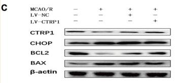
FIGURE 4 CTRP1 protected against cerebral ischemia reperfusion injury via alleviating neuron injury and apoptosis. (A) Neuron injury was analyzed by double labeling immunofluorescence staining. n = 3 per group. The representative images were acquired under × 400 magnification, scale bars = 50 μm. (B) Apoptosis in the cortex was analyzed by TUNEL, n = 3 per group. The representative images were acquired under × 200 magnification, scale bars = 100 μm. (C) Western blot analyzed the expression of CTRP1, BAX, Bcl-2, and CHOP in cortex. n = 4 per group. ****p < 0.0001, ***p < 0.01, **p < 0.01, *p < 0.05 vs. sham group; ####p < 0.0001, ##p < 0.01, #p < 0.05 vs. MCAO/R + LV-NC group.

6). Luo Q et al. Chang qing formula ameliorates colitis-associated colorectal cancer via suppressing IL-17/NF-κB/STAT3 pathway in mice as revealed by network pharmacology study. Front Pharmacol 2022 Aug 3;13:893231. (PubMed: 35991881) [IF=5.810]

Application: WB    Species: Mice    Sample: colon tissue

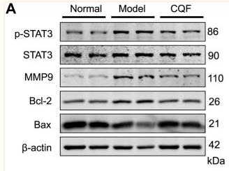
FIGURE 7 CQF suppressed the phosphorylation of STAT3 and altered the expression of its downstream proteins. (A,B) Expression of MMP9, p-STAT3, STAT3, Bcl-2, and Bax proteins in colon tissue (n = 4 per group). (C) Immunofluorescence staining of colon tissue, green: p-STAT3, red: MMP9, blue: DAPI. Values are expressed as mean ± SEM. # p < 0.05, ## p < 0.01, compared with normal mice; * p < 0.05, ** p < 0.01, compared with AOM/DSS-treated mice.

7). Wang L et al. Activation of STING Pathway Contributed to Cisplatin-Induced Cardiac Dysfunction via Promoting the Activation of TNF-α-AP-1 Signal Pathway. Front Pharmacol 2021 Aug 17;12:711238. (PubMed: 34483919) [IF=5.810]

Application: WB    Species: Mice    Sample: HL-1 cells

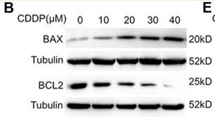
FIGURE 1 Activation of the cGAS-STING pathway was detected in cisplatin-induced HL-1 cells (A) HL-1 cells were incubated with CDDP (10, 20, 30, 40 μM) for 24 h and then the cell viability of HL-1 cells was examined by CCK8 analysis (n = 5 independent experiments; ### p < 0.001, vs. the 0 group). (B) HL-1 cells were incubated with CDDP (10, 20, 30, 40 μM) for 24 h, and then total protein was collected. The protein level of BAX and BCL2 in HL-1 cells was detected by Western blot. (C–D) HL-1 cells were incubated with CDDP (10, 20, 30, 40 μM) for 24 h, and then cell apoptosis induced by CDDP was detected by TUNEL staining. (C) Representative images of TUNEL staining in HL-1 cells (Green: TUNEL positive cell, DAPI: nucleus; Scale Bar: 25 μm, ×400 magnification). (D) Quantification of TUNEL-positive cells (E) HL-1 cells were incubated with CDDP (10, 20, 30, 40 μM) for 1 h, and total protein was collected. The protein level of p-STING, STING, p-TBK1, TBK1 in HL-1 cells was detected by Western blot (n = 3 independent experiments; # p < 0.05, ## p < 0.01, ### p < 0.001, vs. Vehicle group).

8). Wei W et al. Toxicological and physiological effects of successive exposure to ochratoxin A at food regulatory limits. Food Chem Toxicol 2021 Mar 13;112128. (PubMed: 33727177) [IF=5.572]

Application: WB    Species: Rat    Sample: renal tissues

Fig. 4. Effects of OTA on apoptosis in renal tissues of SD rats. Renal tissues were collected from control or OTA-contaminated group of rats on days 1 and 7. The protein levels of bax and bcl-2 were measured by western blotting on day 1 (A) and day 7 (B). The gene expression levels of bax and bcl-2 were measured by qRT-PCR on day 1 (C) and day 7 (D). Data are expressed as mean ± standard deviation (n = 5). *P < 0.05 versus Control (t-test).

9). Cheng J et al. LncRNA PVT1 promotes the malignant progression of acute myeloid leukaemia via sponging miR-29 family to increase WAVE1 expression. Pathology 2021 Feb 6;S0031-3025(21)00021-0. (PubMed: 33558065) [IF=5.335]

Application: WB    Species: Human    Sample: tumour tissues

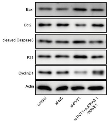
Fig. 6 PVT1/WAVE1 axis regulates xenograft growth in vivo. (A) Photograph of the xenograft tumours from different groups. (B) Tumour volumes at the indicated time points and (C) tumour weight are presented. (D) Apoptosis in tumour tissues was evaluated by TUNEL assay. (E) Expression of Ki-67 in tumour tissues was determined by immunohistochemical staining. (F) The protein levels of Bax, Bcl-2, cleaved Caspase 3, p21, and cyclin D1 in tumour tissues was assessed by western blotting. All data are expressed as mean ± standard deviation. *p<0.05, **p<0.01, ***p<0.001 versus the indicated group.

10). Chen L et al. Hepatocyte nuclear factor 4 gamma (HNF4G) is correlated with poor prognosis and promotes tumor cell growth by inhibiting caspase-dependent intrinsic apoptosis in colorectal cancer. Eur J Pharmacol 2022 Feb 5;916:174727. (PubMed: 34965388) [IF=5.195]

加载更多

限制条款

产品的规格、报价、验证数据请以官网为准,官网链接:www.affbiotech.com | www.affbiotech.cn(简体中文)| www.affbiotech.jp(日本語)

产品的数据信息为Affinity所有,未经授权不得收集Affinity官网数据或资料用于商业用途,对抄袭产品数据的行为我们将保留诉诸法律的权利。

产品相关数据会因产品批次、产品检测情况随时调整,如您已订购该产品,请以订购时随货说明书为准,否则请以官网内容为准,官网内容有改动时恕不另行通知。

Affinity保证所销售产品均经过严格质量检测。如您购买的商品在规定时间内出现问题需要售后时,请您在Affinity官方渠道提交售后申请。

产品仅供科学研究使用。不用于诊断和治疗。 

产品未经授权不得转售。

Affinity Biosciences将不会对在使用我们的产品时可能发生的专利侵权或其他侵权行为负责。Affinity Biosciences, Affinity Biosciences标志和所有其他商标所有权归Affinity Biosciences LTD.