


.
Blue: DAPI.")
 and mouse anti-beta tubulin Ab(T0023 1:200) for 1 hour at 37°C. An AlexaFluor594 conjugated goat anti-rabbit IgG(H+L) Ab(Red) and an AlexaFluor488 conjugated goat anti-mouse IgG(H+L) Ab(Green) were used as the secondary antibody.
The nuclear counter stain is DAPI(blue).")
. a, c, e Group E-IHC; b, d, f group C-IHC. Glo glomerulus, PCT proximal-convoluted tubule, DCT distal-convoluted tubule, CD collecting duct epithelial cells, black arrows thin segment of Henle’s loop")
 Protein expression of meprin a, pro-COL I, TGF-b1, a-SMA, and MCP-1 was detected by western blotting and quantified. (B) Levels of miR-155-5p and Mep1a in rat
lungs. Data are presented as the mean ± SD. n = 5 per group. (C and D) Protein (C) and (D) mRNA (D) levels of meprin a in RAW264.7 cells treated with agomiR-155-5p or
antamiR-155-5p. (E) Luciferase reporter assay demonstrating Mep1a was a target of miR-155-5p. Data are presented as the mean ± SD. n = 3 per group")

 Non-induced IF cells and MF cells showed
different gene expression patterns; Data are mean ± SD of n = 3 replicates, one-way ANOVA, *p<0.05, **p<0.01, ***p<0.001, ****p<0.0001. (B) Non-induced
and induced IF cells and MF cells showed different protein expression patterns. After induction with CLCCM and HERSCM, there were no significant changes of
the expression patterns of tooth eruption-related proteins in IF cells and MF cells. (C) The grey value ratios of Western blotting results; Data are mean ± SD of n = 3
replicates, one-way ANOVA followed by Tukey post hoc test, *p<0.05, **p<0.01, ***p<0.001, ****p<0.0001. (IF1, MF1 represent the cells cultured by α-MEM;
IF2, MF2 represent the cells cultured by α-MEM + CLCCM; IF3, MF3 represent the cells cultured by α-MEM+HERSCM).")
 The intracellular ROS level was detected by flow cytometry after loading with DCFH-DA. Moreover, (c) H2O2 expression, as well as (d) GSH, SOD and MDA
levels were examined. Afterwards, (e-l) the mRNA levels of IL-1β, IL-6, TNF-α, IL-10, IL-4, Arg1, OPN and CD44 were measured by RT-qPCR, and (m) The RT-qPCR of
NOX subunits was analyzed after the exposure of COM crystals (400 μg/ml) or 0.5 mM oxalate with or without kaempferol (40 μM) for 12 h. (n, o) The NOX2, iNOS,
p-NFκB-p65 and MCP-1 expressions in HK-2 cells were measured by WB. Data were expressed as the fold changes of the experimental group to the NC group (except
part b) and were represented as means ± SD of three independent experiments with different cell passages on different days. ∆ p < 0.05; ∆∆ p < 0.01; * p < 0.05 vs. NC
group; # p < 0.05 vs. COM group; ** p < 0.05 as pairwise comparisons of three groups among COM + Kae (10 μM), COM + Kae (20 μM), and COM + Kae (40 μM).")
-,macrophage (Mp)-related molecules, proinflammatory cytokines, and anti-inflammatory cytokines. A: immunohistochemical distribution of the expression of PPAR-, proinflammatory cytokines, and Mp-related molecules in the kidneys as well as the ratio of areas with positive expression. PPAR- expression in pioglitazone (PGZ)-treated groups was significantly higher than in other groups. Expression of monocyte chemotactic protein (MCP)-1, F4/80, inducible nitric oxide synthase (iNOS), arginine 1 (ARG1), interferon regulatory factor 1 (IRF1), and PKNOX1 was significantly higher in 3-day (d) glyoxylic acid (GA)- and 6-day GA-treated groups than in the control group and was significantly lower in PGZ-treated groups than in the 6-day GA group. Original magnification: 200.")
 and Pknox1 are two miR-23 target genes, and miR-23 is important in regulating macrophage polarization and inflammation.B: Western blots were performed in calcium oxalate monohydrate (COM)-stimulated bone marrow-derived macrophages(BMDMs) with miR-23 mimic (MI23) or miR-23 inhibitor (IN23) for 48 h. All Western blot data were quantified. Data are presented as means SE.")
 Placental tissue sections were stained with anti-MCP-1 by immunofluorescence. Nuclei were visualized with DAPI. White dotted lines show the placental cytotrophoblasts (CTBs) areas, the arrowheads indicate positive staining. The immunolocalization results shown in B were semi-quantified in Image J. The immunoreactivity was expressed relative to the data from the control (saline) samples.")

 was
analyzed by western blot in the KPNA2-overexpressing (A, B) or KPNA2-silenced cells (F, G). The nuclear translocation of p65 (nucleus) was detected by immunofluorescence assay in the KPNA2-overexpressing (C, D) or KPNA2-silenced cells (H, I). The NF-κB binding activity was analyzed by EMSA in the KPNA2-
overexpressing (E) or KPNA2-silenced cells (J). Parental, blank control group; NC, negative control group; OV-KPNA2, KPNA2 overexpressed group; siRNA1-KPNA2,
KPNA2-1 silencing group; siRNA2-KPNA2, KPNA2-2 silencing group. The results were obtained in three independent experiments. Mean values were compared by
One-way ANOVA. (**p < 0.01, ***p < 0.001, ****p < 0.001).")
 using tubulin or GAPDH as loading control. Quantitative analysis of protein expression is shown as bar graphs. The mRNA expression levels of ICAM-1 and MCP-1 were determined with RT-qPCR (b). (c, d) Representative images of cellular fluorescence assay for ICAM-1 and MCP-1 (400x magnification) and the quantitative analysis of fluorescence intensity per cell. (e) The representative pictures of BCECF-AM-labeled THP-1 cells attached to HUVECs in different treatments groups are shown (400x magnification); the number of fluorescent cells was counted. The values are mean ± SEM of three independent experiments. ∗P < 0.05 vs. PA group, ∗∗P < 0.001 vs. PA group, and ∗∗∗P < 0.0001 vs. PA group.")
 mRNA levels in THP-1 cells stimulated with COM or ROSI (1 μM) or GW9662 (10 μM) by qRT-PCR. (b) Genetic expression determined by Western blot. COM: calcium oxalate monohydrate; ROSI: rosiglitazone. ∗p < 0.05; ∗∗p < 0.01.")
 PAS staining. PAS staining denotes tubular injury (arrows). Scale bar = 50 μm. (b) Cell apoptosis in the kidneys (arrows). Scale bar = 50 μm. (c) The percentage of damaged tubules displayed in PAS staining. (d) The mean number of apoptotic cells per high-power field (×400; n = 10 fields per section) in the TUNEL assay. (e) Immunohistochemical distribution of genetic expression of PPARγ, Mps-related molecule MCP1, and proinflammatory cytokine IL-1β. Scale bar = 50 μm. (f) The proportion of the IHC-positive area. Gly: glyoxylic acid; ROSI: rosiglitazone; PAS: periodic acid–Schiff; IL-1β: interleukin-1β; MCP1: monocyte chemotactic protein-1; PPARγ: peroxisome proliferator-activated receptor γ; IHC: immunohistochemistry. ∗p < 0.05; ∗∗p < 0.01.")
 was treated with sham operation and injected with 0.9% normal saline; the severe acute pancreatitis (SAP) group (n = 10) was infused with 3% sodium taurocholate (0.1 ml/100 g); and the AMPK agonist (AICAR) group (n = 8) received intraperitoneal injection of AICAR (400 mg/kg) 1 h before the operation. (A,B) The protein expression of IL-6, IL-1β, TNF-α and MCP-1 in liver sections was detected by immunohistochemistry (original magnification ×200, Bar = 50 μm). Image-Pro Plus 6.0 software was used for statistical analysis. (C) The mRNA levels of IL-6, IL-1β, and TNF-α in liver tissues were measured by RT-qPCR. (D) MPO contents in liver tissues. (E) The protein expression of CD68 was assessed by Western blot and quantified by densitometry using VisionWorks imaging software. GAPDH expression was used as a loading control. Each value represents the mean ± SD. # p < 0.05, ## p < 0.01, ### p < 0.001, #### p < 0.0001 vs control group; * p < 0.05, **p < 0.01, ***p < 0.001, ****p < 0.0001 vs. SAP group.")
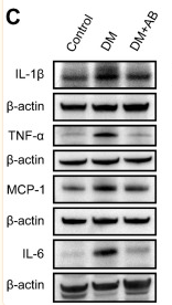
 The mRNA expression levels of IL-6, TNF-α, and MCP-1 in renal tissue
were analyzed by RT-PCR. (B) The protein expression levels of IL-6, TNF-α, and MCP-1were detected by western blot. (C-E) The ratio of IL-6, TNF-α, and MCP-1 to
GAPDH was calculated. Data are expressed as mean ± SEM (n = 6). **** p < 0.0001 vs. control; #### p < 0.0001 vs. model. The ns. means no significance.")
 Heatmap indicating serum chemokine distribution between the two groups (n = 10 in the T group, n = 9 in the TD group). (B) Heatmap indicating tumor tissue chemokine distribution between the two groups (n = 3 in each group). (C) Relative mRNA expression of tissue chemokines tested by qPCR (n = 10 in the T group, n = 9 in the TD group). (D) Relative protein expression of tissue chemokines tested by western blotting (n = 8 in each group). *p < 0.05, **p < 0.01, ***p < 0.001 compared with the T group.")
 Western blot and quantitative results of p65 in the nucleus of macrophages. §")
 Detection of miR-410 expression level in aorta tissues. (B) Arterial impairment and inflammation. (C) The area of aorta impairment. (D) Serum TNF-α, IL-1, and IL-6 levels in mice. (E) VCAM-1, ICAM-1, and MCP-1 expression in aorta tissues; ∗p < 0.05. Measured data are described as mean ± standard deviation, analyzed using independent samples t test between two groups; n = 10.")
 Relative mRNA expression level of MCP1 in the kidneys of rats from each group. (B) Relative mRNA expression level of VCAM1 in the kidneys of rats from each group. (C) Relative mRNA expression level of ICAM1 in the kidneys of rats from each group. (D) Western blot analysis of MCP1 (E), VCAM1 (F) and ICAM1 (G) levels in rats from each group. *P")
 The cell viability of DRGn. After 12 h pretreatment with RS504393 and ZQGCD containing serum (5%, 10%, and 15%), there was no change in cell viability (P>0.05). After adding CCL2 for 24 h, the viability of unpretreated cells decreased significantly (P0.05). (B) Expression of CCR2 and ASIC3 mRNA in DRGn. (C and D) Expression of CCR2 and ASIC3 proteins in DRGn. **P")
 Representative images (magnification: 40x) and semi-quantitative histogram of immunohistochemical staining of CCL2 protein of intervertebral disc. (C and D) Representative images (magnification: 40x) and semi-quantitative histogram of immunohistochemical staining of CCR2 protein of intervertebral disc.")
 Bar graphs showing the expression levels of CCL2 and CCR2 mRNA in DRG. (B and C) Immunoblot and bar graphs showing the expression levels of CCL2 and CCR2 in DRG.")
 and TNF-α, MCP-1 (red) in lung SpC+ (green) alveolar epithelial cells. B Quantitative analysis of fluorescence intensities for IL-6, TNF-α, IL-8 and MCP-1 in SpC+ alveolar epithelial cells. C Levels of pro-inflammatory mediators (IL-6, MCP-1, RANTES and TNF-α) in the lung homogenates were determined by Luminex assay. D Levels of pro-inflammatory mediators (IL-6, IL-8, IP-10, MCP-1, RANTES and TNF-α) were determined by Luminex assay.")
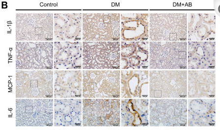
 MCP-1 expression assay using immunohistochemistry staining of paraffin embedded kidney sections (scale bar = 50 μm). (b) Mice were treated as shown and then relative IL-1β and TNF-αmRNA expression levels were detected by RT-PCR. (c) Quantitative results of (b). All quantitative data are shown as means ± SD, ∗∗P < 0.01 vs. the normal control group, ##P < 0.01 vs. the model group.")
 The expression of MCP-1 was detected using Western blot. (C) Cell viability was detected using MTT assay. (D and E) The expression of PCNA was detected using Western blot. (F and G) Cell cycle was detected using flow cytometry. (H and I) Wound healing experiment. (J and K) Transwell migration experiment. ***Represents P < 0.001 vs control group. ##Represents P < 0.01, ###Represents P < 0.001 vs ox-LDL group.")
 Relative expression of TXNIP mRNA in each group; (B) Relative expression of NLRP3 mRNA in each group; (C) Relative expression of MCP-1 mRNA in each group; (D) Western blot bands of TXNIP, NLRP3, and MCP-1 proteins; (E) Relative content of TXNIP protein in each group; (F) Relative content of NLRP3 protein in each group; (G) Relative content of MCP-1 protein in each group. ∗P < 0.05, ∗∗∗P < 0.001, compared with the normal control group; #P < 0.05, ##P < 0.01, ###P < 0.001, compared with the model group, n = 3. The original Western blot bands are shown in Figs. S3(A–D)-S5 (A–D) in the supplemental file.")
 and MPC5 cells (e) were analyzed by RT-PCR. The levels of IL-6, CXCL10, and MCP-1 protein in renal tissues (b) and MPC5 cells (f) were detected by Western blot. The levels of Bcl-2, Bax, and Bcl-xL mRNA in renal tissues (c) and MPC5 cells (g) were analyzed by RT-PCR. The levels of Bcl-2, cleaved caspase-3/caspase-3, Bax, and Bcl-xL protein in renal tissues (d) and MPC5 cells (h) were detected by Western blot. Western blot showed the levels of Beclin-1, LC3-II, Atg5, and p62 protein expression of renal tissues (i) and MPC5 cells (j). All data are represented as the mean ± SD (n = 3). *P < 0.05, **P < 0.01, ***P < 0.001, ****P < 0.0001.")
 The H&E staining in the human control cerebral artery and IA tissues (n = 4; 100 × and 300 ×; bar, 100 µm and 50 µm). (B) Immunohistochemical staining for α-SMA (n = 3; 300 ×; bar, 50 µm). (C) Immunohistochemical staining for SM22α (n = 3; 300 ×; bar, 50 µm). (D) The relative IOD/area of α-SMA and SM22α. (E) The bands of α-SMA and SM22α in the human control cerebral artery and IA tissues (n = 3; Western blot). (F) The relative expressions of α-SMA and SM22α. (G) The bands of MMP2 and MCP-1 in the human control cerebral artery and IA tissues (n = 3; Western blot). (H) The relative expressions of MMP2 and MCP-1. Data were expressed as mean ± SD, * P")

 Representative immunoblot and relative quantification of TLR4 and MyD88 in RAW264.7 cells. (D,E) Representative immunofluorescence images of TLR4-positive and MyD88-positive in RAW264.7 cells. Scale bars: 25 μm. (F–H) Representative immunoblot and relative quantification of p-IκBα (S32/S36), IκBα, p-p65 (S536), and p65 in RAW264.7 cells. (I) Representative immunofluorescence images of NF-κB p65 in RAW264.7 cells. Scale bars: 100 μm. (J–N) Representative immunoblot and relative quantification of CD68, MCP-1, ICAM1, and VCAM1 in RAW264.7 cells. (O–R) mRNA levels of Ccl2, Ccl3, Ccl4, and Cxcl10 in RAW264.7 cells. Data are presented as mean ± SEM (n = 3). * p < 0.05, ** p < 0.01, *** p < 0.001, and **** p < 0.0001 vs. the Con group. # p < 0.05, ## p < 0.01, ### p < 0.001, and #### p < 0.0001 vs. the LPS group.")
 AGS and (B) HGC-27 cells. Western blotting analysis the protein expression levels of VCAM1, ICAM1, PTGS2, IL6 and CCL2 (C) AGS and (D) HGC-27 cells. In order to save and recycle antibodies and save time, membranes were cut in horizontal strips at molecular weight ranges for target proteins and the information was shown in the Supplementary Fig. 1 and Fig. 2. Differences between two groups were assessed by Students t test, and differences among three groups were assessed by one-way analysis of variance (ANOVA).")
 for statistical analysis. Patients were divided into low- and high-expression groups by the median of mean integrated optical density. A Four representative images of CCL2 immunostaining in clinical HCC specimens. B, C The Kaplan–Meier curves for disease-free survival (DFS) and overall survival (OS) of this cohort according to low and high expression of CCL2. D Four representative images of CCR2 immunostaining in clinical HCC specimens. E–F Kaplan–Meier curves for DFS and OS according to the expression levels of CCR2. G Four representative images of CXCL8 immunostaining in clinical HCC specimens. H–I Kaplan–Meier DFS and OS curves according to the expression levels of CXCL8. J Four representative images of CXCR2 immunostaining in clinical HCC specimens. K–L Kaplan–Meier DFS and OS curves according to the expression levels of CXCR2. The log-rank p-value for Kaplan–Meier curve is shown on the Kaplan–Meier curve plot. p")
产品描述
*The optimal dilutions should be determined by the end user. For optimal experimental results, antibody reuse is not recommended.
*Tips:
WB: 适用于变性蛋白样本的免疫印迹检测. IHC: 适用于组织样本的石蜡(IHC-p)或冰冻(IHC-f)切片样本的免疫组化/荧光检测. IF/ICC: 适用于细胞样本的荧光检测. ELISA(peptide): 适用于抗原肽的ELISA检测.
引用格式: Affinity Biosciences Cat# DF7577, RRID:AB_2841069.
展开/折叠
C-C motif chemokine 2; CCL2; CCL2_HUMAN; Chemokine (C C motif) ligand 2; GDCF 2; GDCF-2; GDCF2; HC11; HSMCR30; JE; MCAF; MCP 1; MCP-1; MCP1; MGC9434; Monocyte chemoattractant protein 1; Monocyte chemotactic and activating factor; Monocyte chemotactic protein 1; Monocyte secretory protein JE; SCYA2; Small inducible cytokine A2 (monocyte chemotactic protein 1, homologous to mouse Sig je); Small inducible cytokine A2; Small inducible cytokine subfamily A (Cys Cys), member 2; Small-inducible cytokine A2; SMC CF; SMC-CF; SMCCF;
抗原和靶标
A synthesized peptide derived from human MCP1, corresponding to a region within the internal amino acids.
Expressed in the seminal plasma, endometrial fluid and follicular fluid (at protein level) (PubMed:23765988). Expressed in monocytes (PubMed:2513477).
- P13500 CCL2_HUMAN:
- Protein BLAST With
- NCBI/
- ExPASy/
- Uniprot
MKVSAALLCLLLIAATFIPQGLAQPDAINAPVTCCYNFTNRKISVQRLASYRRITSSKCPKEAVIFKTIVAKEICADPKQKWVQDSMDHLDKQTQTPKT
种属预测
score>80的预测可信度较高,可尝试用于WB检测。*预测模型主要基于免疫原序列比对,结果仅作参考,不作为质保凭据。
High(score>80) Medium(80>score>50) Low(score<50) No confidence
研究背景
Acts as a ligand for C-C chemokine receptor CCR2. Signals through binding and activation of CCR2 and induces a strong chemotactic response and mobilization of intracellular calcium ions. Exhibits a chemotactic activity for monocytes and basophils but not neutrophils or eosinophils. May be involved in the recruitment of monocytes into the arterial wall during the disease process of atherosclerosis.
Processing at the N-terminus can regulate receptor and target cell selectivity. Deletion of the N-terminal residue converts it from an activator of basophil to an eosinophil chemoattractant.
N-Glycosylated.
Secreted.
Expressed in the seminal plasma, endometrial fluid and follicular fluid (at protein level). Expressed in monocytes.
Belongs to the intercrine beta (chemokine CC) family.
研究领域
· Environmental Information Processing > Signaling molecules and interaction > Cytokine-cytokine receptor interaction. (View pathway)
· Environmental Information Processing > Signal transduction > TNF signaling pathway. (View pathway)
· Human Diseases > Infectious diseases: Parasitic > Chagas disease (American trypanosomiasis).
· Human Diseases > Infectious diseases: Parasitic > Malaria.
· Human Diseases > Infectious diseases: Viral > Influenza A.
· Human Diseases > Infectious diseases: Viral > Herpes simplex infection.
· Human Diseases > Immune diseases > Rheumatoid arthritis.
· Organismal Systems > Immune system > Chemokine signaling pathway. (View pathway)
· Organismal Systems > Immune system > NOD-like receptor signaling pathway. (View pathway)
· Organismal Systems > Immune system > IL-17 signaling pathway. (View pathway)
文献引用

Application: IHC Species: Mouse Sample:
Application: IHC Species: Rat Sample:
Application: WB Species: Rat Sample:

Application: WB Species: goat Sample: NPCs

Application: WB Species: Mouse Sample:
Application: WB Species: human Sample: HUVECs
Application: WB Species: Human Sample: HUVECs
限制条款
产品的规格、报价、验证数据请以官网为准,官网链接:www.affbiotech.com | www.affbiotech.cn(简体中文)| www.affbiotech.jp(日本語)产品的数据信息为Affinity所有,未经授权不得收集Affinity官网数据或资料用于商业用途,对抄袭产品数据的行为我们将保留诉诸法律的权利。
产品相关数据会因产品批次、产品检测情况随时调整,如您已订购该产品,请以订购时随货说明书为准,否则请以官网内容为准,官网内容有改动时恕不另行通知。
Affinity保证所销售产品均经过严格质量检测。如您购买的商品在规定时间内出现问题需要售后时,请您在Affinity官方渠道提交售后申请。产品仅供科学研究使用。不用于诊断和治疗。
产品未经授权不得转售。
Affinity Biosciences将不会对在使用我们的产品时可能发生的专利侵权或其他侵权行为负责。Affinity Biosciences, Affinity Biosciences标志和所有其他商标所有权归Affinity Biosciences LTD.