


产品描述
*The optimal dilutions should be determined by the end user.
*Tips:
WB: 适用于变性蛋白样本的免疫印迹检测. IHC: 适用于组织样本的石蜡(IHC-p)或冰冻(IHC-f)切片样本的免疫组化/荧光检测. IF/ICC: 适用于细胞样本的荧光检测. ELISA(peptide): 适用于抗原肽的ELISA检测.
引用格式: Affinity Biosciences Cat# DF9542, RRID:AB_2842738.
展开/折叠
C17orf33; Colony stimulating factor 3 (granulocyte); Colony stimulating factor 3; CSF 3; CSF beta; CSF3; CSF3_HUMAN; CSF3OS; Csfg; Filgrastim; G-CSF; GCSA; GCSF; Granulocyte colony stimulating factor; Granulocyte colony-stimulating factor; Lenograstim; Macrophage granulocyte inducer 2; MGC45931; MGI 2; Pluripoietin;
抗原和靶标
- P09919 CSF3_HUMAN:
- Protein BLAST With
- NCBI/
- ExPASy/
- Uniprot
MAGPATQSPMKLMALQLLLWHSALWTVQEATPLGPASSLPQSFLLKCLEQVRKIQGDGAALQEKLVSECATYKLCHPEELVLLGHSLGIPWAPLSSCPSQALQLAGCLSQLHSGLFLYQGLLQALEGISPELGPTLDTLQLDVADFATTIWQQMEELGMAPALQPTQGAMPAFASAFQRRAGGVLVASHLQSFLEVSYRVLRHLAQP
种属预测
score>80的预测可信度较高,可尝试用于WB检测。*预测模型主要基于免疫原序列比对,结果仅作参考,不作为质保凭据。
High(score>80) Medium(80>score>50) Low(score<50) No confidence
研究背景
Granulocyte/macrophage colony-stimulating factors are cytokines that act in hematopoiesis by controlling the production, differentiation, and function of 2 related white cell populations of the blood, the granulocytes and the monocytes-macrophages. This CSF induces granulocytes.
O-glycan consists of Gal-GalNAc disaccharide which can be modified with up to two sialic acid residues (done in recombinantly expressed G-CSF from CHO cells).
Secreted.
Monomer.
Belongs to the IL-6 superfamily.
研究领域
· Environmental Information Processing > Signaling molecules and interaction > Cytokine-cytokine receptor interaction. (View pathway)
· Environmental Information Processing > Signal transduction > PI3K-Akt signaling pathway. (View pathway)
· Environmental Information Processing > Signal transduction > Jak-STAT signaling pathway. (View pathway)
· Human Diseases > Infectious diseases: Parasitic > Malaria.
· Organismal Systems > Immune system > Hematopoietic cell lineage. (View pathway)
· Organismal Systems > Immune system > IL-17 signaling pathway. (View pathway)
文献引用
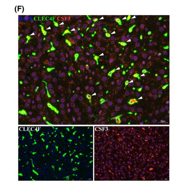
Application: IF/ICC Species: Rat Sample: Kupffer cells
限制条款
产品的规格、报价、验证数据请以官网为准,官网链接:www.affbiotech.com | www.affbiotech.cn(简体中文)| www.affbiotech.jp(日本語)产品的数据信息为Affinity所有,未经授权不得收集Affinity官网数据或资料用于商业用途,对抄袭产品数据的行为我们将保留诉诸法律的权利。
产品相关数据会因产品批次、产品检测情况随时调整,如您已订购该产品,请以订购时随货说明书为准,否则请以官网内容为准,官网内容有改动时恕不另行通知。
Affinity保证所销售产品均经过严格质量检测。如您购买的商品在规定时间内出现问题需要售后时,请您在Affinity官方渠道提交售后申请。产品仅供科学研究使用。不用于诊断和治疗。
产品未经授权不得转售。
Affinity Biosciences将不会对在使用我们的产品时可能发生的专利侵权或其他侵权行为负责。Affinity Biosciences, Affinity Biosciences标志和所有其他商标所有权归Affinity Biosciences LTD.