| 产品: | E-cadherin 抗体 |
| 货号: | AF7718 |
| 描述: | Rabbit polyclonal antibody to E-cadherin |
| 应用: | WB |
| 文献验证: | WB |
| 反应: | Human, Mouse, Rat |
| 预测: | Pig, Zebrafish, Bovine, Horse, Sheep, Rabbit, Dog, Chicken, Xenopus |
| 分子量: | 120kDa; 97kD(Calculated). |
| 蛋白号: | P12830 |
| RRID: | AB_2844082 |
产品描述
*The optimal dilutions should be determined by the end user.
*Tips:
WB: 适用于变性蛋白样本的免疫印迹检测. IHC: 适用于组织样本的石蜡(IHC-p)或冰冻(IHC-f)切片样本的免疫组化/荧光检测. IF/ICC: 适用于细胞样本的荧光检测. ELISA(peptide): 适用于抗原肽的ELISA检测.
引用格式: Affinity Biosciences Cat# AF7718, RRID:AB_2844082.
展开/折叠
Arc 1; CADH1_HUMAN; Cadherin 1; cadherin 1 type 1 E-cadherin; Cadherin1; CAM 120/80; CD 324; CD324; CD324 antigen; cdh1; CDHE; E-Cad/CTF3; E-cadherin; ECAD; Epithelial cadherin; epithelial calcium dependant adhesion protein; LCAM; Liver cell adhesion molecule; UVO; Uvomorulin;
抗原和靶标
- P12830 CADH1_HUMAN:
- Protein BLAST With
- NCBI/
- ExPASy/
- Uniprot
MGPWSRSLSALLLLLQVSSWLCQEPEPCHPGFDAESYTFTVPRRHLERGRVLGRVNFEDCTGRQRTAYFSLDTRFKVGTDGVITVKRPLRFHNPQIHFLVYAWDSTYRKFSTKVTLNTVGHHHRPPPHQASVSGIQAELLTFPNSSPGLRRQKRDWVIPPISCPENEKGPFPKNLVQIKSNKDKEGKVFYSITGQGADTPPVGVFIIERETGWLKVTEPLDRERIATYTLFSHAVSSNGNAVEDPMEILITVTDQNDNKPEFTQEVFKGSVMEGALPGTSVMEVTATDADDDVNTYNAAIAYTILSQDPELPDKNMFTINRNTGVISVVTTGLDRESFPTYTLVVQAADLQGEGLSTTATAVITVTDTNDNPPIFNPTTYKGQVPENEANVVITTLKVTDADAPNTPAWEAVYTILNDDGGQFVVTTNPVNNDGILKTAKGLDFEAKQQYILHVAVTNVVPFEVSLTTSTATVTVDVLDVNEAPIFVPPEKRVEVSEDFGVGQEITSYTAQEPDTFMEQKITYRIWRDTANWLEINPDTGAISTRAELDREDFEHVKNSTYTALIIATDNGSPVATGTGTLLLILSDVNDNAPIPEPRTIFFCERNPKPQVINIIDADLPPNTSPFTAELTHGASANWTIQYNDPTQESIILKPKMALEVGDYKINLKLMDNQNKDQVTTLEVSVCDCEGAAGVCRKAQPVEAGLQIPAILGILGGILALLILILLLLLFLRRRAVVKEPLLPPEDDTRDNVYYYDEEGGGEEDQDFDLSQLHRGLDARPEVTRNDVAPTLMSVPRYLPRPANPDEIGNFIDENLKAADTDPTAPPYDSLLVFDYEGSGSEAASLSSLNSSESDKDQDYDYLNEWGNRFKKLADMYGGGEDD
种属预测
score>80的预测可信度较高,可尝试用于WB检测。*预测模型主要基于免疫原序列比对,结果仅作参考,不作为质保凭据。
High(score>80) Medium(80>score>50) Low(score<50) No confidence
翻译修饰 - P12830 作为底物
| Site | PTM Type | Enzyme | Source |
|---|---|---|---|
| T66 | Phosphorylation | Uniprot | |
| Y68 | Phosphorylation | Uniprot | |
| S70 | Phosphorylation | Uniprot | |
| T211 | Phosphorylation | Uniprot | |
| T217 | O-Glycosylation | Uniprot | |
| T330 | Phosphorylation | Uniprot | |
| N558 | N-Glycosylation | Uniprot | |
| N570 | N-Glycosylation | Uniprot | |
| T576 | Phosphorylation | Uniprot | |
| T599 | Phosphorylation | Uniprot | |
| N622 | N-Glycosylation | Uniprot | |
| N637 | N-Glycosylation | Uniprot | |
| Y663 | Phosphorylation | Uniprot | |
| K738 | Ubiquitination | Uniprot | |
| T748 | Phosphorylation | Uniprot | |
| Y753 | Phosphorylation | Uniprot | |
| Y754 | Phosphorylation | Uniprot | |
| Y755 | Phosphorylation | Uniprot | |
| S770 | Phosphorylation | Uniprot | |
| T790 | Phosphorylation | Q05655 (PRKCD) | Uniprot |
| S793 | Phosphorylation | Uniprot | |
| Y797 | Phosphorylation | Uniprot | |
| S838 | Phosphorylation | Uniprot | |
| S840 | Phosphorylation | Uniprot | |
| S844 | Phosphorylation | P48729 (CSNK1A1) , P49674 (CSNK1E) , P48730 (CSNK1D) | Uniprot |
| S846 | Phosphorylation | Uniprot | |
| S847 | Phosphorylation | P68400 (CSNK2A1) | Uniprot |
| S850 | Phosphorylation | P68400 (CSNK2A1) | Uniprot |
| S851 | Phosphorylation | Uniprot | |
| S853 | Phosphorylation | P68400 (CSNK2A1) | Uniprot |
| K871 | Ubiquitination | Uniprot | |
| Y876 | Phosphorylation | Uniprot |
研究背景
Cadherins are calcium-dependent cell adhesion proteins. They preferentially interact with themselves in a homophilic manner in connecting cells; cadherins may thus contribute to the sorting of heterogeneous cell types. CDH1 is involved in mechanisms regulating cell-cell adhesions, mobility and proliferation of epithelial cells. Has a potent invasive suppressor role. It is a ligand for integrin alpha-E/beta-7.
E-Cad/CTF2 promotes non-amyloidogenic degradation of Abeta precursors. Has a strong inhibitory effect on APP C99 and C83 production.
(Microbial infection) Serves as a receptor for Listeria monocytogenes; internalin A (InlA) binds to this protein and promotes uptake of the bacteria.
During apoptosis or with calcium influx, cleaved by a membrane-bound metalloproteinase (ADAM10), PS1/gamma-secretase and caspase-3. Processing by the metalloproteinase, induced by calcium influx, causes disruption of cell-cell adhesion and the subsequent release of beta-catenin into the cytoplasm. The residual membrane-tethered cleavage product is rapidly degraded via an intracellular proteolytic pathway. Cleavage by caspase-3 releases the cytoplasmic tail resulting in disintegration of the actin microfilament system. The gamma-secretase-mediated cleavage promotes disassembly of adherens junctions. During development of the cochlear organ of Corti, cleavage by ADAM10 at adherens junctions promotes pillar cell separation (By similarity).
N-glycosylation at Asn-637 is essential for expression, folding and trafficking. Addition of bisecting N-acetylglucosamine by MGAT3 modulates its cell membrane location.
Ubiquitinated by a SCF complex containing SKP2, which requires prior phosphorylation by CK1/CSNK1A1. Ubiquitinated by CBLL1/HAKAI, requires prior phosphorylation at Tyr-754.
O-glycosylated. O-manosylated by TMTC1, TMTC2, TMTC3 or TMTC4. Thr-285 and Thr-509 are O-mannosylated by TMTC2 or TMTC4 but not TMTC1 or TMTC3.
Cell junction>Adherens junction. Cell membrane>Single-pass type I membrane protein. Endosome. Golgi apparatus>trans-Golgi network.
Note: Colocalizes with DLGAP5 at sites of cell-cell contact in intestinal epithelial cells. Anchored to actin microfilaments through association with alpha-, beta- and gamma-catenin. Sequential proteolysis induced by apoptosis or calcium influx, results in translocation from sites of cell-cell contact to the cytoplasm. Colocalizes with RAB11A endosomes during its transport from the Golgi apparatus to the plasma membrane.
Non-neural epithelial tissues.
Homodimer; disulfide-linked. Component of an E-cadherin/ catenin adhesion complex composed of at least E-cadherin/CDH1, beta-catenin/CTNNB1 or gamma-catenin/JUP, and potentially alpha-catenin/CTNNA1; the complex is located to adherens junctions. Interacts with the TRPV4 and CTNNB1 complex (By similarity). Interacts with CTNND1. The stable association of CTNNA1 is controversial as CTNNA1 was shown not to bind to F-actin when assembled in the complex (By similarity). Alternatively, the CTNNA1-containing complex may be linked to F-actin by other proteins such as LIMA1 (By similarity). Interaction with PSEN1, cleaves CDH1 resulting in the disassociation of cadherin-based adherens junctions (CAJs). Interacts with AJAP1 and DLGAP5. Interacts with TBC1D2. Interacts with LIMA1. Interacts with CAV1. Interacts with PIP5K1C. Interacts with RAB8B (By similarity). Interacts with RAPGEF2 (By similarity). Interacts with DDR1; this stabilizes CDH1 at the cell surface and inhibits its internalization. Interacts with KLRG1. Forms a ternary complex composed of ADAM10, CADH1 and EPHA4; within the complex, CADH1 is cleaved by ADAM10 which disrupts adherens junctions (By similarity).
(Microbial infection) Interacts with L.monocytogenes InlA. The formation of the complex between InlA and cadherin-1 is calcium-dependent.
Three calcium ions are usually bound at the interface of each cadherin domain and rigidify the connections, imparting a strong curvature to the full-length ectodomain.
研究领域
· Cellular Processes > Cellular community - eukaryotes > Adherens junction. (View pathway)
· Environmental Information Processing > Signal transduction > Rap1 signaling pathway. (View pathway)
· Environmental Information Processing > Signal transduction > Apelin signaling pathway. (View pathway)
· Environmental Information Processing > Signal transduction > Hippo signaling pathway. (View pathway)
· Environmental Information Processing > Signaling molecules and interaction > Cell adhesion molecules (CAMs). (View pathway)
· Human Diseases > Infectious diseases: Bacterial > Bacterial invasion of epithelial cells.
· Human Diseases > Infectious diseases: Bacterial > Pathogenic Escherichia coli infection.
· Human Diseases > Cancers: Overview > Pathways in cancer. (View pathway)
· Human Diseases > Cancers: Specific types > Endometrial cancer. (View pathway)
· Human Diseases > Cancers: Specific types > Thyroid cancer. (View pathway)
· Human Diseases > Cancers: Specific types > Melanoma. (View pathway)
· Human Diseases > Cancers: Specific types > Bladder cancer. (View pathway)
· Human Diseases > Cancers: Specific types > Gastric cancer. (View pathway)
文献引用
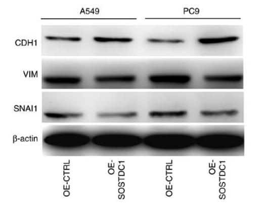
Application: WB Species: human Sample: A549 and PC9 cells
Application: WB Species: Human Sample: MG-63 cells
限制条款
产品的规格、报价、验证数据请以官网为准,官网链接:www.affbiotech.com | www.affbiotech.cn(简体中文)| www.affbiotech.jp(日本語)产品的数据信息为Affinity所有,未经授权不得收集Affinity官网数据或资料用于商业用途,对抄袭产品数据的行为我们将保留诉诸法律的权利。
产品相关数据会因产品批次、产品检测情况随时调整,如您已订购该产品,请以订购时随货说明书为准,否则请以官网内容为准,官网内容有改动时恕不另行通知。
Affinity保证所销售产品均经过严格质量检测。如您购买的商品在规定时间内出现问题需要售后时,请您在Affinity官方渠道提交售后申请。产品仅供科学研究使用。不用于诊断和治疗。
产品未经授权不得转售。
Affinity Biosciences将不会对在使用我们的产品时可能发生的专利侵权或其他侵权行为负责。Affinity Biosciences, Affinity Biosciences标志和所有其他商标所有权归Affinity Biosciences LTD.

