产品: MyoD1 抗体
货号: AF7733
描述: Rabbit polyclonal antibody to MyoD1
应用: WB IHC IF/ICC
反应: Human, Mouse, Rat, Sheep, Monkey
预测: Pig, Zebrafish, Bovine, Sheep, Rabbit, Dog, Chicken, Xenopus
分子量: 40~55kDa; 35kD(Calculated).
蛋白号: P15172
RRID: AB_2844097

浏览相似产品>>

   规格 价格 库存
 50ul RMB¥ 1250 现货
 100ul RMB¥ 2300 现货
 200ul RMB¥ 3000 现货

货期: 当天发货

联系销售

产品描述

来源:
Rabbit
应用:
WB 1:500-1:2000, IHC 1:50-1:200, IF/ICC 1:100-1:500
*The optimal dilutions should be determined by the end user.
*Tips:

WB: 适用于变性蛋白样本的免疫印迹检测. IHC: 适用于组织样本的石蜡(IHC-p)或冰冻(IHC-f)切片样本的免疫组化/荧光检测. IF/ICC: 适用于细胞样本的荧光检测. ELISA(peptide): 适用于抗原肽的ELISA检测.

反应:
Human,Mouse,Rat,Sheep,Monkey
预测:
Pig(100%), Zebrafish(100%), Bovine(100%), Rabbit(100%), Dog(100%), Chicken(100%), Xenopus(100%)
克隆:
Polyclonal
特异性:
MyoD1 Antibody detects endogenous levels of total MyoD1.
RRID:
AB_2844097
引用格式: Affinity Biosciences Cat# AF7733, RRID:AB_2844097.
偶联:
Unconjugated.
纯化:
The antiserum was purified by peptide affinity chromatography using SulfoLink™ Coupling Resin (Thermo Fisher Scientific).
保存:
Rabbit IgG in phosphate buffered saline , pH 7.4, 150mM NaCl, 0.02% sodium azide and 50% glycerol. Store at -20 °C. Stable for 12 months from date of receipt.
别名:

展开/折叠

bHLHc1; Class C basic helix-loop-helix protein 1; MYF 3; Myf-3; MYF3; Myoblast determination protein 1; Myod 1; MYOD; MYOD1; MYOD1_HUMAN; Myogenic differentiation 1; Myogenic factor 3; Myogenic factor MYF 3; Myogenin D1; PUM;

抗原和靶标

免疫原:
Uniprot:
基因/基因ID:
序列:
MELLSPPLRDVDLTAPDGSLCSFATTDDFYDDPCFDSPDLRFFEDLDPRLMHVGALLKPEEHSHFPAAVHPAPGAREDEHVRAPSGHHQAGRCLLWACKACKRKTTNADRRKAATMRERRRLSKVNEAFETLKRCTSSNPNQRLPKVEILRNAIRYIEGLQALLRDQDAAPPGAAAAFYAPGPLPPGRGGEHYSGDSDASSPRSNCSDGMMDYSGPPSGARRRNCYEGAYYNEAPSEPRPGKSAAVSSLDCLSSIVERISTESPAAPALLLADVPSESPPRRQEAAAPSEGESSGDPTQSPDAAPQCPAGANPNPIYQVL

种属预测

种属预测:

score>80的预测可信度较高,可尝试用于WB检测。*预测模型主要基于免疫原序列比对,结果仅作参考,不作为质保凭据。

Species
Results
Score
Pig
100
Bovine
100
Sheep
100
Dog
100
Xenopus
100
Zebrafish
100
Chicken
100
Rabbit
100
Horse
0
Model Confidence:
High(score>80) Medium(80>score>50) Low(score<50) No confidence

翻译修饰 - P15172 作为底物

Site PTM Type Enzyme
S5 Phosphorylation P06493 (CDK1)
Y30 Phosphorylation P00519 (ABL1)
K58 Acetylation
K99 Acetylation
K102 Acetylation
K104 Acetylation
K104 Methylation
T115 Phosphorylation P17252 (PRKCA)
Y156 Phosphorylation Q02750 (MAP2K1)
S200 Phosphorylation P53778 (MAPK12) , P06493 (CDK1) , P24941 (CDK2)
S201 Phosphorylation P53778 (MAPK12)
S204 Phosphorylation
S207 Phosphorylation
Y213 Phosphorylation
S214 Phosphorylation

研究背景

功能:

Acts as a transcriptional activator that promotes transcription of muscle-specific target genes and plays a role in muscle differentiation. Together with MYF5 and MYOG, co-occupies muscle-specific gene promoter core region during myogenesis. Induces fibroblasts to differentiate into myoblasts. Interacts with and is inhibited by the twist protein. This interaction probably involves the basic domains of both proteins (By similarity).

翻译修饰:

Phosphorylated by CDK9. This phosphorylation promotes its function in muscle differentiation.

Acetylated by a complex containing EP300 and PCAF. The acetylation is essential to activate target genes. Conversely, its deacetylation by SIRT1 inhibits its function (By similarity).

Ubiquitinated on the N-terminus; which is required for proteasomal degradation.

Methylation at Lys-104 by EHMT2/G9a inhibits myogenic activity.

细胞定位:

Nucleus.

Extracellular region or secreted Cytosol Plasma membrane Cytoskeleton Lysosome Endosome Peroxisome ER Golgi apparatus Nucleus Mitochondrion Manual annotation Automatic computational assertionSubcellular location
亚基结构:

Efficient DNA binding requires dimerization with another bHLH protein. Seems to form active heterodimers with ITF-2. Interacts with SUV39H1. Interacts with DDX5. Interacts with CHD2. Interacts with TSC22D3 (By similarity). Interacts with SETD3 (By similarity). Interacts with P-TEFB complex; promotes the transcriptional activity of MYOD1 through its CDK9-mediated phosphorylation (By similarity). Interacts with CSRP3. Interacts with NUPR1 (By similarity).

文献引用

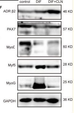
1). Sympathetic activity is correlated with satellite cell aging and myogenesis via β2-adrenoceptor. Stem Cell Research & Therapy, 2021 (PubMed: 34530910) [IF=7.5]

Application: IF/ICC    Species: Mice    Sample: SC cells

Fig. 5 CLB promoted skeletal muscle repair by affecting MyoD and Myf5. A–C CS decreased the number of PAX7+ cells (SCs), but the ratio of MyoD+/MyoD+PAX7+ cells was increased significantly. That is, CS led to the activation of SCs, which hindered their ability to maintain their stemness. CS + CLB treatment prevented this trend. In addition, the PAX7+ cell level and the ratio of MyoD+/MyoD+ PAX7+ cells were not significantly changed by CLB alone in normal mice (F = 15.363 and 5.911, P < 0.001 and = 0.03). D, E On the 10th and 42nd days after injury, the number of Myf5− PAX7+ cells (SCs) in the CS + CLB + injury group was significantly increased compared with that in the CS + injury group (F = 51.219, P < 0.001). F–I On the 10th and 42nd days after injury, the CS + injury group had the lowest number of myonuclei per muscle fiber (transverse section), and this effect was rescued by CLB. The CS + injury group had the largest fibrotic area (F = 11.644, 16.573, both P < 0.001) and the smallest muscle area (F = 11.644, 16.573, both P < 0.001), and this effect was rescued by CLB on both the 10th and 42nd days after injury. One-way ANOVA and Tukey’s test, ns, not significant, # all P < 0.05 compared with all other groups *P < 0.05; **P < 0.001

Application: WB    Species: Mice    Sample: SC cells

Fig. 6 CLB-inhibited myogenic differentiation. A, B After 5 days of differentiation, numerous fused myotubes were observed, while CLB (10 μm, 100 μm) significantly inhibited myotube formation (F = 28.9, P < 0.001). C–E After 48 h of myogenic differentiation, the levels of MyoD+ nuclei and β2-ADR were significantly increased, while CLB significantly decreased the expression of MyoD and further increased the expression of β2-ADR (F = 9.642, 8.157, both P < 0.001). F, G After 48 h of differentiation, β2-ADR expression was increased, and CLB further promoted this increase (F = 115.712, P < 0.001). PAX7 expression was decreased after differentiation, and CLB reversed this trend (F = 18.155, P < 0.001). MyoD, Myf5, and MyoG expressions were increased significantly after differentiation, and CLB significantly mitigated these increments (F = 16.324, 8.647, and 154.647, all P < 0.001). All differences were determined using one-way ANOVA and Tukey’s test. ns, not significant, *P < 0.01; **P < 0.001

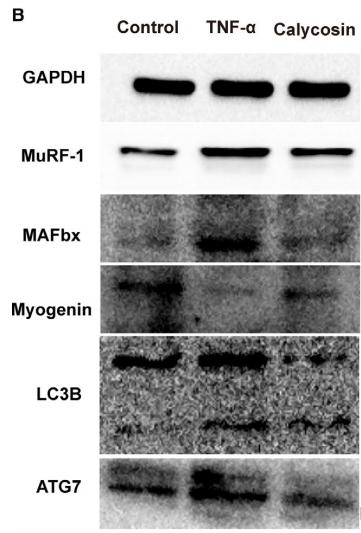
2). Calycosin inhibited autophagy and oxidative stress in chronic kidney disease skeletal muscle atrophy by regulating AMPK/SKP2/CARM1 signalling pathway. JOURNAL OF CELLULAR AND MOLECULAR MEDICINE, 2020 (PubMed: 32910538) [IF=5.3]

Application: WB    Species: mouse    Sample: C2C12 cells

FIGURE 8| Effect of calycosin on the expression of proteins associated with the AMPK/SKP2/CARM1 signalling pathway in TNF-αinduced C2C12 cells in vitro. A, Representative images of H3R17me2a in C2C12 cells treated with TNF-α or calycosin. B-C, Representative immunoblot of key proteins associated with the AMPK/SKP2/CARM1 signalling pathway in C2C12 cells treated with TNF-α or calycosin.

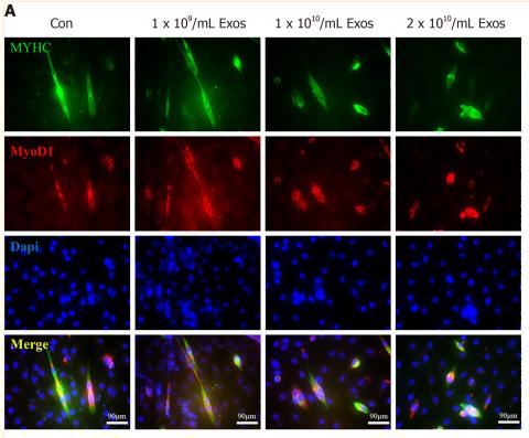
3). Exosomes derived from inflammatory myoblasts promote M1 polarization and break the balance of myoblast proliferation/differentiation. World Journal of Stem Cells, 2021 (PubMed: 34909122) [IF=4.1]

Application: IF/ICC    Species: Human    Sample: macrophages

Figure 7 IF-C2C12-Exos impaired early C2C12 muscle differentiation in vitro. A: Immunofluorescence was used to detect relative expression and distribution of MYHC and Myod1 on day 2 after different treatments. Green, red, and blue signals represent MYHC, Myod1, and nucleus, respectively. Scale bar = 90 μm; B, D and E: Quantification of myotube length, diameter, and fusion rate. Data are presented as mean ± SD. n = 12 or 3. aP < 0.05; C: Representative images of myotube after culture with different concentrations of IF-C2C12-Exosomes for 24 h by Giemsa staining (2 d 2% horse-serum incubation). Scale bar = 100 μm.

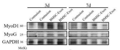
4). Bone Marrow Stromal Cell-Derived Exosomes promote muscle healing following contusion through macrophage polarization. STEM CELLS AND DEVELOPMENT, 2021 (PubMed: 33323007) [IF=4.0]

Application: WB    Species: Mice    Sample:

Figure 5:BMSC-Exos therapy increased the expression of muscle regeneration maker. (A-C) Myod1 and MyoG protein levels in tibialis anterior muscles of contusion and BMSCExos group at 3d and 7d were determined by western blot (n=4). Data are expressed as the means ± standard error of the mean. *P < 0.05. NS: not significant (D) Immunofluorescence was used to detect relative expression and distribution of Pax7 and Myod1 on day 3. Green, red, and blue signal represents Myod1, Pax7, and nucleus, respectively. 200× magnification. Arrows pointed to Pax7 and Myod1 positive cells.

限制条款

产品的规格、报价、验证数据请以官网为准,官网链接:www.affbiotech.com | www.affbiotech.cn(简体中文)| www.affbiotech.jp(日本語)

产品的数据信息为Affinity所有,未经授权不得收集Affinity官网数据或资料用于商业用途,对抄袭产品数据的行为我们将保留诉诸法律的权利。

产品相关数据会因产品批次、产品检测情况随时调整,如您已订购该产品,请以订购时随货说明书为准,否则请以官网内容为准,官网内容有改动时恕不另行通知。

Affinity保证所销售产品均经过严格质量检测。如您购买的商品在规定时间内出现问题需要售后时,请您在Affinity官方渠道提交售后申请。

产品仅供科学研究使用。不用于诊断和治疗。 

产品未经授权不得转售。

Affinity Biosciences将不会对在使用我们的产品时可能发生的专利侵权或其他侵权行为负责。Affinity Biosciences, Affinity Biosciences标志和所有其他商标所有权归Affinity Biosciences LTD.