


 HT-22 cells were treated with ethionine and rapamycin, autophagy related mark-LC3B, P62, Beclin-1 protein level in control and experiment groups were evaluated via Western blotting. (B) PINK1, Parkin protein levels in control and experiment groups were evaluated via Western blotting.")
 Relative protein expression of Parkin. The grey value was analyzed by Image J software.")
 Immunohistochemistry images of ALDH2, PINK1 and Parkin. All images were obtained at identical magnification, ×200, scale bar = 50 μm. (B) Quantitative analysis of ALDH2, PINK1 and Parkin content (n = 3). Data are represented as mean ± SEM. **P")
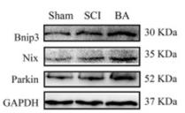
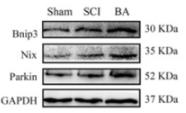
 Immunofluorescence staining for GSDMD (pyroptosis-related marker, green), C-CASP1 (pyroptosis-related marker, red), NIX (mitophagy-related marker, red) and DHE (indicating ROS-positive cells, red) in neurons in the spinal cord (original magnification 30×). Scale bar: 25 μm. (B–E) Quantitative analysis of levels of GSDMD (B), C-CASP1 (C), NIX (D) and DHE (E) in A. (F–H) The levels of 8-OHdG and AOPP in the spinal cord were detected by ELISA, and the levels of MDA were detected by the thiobarbituric acid assay. (I, L) Western blot assay for pyroptosis-related and mitophagy-related proteins. Data were normalized to GAPDH. (M) The levels of mitophagy-related genes in the spinal cord were detected by qPCR and normalized to β-actin. Data are expressed as the mean ± SEM (n = 6 mice per group). *P < 0.05 and **P < 0.01, vs. SCI group; #P < 0.05 and ##P < 0.01, vs. SCI + Bex group (one-way analysis of variance with the least significance difference post hoc test). ASC: Apoptosis-associated speck-like protein containing a CARD; Bex: bexarotene; BNIP3: BCL2/adenovirus E1B 19 kDa interacting protein 3; C-CASP-1: cleaved Caspase 1; DAPI: 4′,6-diamidino-2-phenylindole; DHE: dihydroethidium; ELISA: enzyme-linked immunosorbent assay; GAPDH: glyceraldehyde-3-phosphate dehydrogenase; GSDMD-N: gasdermin D-N; IOD: integrated optical density; MDA: malondialdehyde; NIX/BNIP3L: BCL2/adenovirus E1B 19 kDa interacting protein 3 like; NLRP3: NOD-like receptor thermal protein domain associated protein 3; SCI: spinal cord injury.")
 Immunofluorescence staining for GSDMD (pyroptosis-related marker, green), C-CASP1 (pyroptosis-related marker, red), NIX (mitophagy-related marker, red) and DHE (indicating ROS-positive cells, red) in neurons in the spinal cord (original magnification 30×). Scale bar: 25 μm. (B–E) Quantitative analysis of levels of GSDMD (B), C-CASP1 (C), NIX (D) and DHE (E) in A. (F–H) The levels of 8-OHdG and AOPP in the spinal cord were detected by ELISA, and the levels of MDA were detected by the thiobarbituric acid assay. (I, L) Western blot assay for pyroptosis-related and mitophagy-related proteins. Data were normalized to GAPDH. (M) The levels of mitophagy-related genes in the spinal cord were detected by qPCR and normalized to β-actin. Data are expressed as the mean ± SEM (n = 6 mice per group). *P < 0.05 and **P < 0.01, vs. SCI group; #P < 0.05 and ##P < 0.01, vs. SCI + Bex group (one-way analysis of variance with the least significance difference post hoc test). ASC: Apoptosis-associated speck-like protein containing a CARD; Bex: bexarotene; BNIP3: BCL2/adenovirus E1B 19 kDa interacting protein 3; C-CASP-1: cleaved Caspase 1; DAPI: 4′,6-diamidino-2-phenylindole; DHE: dihydroethidium; ELISA: enzyme-linked immunosorbent assay; GAPDH: glyceraldehyde-3-phosphate dehydrogenase; GSDMD-N: gasdermin D-N; IOD: integrated optical density; MDA: malondialdehyde; NIX/BNIP3L: BCL2/adenovirus E1B 19 kDa interacting protein 3 like; NLRP3: NOD-like receptor thermal protein domain associated protein 3; SCI: spinal cord injury.")
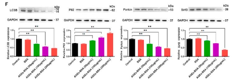
 Expression of autophagy-related genes. (B, C) Expression of autophagy-related proteins in the whole cell lysate. (D, E) Expression of mitophagy-related proteins in the mitochondria protein extract. n=6 per group and the values are the mean ± SEM. ** P")
 Colocalization of mitochondria and LC3. (B) The colocalization of the mitochondria and LC3 was quantitatively analyzed using the Pearson correlation coefficient. (C) Colocalization of mitochondria and lysosomes. (D) The colocalization of the mitochondria and lysosomes was quantitatively analyzed using the Pearson correlation coefficient. (E) Western blotting analysis of mitophagy-related proteins. (F and G) Quantitative analysis of PINK1 and Parkin protein expression. Mean ± SD. n=3. *P")
产品描述
*The optimal dilutions should be determined by the end user.
*Tips:
WB: 适用于变性蛋白样本的免疫印迹检测. IHC: 适用于组织样本的石蜡(IHC-p)或冰冻(IHC-f)切片样本的免疫组化/荧光检测. IF/ICC: 适用于细胞样本的荧光检测. ELISA(peptide): 适用于抗原肽的ELISA检测.
引用格式: Affinity Biosciences Cat# AF0235, RRID:AB_2833410.
展开/折叠
AR JP; E3 ubiquitin ligase; E3 ubiquitin protein ligase parkin; E3 ubiquitin-protein ligase parkin; FRA6E; LPRS 2; LPRS2; PARK 2; Park2; Parkin 2; Parkinson disease (autosomal recessive juvenile) 2; Parkinson disease (autosomal recessive, juvenile) 2, parkin; Parkinson disease protein 2; Parkinson juvenile disease protein 2; Parkinson protein 2 E3 ubiquitin protein ligase; Parkinson protein 2, E3 ubiquitin protein ligase (parkin); PDJ; PRKN 2; PRKN; PRKN2; PRKN2_HUMAN; Ubiquitin E3 ligase PRKN;
抗原和靶标
Highly expressed in the brain including the substantia nigra. Expressed in heart, testis and skeletal muscle. Expression is down-regulated or absent in tumor biopsies, and absent in the brain of PARK2 patients. Overexpression protects dopamine neurons from kainate-mediated apoptosis. Found in serum (at protein level).
- O60260 PRKN_HUMAN:
- Protein BLAST With
- NCBI/
- ExPASy/
- Uniprot
MIVFVRFNSSHGFPVEVDSDTSIFQLKEVVAKRQGVPADQLRVIFAGKELRNDWTVQNCDLDQQSIVHIVQRPWRKGQEMNATGGDDPRNAAGGCEREPQSLTRVDLSSSVLPGDSVGLAVILHTDSRKDSPPAGSPAGRSIYNSFYVYCKGPCQRVQPGKLRVQCSTCRQATLTLTQGPSCWDDVLIPNRMSGECQSPHCPGTSAEFFFKCGAHPTSDKETSVALHLIATNSRNITCITCTDVRSPVLVFQCNSRHVICLDCFHLYCVTRLNDRQFVHDPQLGYSLPCVAGCPNSLIKELHHFRILGEEQYNRYQQYGAEECVLQMGGVLCPRPGCGAGLLPEPDQRKVTCEGGNGLGCGFAFCRECKEAYHEGECSAVFEASGTTTQAYRVDERAAEQARWEAASKETIKKTTKPCPRCHVPVEKNGGCMHMKCPQPQCRLEWCWNCGCEWNRVCMGDHWFDV
研究背景
Functions within a multiprotein E3 ubiquitin ligase complex, catalyzing the covalent attachment of ubiquitin moieties onto substrate proteins, such as BCL2, SYT11, CCNE1, GPR37, RHOT1/MIRO1, MFN1, MFN2, STUB1, SNCAIP, SEPTIN5, TOMM20, USP30, ZNF746 and AIMP2. Mediates monoubiquitination as well as 'Lys-6', 'Lys-11', 'Lys-48'-linked and 'Lys-63'-linked polyubiquitination of substrates depending on the context. Participates in the removal and/or detoxification of abnormally folded or damaged protein by mediating 'Lys-63'-linked polyubiquitination of misfolded proteins such as PARK7: 'Lys-63'-linked polyubiquitinated misfolded proteins are then recognized by HDAC6, leading to their recruitment to aggresomes, followed by degradation. Mediates 'Lys-63'-linked polyubiquitination of a 22 kDa O-linked glycosylated isoform of SNCAIP, possibly playing a role in Lewy-body formation. Mediates monoubiquitination of BCL2, thereby acting as a positive regulator of autophagy. Promotes the autophagic degradation of dysfunctional depolarized mitochondria (mitophagy) by promoting the ubiquitination of mitochondrial proteins such as TOMM20, RHOT1/MIRO1 and USP30. Preferentially assembles 'Lys-6'-, 'Lys-11'- and 'Lys-63'-linked polyubiquitin chains following mitochondrial damage, leading to mitophagy. Mediates 'Lys-48'-linked polyubiquitination of ZNF746, followed by degradation of ZNF746 by the proteasome; possibly playing a role in the regulation of neuron death. Limits the production of reactive oxygen species (ROS). Regulates cyclin-E during neuronal apoptosis. In collaboration with CHPF isoform 2, may enhance cell viability and protect cells from oxidative stress. Independently of its ubiquitin ligase activity, protects from apoptosis by the transcriptional repression of p53/TP53. May protect neurons against alpha synuclein toxicity, proteasomal dysfunction, GPR37 accumulation, and kainate-induced excitotoxicity. May play a role in controlling neurotransmitter trafficking at the presynaptic terminal and in calcium-dependent exocytosis. May represent a tumor suppressor gene.
Auto-ubiquitinates in an E2-dependent manner leading to its own degradation. Also polyubiquitinated by RNF41 for proteasomal degradation.
S-nitrosylated. The inhibition of PRKN ubiquitin E3 ligase activity by S-nitrosylation could contribute to the degenerative process in PD by impairing the ubiquitination of PRKN substrates.
Phosphorylation at Ser-65 by PINK1 contributes to activate PRKN activity. It is however not sufficient and requires binding to phosphorylated ubiquitin as well.
Cytoplasm>Cytosol. Nucleus. Endoplasmic reticulum. Mitochondrion.
Note: Mainly localizes in the cytosol. Co-localizes with SYT11 in neutrites. Co-localizes with SNCAIP in brainstem Lewy bodies. Mitochondrial localization gradually increases with cellular growth. Also relocates to dysfunctional mitochondria that have lost the mitochondrial membrane potential; recruitment to mitochondria is PINK1-dependent.
Highly expressed in the brain including the substantia nigra. Expressed in heart, testis and skeletal muscle. Expression is down-regulated or absent in tumor biopsies, and absent in the brain of PARK2 patients. Overexpression protects dopamine neurons from kainate-mediated apoptosis. Found in serum (at protein level).
Forms an E3 ubiquitin ligase complex with UBE2L3 or UBE2L6. Mediates 'Lys-63'-linked polyubiquitination by associating with UBE2V1. Part of a SCF-like complex, consisting of PRKN, CUL1 and FBXW7. Interacts with SNCAIP. Binds to the C2A and C2B domains of SYT11. Interacts and regulates the turnover of SEPTIN5. Part of a complex, including STUB1, HSP70 and GPR37. The amount of STUB1 in the complex increases during ER stress. STUB1 promotes the dissociation of HSP70 from PRKN and GPR37, thus facilitating PRKN-mediated GPR37 ubiquitination. HSP70 transiently associates with unfolded GPR37 and inhibits the E3 activity of PRKN, whereas, STUB1 enhances the E3 activity of PRKN through promotion of dissociation of HSP70 from PRKN-GPR37 complexes. Interacts with PSMD4 and PACRG. Interacts with LRRK2. Interacts with RANBP2. Interacts with SUMO1 but not SUMO2, which promotes nuclear localization and autoubiquitination. Interacts (via first RING-type domain) with AIMP2 (via N-terminus). Interacts with PSMA7 and RNF41. Interacts with PINK1. Interacts with CHPF, the interaction with isoform 2 may facilitate PRKN transport into the mitochondria. Interacts with MFN2 (phosphorylated), promotes PRKN localization in dysfunctional depolarized mitochondria. Interacts with FBXO7; this promotes translocation to dysfunctional depolarized mitochondria. Interacts with heat shock protein 70 family members, including HSPA1L, HSPA1A and HSPA8; interaction HSPA1L promotes translocation to damaged mitochondria. Interacts with BAG4 and, to a lesser extent, BAG5; interaction with BAG4 inhibits translocation to damaged mitochondria. Forms a complex with PINK1 and PARK7.
The ubiquitin-like domain binds the PSMD4 subunit of 26S proteasomes.
The RING-type 1 zinc finger domain is required to repress p53/TP53 transcription.
Members of the RBR family are atypical E3 ligases. They interact with the E2 conjugating enzyme UBE2L3 and function like HECT-type E3 enzymes: they bind E2s via the first RING domain, but require an obligate trans-thiolation step during the ubiquitin transfer, requiring a conserved cysteine residue in the second RING domain (PubMed:21532592).
Belongs to the RBR family. Parkin subfamily.
研究领域
· Genetic Information Processing > Folding, sorting and degradation > Ubiquitin mediated proteolysis. (View pathway)
· Genetic Information Processing > Folding, sorting and degradation > Protein processing in endoplasmic reticulum. (View pathway)
· Human Diseases > Neurodegenerative diseases > Parkinson's disease.
文献引用
Application: WB Species: mice Sample: bone marrow mesenchymal stem (BMSCs)
Application: WB Species: Mice Sample: spinal cords
Application: WB Species: Mice Sample: spinal cords
限制条款
产品的规格、报价、验证数据请以官网为准,官网链接:www.affbiotech.com | www.affbiotech.cn(简体中文)| www.affbiotech.jp(日本語)产品的数据信息为Affinity所有,未经授权不得收集Affinity官网数据或资料用于商业用途,对抄袭产品数据的行为我们将保留诉诸法律的权利。
产品相关数据会因产品批次、产品检测情况随时调整,如您已订购该产品,请以订购时随货说明书为准,否则请以官网内容为准,官网内容有改动时恕不另行通知。
Affinity保证所销售产品均经过严格质量检测。如您购买的商品在规定时间内出现问题需要售后时,请您在Affinity官方渠道提交售后申请。产品仅供科学研究使用。不用于诊断和治疗。
产品未经授权不得转售。
Affinity Biosciences将不会对在使用我们的产品时可能发生的专利侵权或其他侵权行为负责。Affinity Biosciences, Affinity Biosciences标志和所有其他商标所有权归Affinity Biosciences LTD.