产品: SYVN1 抗体
货号: DF12235
描述: Rabbit polyclonal antibody to SYVN1
应用: WB IHC IF/ICC
反应: Human, Mouse, Rat
预测: Pig, Bovine, Horse, Sheep, Rabbit, Dog
分子量: 68-76 kDa; 68kD(Calculated).
蛋白号: Q86TM6
RRID: AB_2845040

浏览相似产品>>

   规格 价格 库存
 50ul RMB¥ 1250 现货
 100ul RMB¥ 2300 现货
 200ul RMB¥ 3000 现货

货期: 当天发货

联系销售

产品描述

来源:
Rabbit
应用:
WB 1:500-1:2000, IHC 1:50-1:200, IF/ICC 1:100-1:500
*The optimal dilutions should be determined by the end user.
*Tips:

WB: 适用于变性蛋白样本的免疫印迹检测. IHC: 适用于组织样本的石蜡(IHC-p)或冰冻(IHC-f)切片样本的免疫组化/荧光检测. IF/ICC: 适用于细胞样本的荧光检测. ELISA(peptide): 适用于抗原肽的ELISA检测.

反应:
Human,Mouse,Rat
预测:
Pig(100%), Bovine(100%), Horse(100%), Sheep(100%), Rabbit(100%), Dog(100%)
克隆:
Polyclonal
特异性:
SYVN1 Antibody detects endogenous levels of total SYVN1.
RRID:
AB_2845040
引用格式: Affinity Biosciences Cat# DF12235, RRID:AB_2845040.
偶联:
Unconjugated.
纯化:
The antiserum was purified by peptide affinity chromatography using SulfoLink™ Coupling Resin (Thermo Fisher Scientific).
保存:
Rabbit IgG in phosphate buffered saline , pH 7.4, 150mM NaCl, 0.02% sodium azide and 50% glycerol. Store at -20 °C. Stable for 12 months from date of receipt.
别名:

展开/折叠

1200010C09Rik; DER3; E3 ubiquitin-protein ligase synoviolin; HMG coA reductase degradation 1 homolog; HRD1; KIAA1810; MGC40372; OTTHUMP00000230429; OTTHUMP00000230430; OTTHUMP00000230431; OTTHUMP00000230432; Synovial apoptosis inhibitor 1; Synovial apoptosis inhibitor 1, synoviolin; Synoviolin 1 isoform b; SYNOVIOLIN; SYVN1; SYVN1_HUMAN;

抗原和靶标

免疫原:
Uniprot:
基因/基因ID:
表达:
Q86TM6 SYVN1_HUMAN:

Ubiquitously expressed, with highest levels in liver and kidney (at protein level). Up-regulated in synovial tissues from patients with rheumatoid arthritis (at protein level).

序列:
MFRTAVMMAASLALTGAVVAHAYYLKHQFYPTVVYLTKSSPSMAVLYIQAFVLVFLLGKVMGKVFFGQLRAAEMEHLLERSWYAVTETCLAFTVFRDDFSPRFVALFTLLLFLKCFHWLAEDRVDFMERSPNISWLFHCRIVSLMFLLGILDFLFVSHAYHSILTRGASVQLVFGFEYAILMTMVLTIFIKYVLHSVDLQSENPWDNKAVYMLYTELFTGFIKVLLYMAFMTIMIKVHTFPLFAIRPMYLAMRQFKKAVTDAIMSRRAIRNMNTLYPDATPEELQAMDNVCIICREEMVTGAKRLPCNHIFHTSCLRSWFQRQQTCPTCRMDVLRASLPAQSPPPPEPADQGPPPAPHPPPLLPQPPNFPQGLLPPFPPGMFPLWPPMGPFPPVPPPPSSGEAVAPPSTSAAALSRPSGAATTTAAGTSATAASATASGPGSGSAPEAGPAPGFPFPPPWMGMPLPPPFAFPPMPVPPAGFAGLTPEELRALEGHERQHLEARLQSLRNIHTLLDAAMLQINQYLTVLASLGPPRPATSVNSTEETATTVVAAASSTSIPSSEATTPTPGASPPAPEMERPPAPESVGTEEMPEDGEPDAAELRRRRLQKLESPVAH

种属预测

种属预测:

score>80的预测可信度较高,可尝试用于WB检测。*预测模型主要基于免疫原序列比对,结果仅作参考,不作为质保凭据。

Species
Results
Score
Pig
100
Horse
100
Bovine
100
Sheep
100
Dog
100
Rabbit
100
Xenopus
67
Zebrafish
67
Chicken
0
Model Confidence:
High(score>80) Medium(80>score>50) Low(score<50) No confidence

翻译修饰 - Q86TM6 作为底物

Site PTM Type Enzyme
T93 Phosphorylation
K191 Ubiquitination
K257 Ubiquitination
T260 Phosphorylation
S265 Phosphorylation
K303 Ubiquitination
T557 Phosphorylation
S558 Phosphorylation
S562 Phosphorylation
T565 Phosphorylation
S572 Phosphorylation
S613 Phosphorylation

研究背景

功能:

Acts as an E3 ubiquitin-protein ligase which accepts ubiquitin specifically from endoplasmic reticulum-associated UBC7 E2 ligase and transfers it to substrates, promoting their degradation. Component of the endoplasmic reticulum quality control (ERQC) system also called ER-associated degradation (ERAD) involved in ubiquitin-dependent degradation of misfolded endoplasmic reticulum proteins. Also promotes the degradation of normal but naturally short-lived proteins such as SGK. Protects cells from ER stress-induced apoptosis. Protects neurons from apoptosis induced by polyglutamine-expanded huntingtin (HTT) or unfolded GPR37 by promoting their degradation. Sequesters p53/TP53 in the cytoplasm and promotes its degradation, thereby negatively regulating its biological function in transcription, cell cycle regulation and apoptosis. Mediates the ubiquitination and subsequent degradation of cytoplasmic NFE2L1 (By similarity).

翻译修饰:

Not N-glycosylated.

Auto-ubiquitinated.

细胞定位:

Endoplasmic reticulum membrane>Multi-pass membrane protein.

Extracellular region or secreted Cytosol Plasma membrane Cytoskeleton Lysosome Endosome Peroxisome ER Golgi apparatus Nucleus Mitochondrion Manual annotation Automatic computational assertionSubcellular location
组织特异性:

Ubiquitously expressed, with highest levels in liver and kidney (at protein level). Up-regulated in synovial tissues from patients with rheumatoid arthritis (at protein level).

亚基结构:

Homodimer. Interacts with p53/TP53 and HTT. Interacts with VCP, HERPUD1 and DERL1. Part of a complex containing SYVN1, HERPUD1, VIMP and DERL1; which probably transfer misfolded proteins from the ER to VCP. Part of a complex containing SYVN1, SEL1L and DERL2. Interacts with UBXN6. Interacts (via N-terminal loop) with SEL1L; recruits ERLEC1 and OS9. May form a complex with ERLEC1; HSPA5; OS9 AND SEL1L. Interacts with BAG6. Interacts with NFE2L1 (By similarity).

蛋白家族:

The RING-type zinc finger is required for E3 ligase activity.

Belongs to the HRD1 family.

研究领域

· Genetic Information Processing > Folding, sorting and degradation > Ubiquitin mediated proteolysis.   (View pathway)

· Genetic Information Processing > Folding, sorting and degradation > Protein processing in endoplasmic reticulum.   (View pathway)

文献引用

1). Endoplasmic reticulum stress-triggered ferroptosis via the XBP1-Hrd1-Nrf2 pathway induces EMT progression in diabetic nephropathy.. Biomedicine & Pharmacotherapy (PubMed: 37224754) [IF=7.5]

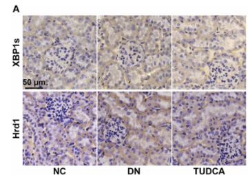
Application: IHC    Species: Mouse    Sample: kidney tissues

Fig. 6. The XBP1-Hrd1 arm of the ERS pathway was triggered by high glucose. The XBP1s and Hrd1 expression in tissue samples was detected by immunohistochemical staining (n = 3) (A). Semi-quantitative analysis of XBP1s and Hrd1 expression in each group (n = 3) (B). The qRT-PCR results of Hrd1 in NC, DM, TUDCA, and PBS group (n = 3) (C). The protein expression of Hrd1 in each group of mice (n = 3) (D - E). The fluorescence intensity of XBP1s and Hrd1 in HK-2 cells was measured by the confocal fluorescence microscope. Blue represented the cell nucleus and red represented XBP1s or Hrd1 (n = 3) (F - G). Fluorescence intensity was quantified using Image J (n = 3) (H - I). The mRNA level of Hrd1 among Ctrl, HG, Mannitol, TUDCA, and DMSO groups (n = 3) (J). Immunoblot and semi-quantitative analysis of Hrd1 in each group cells (n = 3) (K - L). The qRT-PCR results of XBP1s and Hrd1 in cells transfected with empty vectors or XBP1 overexpression plasmids. At 6 h post-transfection, cells were changed to a medium containing 30 mmol/L glucose with or without TUDCA (200 μM) for 48 h (n = 3) (M). Immunoblot and quantitative analysis of XBP1s and Hrd1 under the same conditions (n = 3) (N - O). Data are expressed as the mean ± SEM.

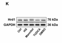
Application: WB    Species: Human    Sample: HK-2 cells

Fig. 6. The XBP1-Hrd1 arm of the ERS pathway was triggered by high glucose. The XBP1s and Hrd1 expression in tissue samples was detected by immunohistochemical staining (n = 3) (A). Semi-quantitative analysis of XBP1s and Hrd1 expression in each group (n = 3) (B). The qRT-PCR results of Hrd1 in NC, DM, TUDCA, and PBS group (n = 3) (C). The protein expression of Hrd1 in each group of mice (n = 3) (D - E). The fluorescence intensity of XBP1s and Hrd1 in HK-2 cells was measured by the confocal fluorescence microscope. Blue represented the cell nucleus and red represented XBP1s or Hrd1 (n = 3) (F - G). Fluorescence intensity was quantified using Image J (n = 3) (H - I). The mRNA level of Hrd1 among Ctrl, HG, Mannitol, TUDCA, and DMSO groups (n = 3) (J). Immunoblot and semi-quantitative analysis of Hrd1 in each group cells (n = 3) (K - L). The qRT-PCR results of XBP1s and Hrd1 in cells transfected with empty vectors or XBP1 overexpression plasmids. At 6 h post-transfection, cells were changed to a medium containing 30 mmol/L glucose with or without TUDCA (200 μM) for 48 h (n = 3) (M). Immunoblot and quantitative analysis of XBP1s and Hrd1 under the same conditions (n = 3) (N - O). Data are expressed as the mean ± SEM.

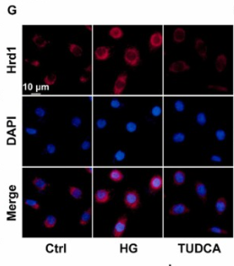
Application: IF/ICC    Species: Human    Sample: HK-2 cells

Fig. 6. The XBP1-Hrd1 arm of the ERS pathway was triggered by high glucose. The XBP1s and Hrd1 expression in tissue samples was detected by immunohistochemical staining (n = 3) (A). Semi-quantitative analysis of XBP1s and Hrd1 expression in each group (n = 3) (B). The qRT-PCR results of Hrd1 in NC, DM, TUDCA, and PBS group (n = 3) (C). The protein expression of Hrd1 in each group of mice (n = 3) (D - E). The fluorescence intensity of XBP1s and Hrd1 in HK-2 cells was measured by the confocal fluorescence microscope. Blue represented the cell nucleus and red represented XBP1s or Hrd1 (n = 3) (F - G). Fluorescence intensity was quantified using Image J (n = 3) (H - I). The mRNA level of Hrd1 among Ctrl, HG, Mannitol, TUDCA, and DMSO groups (n = 3) (J). Immunoblot and semi-quantitative analysis of Hrd1 in each group cells (n = 3) (K - L). The qRT-PCR results of XBP1s and Hrd1 in cells transfected with empty vectors or XBP1 overexpression plasmids. At 6 h post-transfection, cells were changed to a medium containing 30 mmol/L glucose with or without TUDCA (200 μM) for 48 h (n = 3) (M). Immunoblot and quantitative analysis of XBP1s and Hrd1 under the same conditions (n = 3) (N - O). Data are expressed as the mean ± SEM.

限制条款

产品的规格、报价、验证数据请以官网为准,官网链接:www.affbiotech.com | www.affbiotech.cn(简体中文)| www.affbiotech.jp(日本語)

产品的数据信息为Affinity所有,未经授权不得收集Affinity官网数据或资料用于商业用途,对抄袭产品数据的行为我们将保留诉诸法律的权利。

产品相关数据会因产品批次、产品检测情况随时调整,如您已订购该产品,请以订购时随货说明书为准,否则请以官网内容为准,官网内容有改动时恕不另行通知。

Affinity保证所销售产品均经过严格质量检测。如您购买的商品在规定时间内出现问题需要售后时,请您在Affinity官方渠道提交售后申请。

产品仅供科学研究使用。不用于诊断和治疗。 

产品未经授权不得转售。

Affinity Biosciences将不会对在使用我们的产品时可能发生的专利侵权或其他侵权行为负责。Affinity Biosciences, Affinity Biosciences标志和所有其他商标所有权归Affinity Biosciences LTD.