产品描述
*The optimal dilutions should be determined by the end user.
*Tips:
WB: 适用于变性蛋白样本的免疫印迹检测. IHC: 适用于组织样本的石蜡(IHC-p)或冰冻(IHC-f)切片样本的免疫组化/荧光检测. IF/ICC: 适用于细胞样本的荧光检测. ELISA(peptide): 适用于抗原肽的ELISA检测.
引用格式: Affinity Biosciences Cat# DF12589, RRID:AB_2845551.
展开/折叠
4732461B14Rik; AN2; AN2 proteoglycan; Chondroitin sulfate proteoglycan 4 (melanoma-associated); Chondroitin sulfate proteoglycan 4; Chondroitin sulfate proteoglycan NG2; Cspg4; Cspg4 chondroitin sulfate proteoglycan 4; CSPG4_HUMAN; HMW-MAA; HSN tumor-specific antigen; Kiaa4232; MCSP; MCSPG; MEL-CSPG; Melanoma chondroitin sulfate proteoglycan; Melanoma-associated chondroitin sulfate proteoglycan; MELCSPG; MSK16; NG2;
抗原和靶标
- Q6UVK1 CSPG4_HUMAN:
- Protein BLAST With
- NCBI/
- ExPASy/
- Uniprot
MQSGPRPPLPAPGLALALTLTMLARLASAASFFGENHLEVPVATALTDIDLQLQFSTSQPEALLLLAAGPADHLLLQLYSGRLQVRLVLGQEELRLQTPAETLLSDSIPHTVVLTVVEGWATLSVDGFLNASSAVPGAPLEVPYGLFVGGTGTLGLPYLRGTSRPLRGCLHAATLNGRSLLRPLTPDVHEGCAEEFSASDDVALGFSGPHSLAAFPAWGTQDEGTLEFTLTTQSRQAPLAFQAGGRRGDFIYVDIFEGHLRAVVEKGQGTVLLHNSVPVADGQPHEVSVHINAHRLEISVDQYPTHTSNRGVLSYLEPRGSLLLGGLDAEASRHLQEHRLGLTPEATNASLLGCMEDLSVNGQRRGLREALLTRNMAAGCRLEEEEYEDDAYGHYEAFSTLAPEAWPAMELPEPCVPEPGLPPVFANFTQLLTISPLVVAEGGTAWLEWRHVQPTLDLMEAELRKSQVLFSVTRGARHGELELDIPGAQARKMFTLLDVVNRKARFIHDGSEDTSDQLVLEVSVTARVPMPSCLRRGQTYLLPIQVNPVNDPPHIIFPHGSLMVILEHTQKPLGPEVFQAYDPDSACEGLTFQVLGTSSGLPVERRDQPGEPATEFSCRELEAGSLVYVHRGGPAQDLTFRVSDGLQASPPATLKVVAIRPAIQIHRSTGLRLAQGSAMPILPANLSVETNAVGQDVSVLFRVTGALQFGELQKQGAGGVEGAEWWATQAFHQRDVEQGRVRYLSTDPQHHAYDTVENLALEVQVGQEILSNLSFPVTIQRATVWMLRLEPLHTQNTQQETLTTAHLEATLEEAGPSPPTFHYEVVQAPRKGNLQLQGTRLSDGQGFTQDDIQAGRVTYGATARASEAVEDTFRFRVTAPPYFSPLYTFPIHIGGDPDAPVLTNVLLVVPEGGEGVLSADHLFVKSLNSASYLYEVMERPRHGRLAWRGTQDKTTMVTSFTNEDLLRGRLVYQHDDSETTEDDIPFVATRQGESSGDMAWEEVRGVFRVAIQPVNDHAPVQTISRIFHVARGGRRLLTTDDVAFSDADSGFADAQLVLTRKDLLFGSIVAVDEPTRPIYRFTQEDLRKRRVLFVHSGADRGWIQLQVSDGQHQATALLEVQASEPYLRVANGSSLVVPQGGQGTIDTAVLHLDTNLDIRSGDEVHYHVTAGPRWGQLVRAGQPATAFSQQDLLDGAVLYSHNGSLSPRDTMAFSVEAGPVHTDATLQVTIALEGPLAPLKLVRHKKIYVFQGEAAEIRRDQLEAAQEAVPPADIVFSVKSPPSAGYLVMVSRGALADEPPSLDPVQSFSQEAVDTGRVLYLHSRPEAWSDAFSLDVASGLGAPLEGVLVELEVLPAAIPLEAQNFSVPEGGSLTLAPPLLRVSGPYFPTLLGLSLQVLEPPQHGALQKEDGPQARTLSAFSWRMVEEQLIRYVHDGSETLTDSFVLMANASEMDRQSHPVAFTVTVLPVNDQPPILTTNTGLQMWEGATAPIPAEALRSTDGDSGSEDLVYTIEQPSNGRVVLRGAPGTEVRSFTQAQLDGGLVLFSHRGTLDGGFRFRLSDGEHTSPGHFFRVTAQKQVLLSLKGSQTLTVCPGSVQPLSSQTLRASSSAGTDPQLLLYRVVRGPQLGRLFHAQQDSTGEALVNFTQAEVYAGNILYEHEMPPEPFWEAHDTLELQLSSPPARDVAATLAVAVSFEAACPQRPSHLWKNKGLWVPEGQRARITVAALDASNLLASVPSPQRSEHDVLFQVTQFPSRGQLLVSEEPLHAGQPHFLQSQLAAGQLVYAHGGGGTQQDGFHFRAHLQGPAGASVAGPQTSEAFAITVRDVNERPPQPQASVPLRLTRGSRAPISRAQLSVVDPDSAPGEIEYEVQRAPHNGFLSLVGGGLGPVTRFTQADVDSGRLAFVANGSSVAGIFQLSMSDGASPPLPMSLAVDILPSAIEVQLRAPLEVPQALGRSSLSQQQLRVVSDREEPEAAYRLIQGPQYGHLLVGGRPTSAFSQFQIDQGEVVFAFTNFSSSHDHFRVLALARGVNASAVVNVTVRALLHVWAGGPWPQGATLRLDPTVLDAGELANRTGSVPRFRLLEGPRHGRVVRVPRARTEPGGSQLVEQFTQQDLEDGRLGLEVGRPEGRAPGPAGDSLTLELWAQGVPPAVASLDFATEPYNAARPYSVALLSVPEAARTEAGKPESSTPTGEPGPMASSPEPAVAKGGFLSFLEANMFSVIIPMCLVLLLLALILPLLFYLRKRNKTGKHDVQVLTAKPRNGLAGDTETFRKVEPGQAIPLTAVPGQGPPPGGQPDPELLQFCRTPNPALKNGQYWV
种属预测
score>80的预测可信度较高,可尝试用于WB检测。*预测模型主要基于免疫原序列比对,结果仅作参考,不作为质保凭据。
High(score>80) Medium(80>score>50) Low(score<50) No confidence
翻译修饰 - Q6UVK1 作为底物
| Site | PTM Type | Enzyme | Source |
|---|---|---|---|
| T21 | Phosphorylation | Uniprot | |
| S163 | Phosphorylation | Uniprot | |
| S643 | Phosphorylation | Uniprot | |
| Y743 | Phosphorylation | Uniprot | |
| T1591 | Phosphorylation | Uniprot | |
| S2036 | Phosphorylation | Uniprot | |
| T2042 | Phosphorylation | Uniprot | |
| N2075 | N-Glycosylation | Uniprot | |
| T2077 | Phosphorylation | Uniprot | |
| S2079 | Phosphorylation | Uniprot | |
| T2252 | Phosphorylation | P17252 (PRKCA) | Uniprot |
| T2261 | Phosphorylation | Uniprot | |
| K2263 | Ubiquitination | Uniprot | |
| T2274 | Phosphorylation | Uniprot | |
| K2277 | Ubiquitination | Uniprot | |
| T2310 | Phosphorylation | Uniprot | |
| K2316 | Ubiquitination | Uniprot | |
| Y2320 | Phosphorylation | Uniprot |
研究背景
Proteoglycan playing a role in cell proliferation and migration which stimulates endothelial cells motility during microvascular morphogenesis. May also inhibit neurite outgrowth and growth cone collapse during axon regeneration. Cell surface receptor for collagen alpha 2(VI) which may confer cells ability to migrate on that substrate. Binds through its extracellular N-terminus growth factors, extracellular matrix proteases modulating their activity. May regulate MPP16-dependent degradation and invasion of type I collagen participating in melanoma cells invasion properties. May modulate the plasminogen system by enhancing plasminogen activation and inhibiting angiostatin. Functions also as a signal transducing protein by binding through its cytoplasmic C-terminus scaffolding and signaling proteins. May promote retraction fiber formation and cell polarization through Rho GTPase activation. May stimulate alpha-4, beta-1 integrin-mediated adhesion and spreading by recruiting and activating a signaling cascade through CDC42, ACK1 and BCAR1. May activate FAK and ERK1/ERK2 signaling cascades.
O-glycosylated; contains glycosaminoglycan chondroitin sulfate which are required for proper localization and function in stress fiber formation (By similarity). Involved in interaction with MMP16 and ITGA4.
Phosphorylation by PRKCA regulates its subcellular location and function in cell motility.
Cell membrane>Single-pass type I membrane protein>Extracellular side. Apical cell membrane>Single-pass type I membrane protein>Extracellular side. Cell projection>Lamellipodium membrane>Single-pass type I membrane protein>Extracellular side. Cell surface.
Note: Localized at the apical plasma membrane it relocalizes to the lamellipodia of astrocytoma upon phosphorylation by PRKCA. Localizes to the retraction fibers. Localizes to the plasma membrane of oligodendrocytes (By similarity).
Detected only in malignant melanoma cells.
Interacts with the first PDZ domain of MPDZ. Interacts with PRKCA. Binds TNC, laminin-1, COL5A1 and COL6A2. Interacts with PLG and angiostatin. Binds FGF2 and PDGFA. Interacts with GRIP1, GRIP2 and GRIA2. Forms a ternary complex with GRIP1 and GRIA2 (By similarity). Interacts with LGALS3 and the integrin composed of ITGB1 and ITGA3. Interacts with ITGA4 through its chondroitin sulfate glycosaminoglycan. Interacts with BCAR1, CDC42 and ACK1. Interacts with MMP16.
文献引用
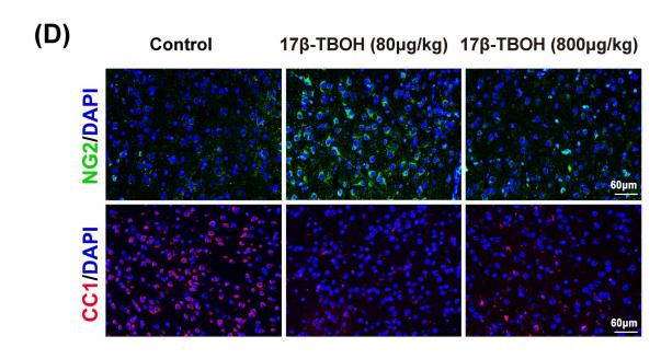
Application: IF/ICC Species: mice Sample: medial prefrontal cortex (mPFC)
Application: WB Species: Mouse Sample:
Application: IF/ICC Species: Mouse Sample:
Application: IF/ICC Species: Mice Sample: retinas
限制条款
产品的规格、报价、验证数据请以官网为准,官网链接:www.affbiotech.com | www.affbiotech.cn(简体中文)| www.affbiotech.jp(日本語)产品的数据信息为Affinity所有,未经授权不得收集Affinity官网数据或资料用于商业用途,对抄袭产品数据的行为我们将保留诉诸法律的权利。
产品相关数据会因产品批次、产品检测情况随时调整,如您已订购该产品,请以订购时随货说明书为准,否则请以官网内容为准,官网内容有改动时恕不另行通知。
Affinity保证所销售产品均经过严格质量检测。如您购买的商品在规定时间内出现问题需要售后时,请您在Affinity官方渠道提交售后申请。产品仅供科学研究使用。不用于诊断和治疗。
产品未经授权不得转售。
Affinity Biosciences将不会对在使用我们的产品时可能发生的专利侵权或其他侵权行为负责。Affinity Biosciences, Affinity Biosciences标志和所有其他商标所有权归Affinity Biosciences LTD.


