产品: Vitamin D Receptor 抗体
货号: AF6159
描述: Rabbit polyclonal antibody to Vitamin D Receptor
应用: WB IHC IF/ICC
反应: Human, Mouse
预测: Pig, Horse, Chicken
分子量: 48kDa; 48kD(Calculated).
蛋白号: P11473
RRID: AB_2835028

浏览相似产品>>

   规格 价格 库存
 50ul RMB¥ 1250 现货
 100ul RMB¥ 2300 现货
 200ul RMB¥ 3000 现货

货期: 当天发货

联系销售

产品描述

来源:
Rabbit
应用:
WB 1:500-1:2000, IHC 1:50-1:200, IF/ICC 1:100-1:500
*The optimal dilutions should be determined by the end user.
*Tips:

WB: 适用于变性蛋白样本的免疫印迹检测. IHC: 适用于组织样本的石蜡(IHC-p)或冰冻(IHC-f)切片样本的免疫组化/荧光检测. IF/ICC: 适用于细胞样本的荧光检测. ELISA(peptide): 适用于抗原肽的ELISA检测.

反应:
Human,Mouse
预测:
Pig(86%), Horse(86%), Chicken(100%)
克隆:
Polyclonal
特异性:
Vitamin D Receptor Antibody detects endogenous levels of total Vitamin D Receptor.
RRID:
AB_2835028
引用格式: Affinity Biosciences Cat# AF6159, RRID:AB_2835028.
偶联:
Unconjugated.
纯化:
The antiserum was purified by peptide affinity chromatography using SulfoLink™ Coupling Resin (Thermo Fisher Scientific).
保存:
Rabbit IgG in phosphate buffered saline , pH 7.4, 150mM NaCl, 0.02% sodium azide and 50% glycerol. Store at -20 °C. Stable for 12 months from date of receipt.
别名:

展开/折叠

1 25 dihydroxyvitamin D3 receptor; 1; 1,25 dihydroxyvitamin D3 receptor; 1,25-@dihydroxyvitamin D3 receptor; 25-dihydroxyvitamin D3 receptor; Member 1; NR1I1; Nuclear receptor subfamily 1 group I member 1; PPP1R163; Protein phosphatase 1, regulatory subunit 163; VDR; VDR_HUMAN; Vitamin D (1,25- dihydroxyvitamin D3) receptor; Vitamin D hormone receptor; Vitamin D nuclear receptor variant 1; Vitamin D receptor; Vitamin D3 receptor;

抗原和靶标

免疫原:
Uniprot:
基因/基因ID:
描述:
Nuclear hormone receptor. VDR mediates the action of vitamin D3 by controlling the expression of hormone sensitive genes.
序列:
MEAMAASTSLPDPGDFDRNVPRICGVCGDRATGFHFNAMTCEGCKGFFRRSMKRKALFTCPFNGDCRITKDNRRHCQACRLKRCVDIGMMKEFILTDEEVQRKREMILKRKEEEALKDSLRPKLSEEQQRIIAILLDAHHKTYDPTYSDFCQFRPPVRVNDGGGSHPSRPNSRHTPSFSGDSSSSCSDHCITSSDMMDSSSFSNLDLSEEDSDDPSVTLELSQLSMLPHLADLVSYSIQKVIGFAKMIPGFRDLTSEDQIVLLKSSAIEVIMLRSNESFTMDDMSWTCGNQDYKYRVSDVTKAGHSLELIEPLIKFQVGLKKLNLHEEEHVLLMAICIVSPDRPGVQDAALIEAIQDRLSNTLQTYIRCRHPPPGSHLLYAKMIQKLADLRSLNEEHSKQYRCLSFQPECSMKLTPLVLEVFGNEIS

种属预测

种属预测:

score>80的预测可信度较高,可尝试用于WB检测。*预测模型主要基于免疫原序列比对,结果仅作参考,不作为质保凭据。

Species
Results
Score
Chicken
100
Pig
86
Horse
86
Bovine
0
Sheep
0
Dog
0
Xenopus
0
Zebrafish
0
Rabbit
0
Model Confidence:
High(score>80) Medium(80>score>50) Low(score<50) No confidence

翻译修饰 - P11473 作为底物

Site PTM Type Enzyme
S51 Phosphorylation P17252 (PRKCA) , P05771 (PRKCB)
K117 Ubiquitination
S119 Phosphorylation
K123 Ubiquitination
S125 Phosphorylation
S172 Phosphorylation
T175 Phosphorylation
S182 Phosphorylation P17612 (PRKACA)
S208 Phosphorylation P68400 (CSNK2A1)
Y293 Phosphorylation
K302 Ubiquitination
Y380 Phosphorylation
K386 Ubiquitination
S392 Phosphorylation
K399 Ubiquitination

研究背景

功能:

Nuclear receptor for calcitriol, the active form of vitamin D3 which mediates the action of this vitamin on cells. Enters the nucleus upon vitamin D3 binding where it forms heterodimers with the retinoid X receptor/RXR. The VDR-RXR heterodimers bind to specific response elements on DNA and activate the transcription of vitamin D3-responsive target genes. Plays a central role in calcium homeostasis (By similarity).

细胞定位:

Nucleus. Cytoplasm.
Note: Localizes mainly to the nucleus (PubMed:28698609, PubMed:12145331). Localization to the nucleus is enhanced by vitamin D3.

Extracellular region or secreted Cytosol Plasma membrane Cytoskeleton Lysosome Endosome Peroxisome ER Golgi apparatus Nucleus Mitochondrion Manual annotation Automatic computational assertionSubcellular location
亚基结构:

Homodimer in the absence of bound vitamin D3. Heterodimer with RXRA after vitamin D3 binding. Interacts with MED1, NCOA1, NCOA2, NCOA3 and NCOA6 coactivators, leading to a strong increase of transcription of target genes. Interacts with the corepressor NCOR1. Interacts with SNW1. Interacts with IRX4, the interaction does not affect its transactivation activity. Interacts with CRY1 (By similarity). Interacts with CRY2 in a ligand-dependent manner (By similarity).

蛋白家族:

Composed of three domains: a modulating N-terminal domain, a DNA-binding domain and a C-terminal ligand-binding domain.

The 9aaTAD motif is a transactivation domain present in a large number of yeast and animal transcription factors.

Belongs to the nuclear hormone receptor family. NR1 subfamily.

研究领域

· Human Diseases > Infectious diseases: Bacterial > Tuberculosis.

· Organismal Systems > Excretory system > Endocrine and other factor-regulated calcium reabsorption.

· Organismal Systems > Digestive system > Mineral absorption.

文献引用

1). VDR activation attenuates osteoblastic ferroptosis and senescence by stimulating the Nrf2/GPX4 pathway in age-related osteoporosis. Free Radical Biology and Medicine (PubMed: 36402439) [IF=7.4]

2). Eriodictyol ameliorates cognitive dysfunction in APP/PS1 mice by inhibiting ferroptosis via vitamin D receptor-mediated Nrf2 activation. Molecular Medicine (PubMed: 35093024) [IF=5.7]

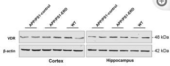
Application: WB    Species: Mouse    Sample: brain

Fig. 7 Eriodictyol up-regulates VDR expression and activates the Nrf2/HO-1 signaling pathway. A VDR expression in the mouse brain was detected using immunohistochemical staining. Then, B the VDR protein expression level in the mouse cortex and hippocampus was measured using Western blotting. C The VDR protein expression level in HT-22 cells was measured using Western blotting, and D its relative mRNA expression level was detected using q-PCR. E The levels of Nrf2, p-Nrf2 and HO-1 in the mouse cortex and hippocampus were measured using Western blotting. F The levels of Nrf2, p-Nrf2 and HO-1 in HT-22 cells were measured using Western blotting. G The level of Nrf2 in the nucleus and cytoplasm of HT-22 cells was detected using Western blotting, and Lamin B1 and β-actin were used as loading controls for the nuclear and cytoplasmic proteins, respectively. The data are presented as the means ± SD of three experiments. **P < 0.01 and ***P < 0.001 compared with the Aβ1–42 oligomer group. ##P < 0.01 compared with the control group

3). Monotropein inhibits colitis associated cancer through VDR/JAK1/STAT1 regulation of macrophage polarization. International Immunopharmacology (PubMed: 37633235) [IF=5.6]

Application: WB    Species: Mouse    Sample:

FIGURE 2 PTS reduces reduction of NLRP3 inflammasome and apoptosis in DOX-treated mice. (A) WT C57/BL6J mice were pretreated with PTS (10 mg/kg) and afterwards with DOX (20 mg/kg cumulative dose) for 6 days. Immunochemistry staining of the liver sections with anti-NLRP3 (left, scale bar = 50 μm), the quantification of NLRP3 positive area in each group (right, n = 6); (B) qPCR analyses of NLRP3, IL-1β and IL-18 mRNA levels in each group (n = 6); (C) Western blot analyses of NLRP3, ASC, Caspase-1 p20, IL-1β, and IL-18 proteins and the quantification of the blots in each group (n = 4 per group); (D) Western blot analyses of Cleaved Caspase-3, BAX, and BCL-2 proteins (left) and quantification of the blots in each group (right, n = 4 per group).

4). Vitamin D stimulates placental L-type amino acid transporter 1 (LAT1) in preeclampsia. Scientific Reports (PubMed: 35301401) [IF=4.6]

Application: IHC    Species: Human    Sample: trophoblast cells

Figure 1 Expression of LAT1 and VDR in normal and preeclamptic placentas. (A) Representative immunostaining images of LAT1 and VDR expressions in tissue sections from normal and preeclamptic (PE) placentas. Bar = 100 micron. (B) Representative blots of LAT1 and VDR expression in placenta from normotensive and PE pregnancies. (C,D) The bar graphs show the relative density of protein expression for LAT1 and VDR after normalization with β-actin expression in each sample. n = 5, #P < 0.05, PE vs Normal.

Application: WB    Species: Humna    Sample: trophoblast cells

Figure 2 Effects of 1,25(OH)2D3 and CoCl2 on LAT1 and VDR expression in placental trophoblasts. (A) Protein expression for LAT1 and VDR in HTR-8/SVneo trophoblast cells treated with different concentrations of CoCl2. The bar graphs show relative protein expression for LAT1 and VDR after normalized against β-actin in each sample from three independent experiments. CoCl2 induced a dose-dependent decrease in both LAT1 and VDR expression in trophoblasts. *P < 0.05, **P < 0.01, ***P < 0.001, CoCl2 treated vs control cells. (B) Protein expression for LAT1 and VDR in HTR-8/SVneo trophoblast cells treated with different concentrations of 1,25(OH)2D3. The bar graphs show relative protein expression for LAT1 and VDR after normalization with β-actin in each sample from four independent experiments. In contrast to CoCl2, 1,25(OH)2D3 stimulated LAT1 and VDR expression in a dose-dependent manner in trophoblast cells. #P < 0.05, ##P < 0.01, 1,25(OH)2D3 treated vs control cells.

Application: WB    Species: human    Sample: trophoblast cells

Figure 3.| Efects of 1,25(OH)2D3 on CoCl2-induced LAT1 expression in placental trophoblasts. (C) Protein expression of VDR and LAT1 expression in trophoblasts treated with VDR siRNA in the presence or absence of 1,25(OH)2D3. Inhibition of VDR expression attenuates 1,25(OH)2D3-stimulated LAT1 expression in trophoblast cells. Te bar graphs show relative VDR and LAT1 expression afer being normalized by β-actin in each sample from six independent experiments, *P<0.05, **P<0.01, treated vs control; ##P<0.01, ###P<0.001, 1,25(OH)2D3+VDR siRNA vs 1,25(OH)2D3 alone.

5). Vitamin D stimulates miR-26b-5p to inhibit placental COX-2 expression in preeclampsia. Scientific Reports (PubMed: 34045549) [IF=4.6]

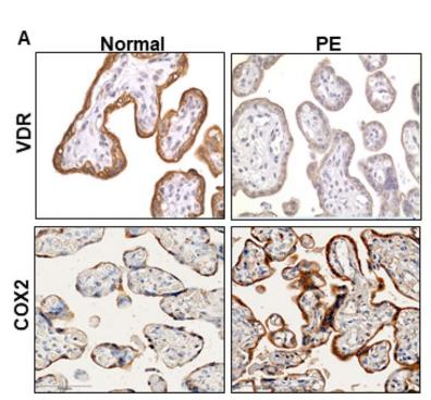
Application: IHC    Species: human    Sample: placental
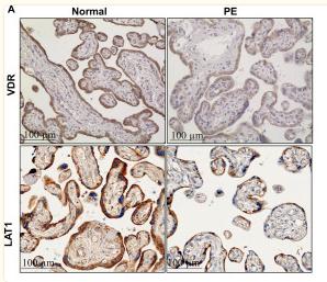
Figure 1.| Representative VDR, COX-2, and miR-26b-5p expression in placenta and trophoblasts from normotensive pregnant women and women complicated with preeclampsia (PE). (A) VDR expression and COX-2 expression in placental tissues from 12 pregnant women (6 from normotensive and 6 from PE). Strong VDR staining was seen in the placentas from normotensive pregnant women compared to those from women with PE. In contrast, intensive COX-2 expression was detected in placentas from PE specimens. Bar=100 micron.

6). Wnt/PCP-YAP-BIRC2 axis maintains cartilage stem/progenitor cell homeostasis in osteoarthritis. Research Square

Application: WB    Species: Human    Sample: CSPCs

Figure 3. Wnt5A/B upregulates YAP in OA-CSPCs. a The mRNA expression of the genes associated with stem cell function in OA-CSPCs. b The protein levels of YAP, FAP, VDR, TERT, and MMP3 in OA-CSPCs. c Immunofluorescence staining of YAP (red) in OA-CSPCs. Nuclei were stained by 4,6-diamidina-2-phenylin (DAPI,blue), Bars=100 μm. d The protein levels of YAP in OA-CSPCs derived from OA patients.

限制条款

产品的规格、报价、验证数据请以官网为准,官网链接:www.affbiotech.com | www.affbiotech.cn(简体中文)| www.affbiotech.jp(日本語)

产品的数据信息为Affinity所有,未经授权不得收集Affinity官网数据或资料用于商业用途,对抄袭产品数据的行为我们将保留诉诸法律的权利。

产品相关数据会因产品批次、产品检测情况随时调整,如您已订购该产品,请以订购时随货说明书为准,否则请以官网内容为准,官网内容有改动时恕不另行通知。

Affinity保证所销售产品均经过严格质量检测。如您购买的商品在规定时间内出现问题需要售后时,请您在Affinity官方渠道提交售后申请。

产品仅供科学研究使用。不用于诊断和治疗。 

产品未经授权不得转售。

Affinity Biosciences将不会对在使用我们的产品时可能发生的专利侵权或其他侵权行为负责。Affinity Biosciences, Affinity Biosciences标志和所有其他商标所有权归Affinity Biosciences LTD.