产品: p27 Kip1 抗体
货号: AF6324
描述: Rabbit polyclonal antibody to p27 Kip1
应用: WB IF/ICC
反应: Human, Mouse, Rat
预测: Bovine, Sheep, Rabbit, Dog, Chicken
分子量: 27kDa; 22kD(Calculated).
蛋白号: P46527
RRID: AB_2835182

浏览相似产品>>

   规格 价格 库存
 50ul RMB¥ 1250 现货
 100ul RMB¥ 2300 现货
 200ul RMB¥ 3000 现货

货期: 当天发货

联系销售

产品描述

来源:
Rabbit
应用:
WB 1:500-1:2000, IF/ICC 1:100-1:500
*The optimal dilutions should be determined by the end user.
*Tips:

WB: 适用于变性蛋白样本的免疫印迹检测. IHC: 适用于组织样本的石蜡(IHC-p)或冰冻(IHC-f)切片样本的免疫组化/荧光检测. IF/ICC: 适用于细胞样本的荧光检测. ELISA(peptide): 适用于抗原肽的ELISA检测.

反应:
Human,Mouse,Rat
预测:
Bovine(100%), Sheep(100%), Rabbit(100%), Dog(100%), Chicken(90%)
克隆:
Polyclonal
特异性:
p27 Kip1 Antibody detects endogenous levels of total p27 Kip1.
RRID:
AB_2835182
引用格式: Affinity Biosciences Cat# AF6324, RRID:AB_2835182.
偶联:
Unconjugated.
纯化:
The antiserum was purified by peptide affinity chromatography using SulfoLink™ Coupling Resin (Thermo Fisher Scientific).
保存:
Rabbit IgG in phosphate buffered saline , pH 7.4, 150mM NaCl, 0.02% sodium azide and 50% glycerol. Store at -20 °C. Stable for 12 months from date of receipt.
别名:

展开/折叠

AA408329; AI843786; Cdki1b; CDKN 1B; CDKN 4; CDKN1B; CDKN4; CDN1B_HUMAN; Cyclin Dependent Kinase Inhibitor 1B; Cyclin dependent kinase inhibitor p27; Cyclin-dependent kinase inhibitor 1B (p27, Kip1); Cyclin-dependent kinase inhibitor 1B; Cyclin-dependent kinase inhibitor p27; Cyclin-dependent kinase inhibitor p27 Kip1; KIP 1; KIP1; MEN1B; MEN4; OTTHUMP00000195098; OTTHUMP00000195099; p27; p27 Kip1; P27-like cyclin-dependent kinase inhibitor; p27Kip1;

抗原和靶标

免疫原:
Uniprot:
基因/基因ID:
表达:
P46527 CDN1B_HUMAN:

Expressed in all tissues tested. Highest levels in skeletal muscle, lowest in liver and kidney.

描述:
This gene encodes a cyclin-dependent kinase inhibitor, which shares a limited similarity with CDK inhibitor CDKN1A/p21. The encoded protein binds to and prevents the activation of cyclin E-CDK2 or cyclin D-CDK4 complexes, and thus controls the cell cycle progression at G1.
序列:
MSNVRVSNGSPSLERMDARQAEHPKPSACRNLFGPVDHEELTRDLEKHCRDMEEASQRKWNFDFQNHKPLEGKYEWQEVEKGSLPEFYYRPPRPPKGACKVPAQESQDVSGSRPAAPLIGAPANSEDTHLVDPKTDPSDSQTGLAEQCAGIRKRPATDDSSTQNKRANRTEENVSDGSPNAGSVEQTPKKPGLRRRQT

种属预测

种属预测:

score>80的预测可信度较高,可尝试用于WB检测。*预测模型主要基于免疫原序列比对,结果仅作参考,不作为质保凭据。

Species
Results
Score
Bovine
100
Sheep
100
Dog
100
Rabbit
100
Chicken
90
Pig
0
Horse
0
Xenopus
0
Zebrafish
0
Model Confidence:
High(score>80) Medium(80>score>50) Low(score<50) No confidence

翻译修饰 - P46527 作为底物

Site PTM Type Enzyme
S2 O-Glycosylation
S10 Phosphorylation Q9UQM7 (CAMK2A) , Q00534 (CDK6) , Q9H2X6 (HIPK2) , Q8TAS1 (UHMK1) , Q00536 (CDK16) , P31749 (AKT1) , P28482 (MAPK1) , Q9Y463 (DYRK1B)
S12 Phosphorylation
R15 Methylation
K25 Ubiquitination
K68 Ubiquitination
Y74 Phosphorylation P07947 (YES1) , P12931 (SRC)
K81 Acetylation
K81 Ubiquitination
S83 Phosphorylation P68400 (CSNK2A1)
Y88 Phosphorylation O60674 (JAK2) , P12931 (SRC) , P00519 (ABL1) , P07947 (YES1) , P07948 (LYN)
Y89 Phosphorylation P12931 (SRC) , P00519 (ABL1) , P07947 (YES1)
S106 O-Glycosylation
S110 O-Glycosylation
K134 Ubiquitination
S140 Phosphorylation Q13315 (ATM)
K153 Ubiquitination
R154 Methylation
T157 O-Glycosylation
T157 Phosphorylation Q14012 (CAMK1) , P31749 (AKT1) , P31751 (AKT2) , P11309-2 (PIM1) , O00141 (SGK1) , Q86V86 (PIM3) , Q9P1W9 (PIM2)
K165 Ubiquitination
S175 Phosphorylation
S178 Phosphorylation P28482 (MAPK1)
S183 Phosphorylation
T187 Phosphorylation Q00534 (CDK6) , P31749 (AKT1) , P28482 (MAPK1) , Q00535 (CDK5) , P27361 (MAPK3) , P24941 (CDK2)
T198 O-Glycosylation
T198 Phosphorylation P11309-2 (PIM1) , P31749 (AKT1) , Q14012 (CAMK1) , P51812 (RPS6KA3) , Q13131 (PRKAA1) , Q86V86 (PIM3) , Q15418 (RPS6KA1) , O00141 (SGK1) , Q9P1W9 (PIM2)

研究背景

功能:

Important regulator of cell cycle progression. Inhibits the kinase activity of CDK2 bound to cyclin A, but has little inhibitory activity on CDK2 bound to SPDYA. Involved in G1 arrest. Potent inhibitor of cyclin E- and cyclin A-CDK2 complexes. Forms a complex with cyclin type D-CDK4 complexes and is involved in the assembly, stability, and modulation of CCND1-CDK4 complex activation. Acts either as an inhibitor or an activator of cyclin type D-CDK4 complexes depending on its phosphorylation state and/or stoichometry.

翻译修饰:

Phosphorylated; phosphorylation occurs on serine, threonine and tyrosine residues. Phosphorylation on Ser-10 is the major site of phosphorylation in resting cells, takes place at the G(0)-G(1) phase and leads to protein stability. Phosphorylation on other sites is greatly enhanced by mitogens, growth factors, cMYC and in certain cancer cell lines. The phosphorylated form found in the cytoplasm is inactivate. Phosphorylation on Thr-198 is required for interaction with 14-3-3 proteins. Phosphorylation on Thr-187, by CDK1 and CDK2 leads to protein ubiquitination and proteasomal degradation. Tyrosine phosphorylation promotes this process. Phosphorylation by PKB/AKT1 can be suppressed by LY294002, an inhibitor of the catalytic subunit of PI3K. Phosphorylation on Tyr-88 and Tyr-89 has no effect on binding CDK2, but is required for binding CDK4. Dephosphorylated on tyrosine residues by G-CSF.

Ubiquitinated; in the cytoplasm by the KPC complex (composed of RNF123/KPC1 and UBAC1/KPC2) and, in the nucleus, by SCF(SKP2). The latter requires prior phosphorylation on Thr-187. Ubiquitinated; by a TRIM21-containing SCF(SKP2)-like complex; leads to its degradation.

Subject to degradation in the lysosome. Interaction with SNX6 promotes lysosomal degradation (By similarity).

细胞定位:

Nucleus. Cytoplasm. Endosome.
Note: Nuclear and cytoplasmic in quiescent cells. AKT- or RSK-mediated phosphorylation on Thr-198, binds 14-3-3, translocates to the cytoplasm and promotes cell cycle progression. Mitogen-activated UHMK1 phosphorylation on Ser-10 also results in translocation to the cytoplasm and cell cycle progression. Phosphorylation on Ser-10 facilitates nuclear export. Translocates to the nucleus on phosphorylation of Tyr-88 and Tyr-89. Colocalizes at the endosome with SNX6; this leads to lysosomal degradation (By similarity).

Extracellular region or secreted Cytosol Plasma membrane Cytoskeleton Lysosome Endosome Peroxisome ER Golgi apparatus Nucleus Mitochondrion Manual annotation Automatic computational assertionSubcellular location
组织特异性:

Expressed in all tissues tested. Highest levels in skeletal muscle, lowest in liver and kidney.

亚基结构:

Forms a ternary complex composed of CCNE1, CDK2 and CDKN1B. Interacts directly with CCNE1; the interaction is inhibited by CDK2-dependent phosphorylation on Thr-187. Interacts with COPS5, subunit of the COP9 signalosome complex; the interaction leads to CDKN1B degradation. Interacts with NUP50; the interaction leads to nuclear import and degradation of phosphorylated CDKN1B. Interacts with CCND1 and SNX6 (By similarity). Interacts (Thr-198-phosphorylated form) with 14-3-3 proteins, binds strongly YWHAQ, weakly YWHAE and YWHAH, but not YWHAB nor YWHAZ; the interaction with YWHAQ results in translocation to the cytoplasm. Interacts with AKT1 and LYN; the interactions lead to cytoplasmic mislocation, phosphorylation of CDKN1B and inhibition of cell cycle arrest. Forms a ternary complex with CCNA2 and CDK2; CDKN1B inhibits the kinase activity of CDK2 through conformational rearrangements. Interacts (unphosphorylated form) with CDK2. Forms a complex with CDK2 and SPDYA, but does not directly interact with SPDYA. Forms a ternary complex composed of cyclin D, CDK4 and CDKN1B. Interacts (phosphorylated on Tyr-88 and Tyr-89) with CDK4; the interaction is required for cyclin D and CDK4 complex assembly, induces nuclear translocation and activates the CDK4 kinase activity. Interacts with GRB2. Interacts with PIM1. Identified in a complex with SKP1, SKP2 and CKS1B. Interacts with UHMK1; the interaction leads to cytoplasmic mislocation, phosphorylation of CDKN1B and inhibition of cell cycle arrest. Interacts also with CDK1. Dephosphorylated on Thr-187 by PPM1H, leading to CDKN1B stability.

蛋白家族:

A peptide sequence containing only AA 28-79 retains substantial Kip1 cyclin A/CDK2 inhibitory activity.

Belongs to the CDI family.

研究领域

· Cellular Processes > Cell growth and death > Cell cycle.   (View pathway)

· Environmental Information Processing > Signal transduction > ErbB signaling pathway.   (View pathway)

· Environmental Information Processing > Signal transduction > HIF-1 signaling pathway.   (View pathway)

· Environmental Information Processing > Signal transduction > FoxO signaling pathway.   (View pathway)

· Environmental Information Processing > Signal transduction > PI3K-Akt signaling pathway.   (View pathway)

· Human Diseases > Drug resistance: Antineoplastic > Endocrine resistance.

· Human Diseases > Infectious diseases: Viral > Hepatitis B.

· Human Diseases > Infectious diseases: Viral > Measles.

· Human Diseases > Infectious diseases: Viral > Human papillomavirus infection.

· Human Diseases > Infectious diseases: Viral > Epstein-Barr virus infection.

· Human Diseases > Cancers: Overview > Pathways in cancer.   (View pathway)

· Human Diseases > Cancers: Overview > Transcriptional misregulation in cancer.

· Human Diseases > Cancers: Overview > Viral carcinogenesis.

· Human Diseases > Cancers: Overview > MicroRNAs in cancer.

· Human Diseases > Cancers: Specific types > Prostate cancer.   (View pathway)

· Human Diseases > Cancers: Specific types > Chronic myeloid leukemia.   (View pathway)

· Human Diseases > Cancers: Specific types > Small cell lung cancer.   (View pathway)

· Human Diseases > Cancers: Specific types > Gastric cancer.   (View pathway)

文献引用

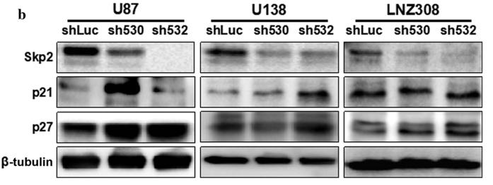
1). Skp2 modulates proliferation, senescence and tumorigenesis of glioma. Cancer Cell International, 2020 (PubMed: 32165861) [IF=5.8]

Application: WB    Species: mouse    Sample: U87, U138, and LNZ308 cells

Fig. 2| Downregulation of Skp2 attenuated in vitro and in vivo cell proliferation. a On Western blot analyses, the protein level of Skp2 in glioma cell lines (A172, U87, U118, U373, LNZ308, U138 and U343), as compared to normal astrocytes (AST), was enhanced. b Skp2 was successfully knocked-down by two shRNA fragments in U87, U138, and LNZ308 cells compared with the negative control shLuc. The protein levels of the downstream molecules ­p21Cip1/Waf1 and ­p27Kip1 were increased upon the knockdown of Skp2 in all three cell lines.

2). Compound Kushen injection attenuates angiotensin II‑mediated heart failure by inhibiting the PI3K/Akt pathway. International Journal of Molecular Medicine, 2023 (PubMed: 36734284) [IF=5.7]

Application: WB    Species: Rat    Sample: H9C2 cells

Figure 7 Expression of apoptosis-related proteins in vitro. (A) The results of western blot analysis for each protein. (B-I) The expression of representative proteins. (B) p-Akt expression, (C) t-Akt expression, (D) p-Akt/t-Akt ratio, (E) Bcl-2 expression, (F) cyclin D1 expression, (G) PI3K expression, (H) Bax expression, and (I) p27 expression in the different groups. Data are presented as the mean ± SEM. One-way ANOVA and post-hoc Tukey's multiple comparisons test were used to determine statistically significant differences (n=3 per group). *P

3). Protective effect of Huanglian Pingwei San on DSS-induced ulcerative colitis in mice through amelioration of the inflammatory response and oxidative stress. Frontiers in pharmacology, 2024 (PubMed: 39697546) [IF=5.6]

Application: WB    Species: Mouse    Sample: colon tissues

FIGURE 5. HLPWS inhibited apoptosis-related proteins. (A) Protein expression of P53, P21, cleaved caspase 3, p-H2AX, and P27 in colon tissues was determined by Western blotting. (B) Quantitative analysis of the Western blot results (n = 3). (C) Expression of cleaved caspase three and p-H2AX in the colon tissues of UC mice detected by IHC (original magnifications, ×200; scale bar = 100 μm). The results are expressed as the means ± SEM. ## p < 0.01, # p < 0.05 vs. the control group; ** p < 0.01, * p < 0.05 vs. the DSS group.

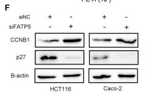
4). Over-Expression and Prognostic Significance of FATP5, as a New Biomarker, in Colorectal Carcinoma. Frontiers in Molecular Biosciences, 2022 (PubMed: 35155561) [IF=5.0]

Application: WB    Species: Human    Sample: CRC cells

FIGURE 6 siFATP5 facilitates cell growth and cell cycle G2/M phase transition. FATP5 expression was detected after transfection in CRC cells by qRT-PCR (A) and western blotting (B,C) CCK-8 assays were used to detect cell viability of CRC cells after transfection. (D,E) Flow cytometric analysis was used to detect cell cycle distribution of CRC cells after transfection. (F) The expression of cycle-related protein. *p < 0.05, **p < 0.01.

5). Ligustrazine Suppresses Platelet-Derived Growth Factor-BB-Induced Pulmonary Artery Smooth Muscle Cell Proliferation and Inflammation by Regulating the PI3K/AKT Signaling Pathway. The American journal of Chinese medicine, 2021 (PubMed: 33622214) [IF=4.8]

6). IDH1 gene mutation activates Smad signaling molecules to regulate the expression levels of cell cycle and biological rhythm genes in human glioma U87‑MG cells. Molecular Medicine Reports, 2021 (PubMed: 33760141) [IF=3.4]

7). Regulation of the PTEN/PI3K/AKT pathway in RCC using the active compounds of natural products in vitro. Molecular Medicine Reports, 2021 (PubMed: 34490473) [IF=3.4]

Application: WB    Species: Human    Sample: RCC cells

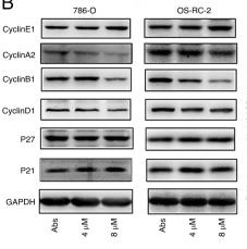
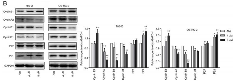
Figure 3. Naringenin arrests cell cycle progression of renal cell carcinoma cells in the G2 phase. 786-O and OS-RC-2 cells were treated with naringenin (0, 4 or 8 µM) for 48 h. (A) Cell cycle distribution was determined using flow cytometry. (B) Western blot analysis of cyclin E1, cyclin A2, cyclin B1, cyclin D1, P27 and P21 expression levels. Data are presented as the mean ± SD. *P<0.05, **P<0.01 vs. Abs. Abs, absolute ethanol.

Application: WB    Species: human    Sample: 786‑O and OS‑RC‑2 cells

Figure 3.| Naringenin arrests cell cycle progression of renal cell carcinoma cells in the G2 phase. 786‑O and OS‑RC‑2 cells were treated with naringenin (0, 4 or 8 µM) for 48 h. (A) Cell cycle distribution was determined using flow cytometry. (B) Western blot analysis of cyclin E1, cyclin A2, cyclin B1, cyclin D1, P27 and P21 expression levels. Data are presented as the mean ± SD. * P<0.05, **P<0.01 vs. Abs. Abs, absolute ethanol.

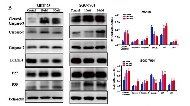
8). Dihydroartemisinin inhibits the tumorigenesis and invasion of gastric cancer by regulating STAT1/KDR/MMP9 and P53/BCL2L1/CASP3/7 pathways. PATHOLOGY RESEARCH AND PRACTICE, 2021 (PubMed: 33370709) [IF=2.9]

Application: WB    Species: mice    Sample: GC cells

Fig. 6. DHA regulated p53/BCL2L1/Caspase-3/-7 and STAT1/KDR/MMP9 signaling pathways. (A) qRT-PCR analysis of the effects of DHA on the expression levels of BCL2, BAX, CKD4, p53/BCL2L1/Caspase-3/-7 and STAT1/KDR/MMP9 in GC cells. (B) WB analysis of the effects of DHA on the expression of apoptosis and cycle-related proteins including cleaved-caspase 3, caspase 3, caspase 7, BCL2L1, p27 and p53. (C) WB analysis of the effects of DHA on the expression of p-STAT1, STAT1, p-KDR, KDR and MMP9.

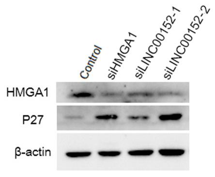
9). LINC00152 acts as a competing endogenous RNA of HMGA1 to promote the growth of gastric cancer cells. Journal of Clinical Laboratory Analysis, 2022 (PubMed: 35014092) [IF=2.6]

Application: WB    Species: Human    Sample: GC cells

FIGURE 4 HMGA1 and LINC00152 regulate cell cycle progression. (A) The relative expression of P27 was measured after siRNA transfection. (B) Western blot analysis of HMGA1, P27, and β‐actin 48 h after siRNA transfection. (C) The relative HMGA1 and P27 protein levels were quantified from B. Data represent the mean ± S.D. (n = 3). *p < 0.05; **p < 0.01; ***p < 0.001

10). LncRNA GACAT3 acts as a competing endogenous RNA of HMGA1 and alleviates cucurbitacin B-induced apoptosis of gastric cancer cells. GENE, 2018 (PubMed: 30098426) [IF=2.6]

Application: WB    Species: human    Sample: SGC-7901 cells

Fig. 3. |GACAT3 and HMGA1 promote cell growth by inhibiting p21 and p27.qRT-PCR and western blot analyses of p27 and p21 levels in gastric cancer cells after GACAT3 or HMGA1 siRNA-mediated knockdown. The mRNA expression levels of p27 and p21 in SGC-7901 (A) and BGC-823 (B) cells. Western blotting results and relative protein levels of p27, p21, and HMGA1 in SGC-7901 (C) and BGC-823 (D)cells. Data represent the mean ± S.D. of three biological replicates. *p < 0.05; **p < 0.01.

加载更多

限制条款

产品的规格、报价、验证数据请以官网为准,官网链接:www.affbiotech.com | www.affbiotech.cn(简体中文)| www.affbiotech.jp(日本語)

产品的数据信息为Affinity所有,未经授权不得收集Affinity官网数据或资料用于商业用途,对抄袭产品数据的行为我们将保留诉诸法律的权利。

产品相关数据会因产品批次、产品检测情况随时调整,如您已订购该产品,请以订购时随货说明书为准,否则请以官网内容为准,官网内容有改动时恕不另行通知。

Affinity保证所销售产品均经过严格质量检测。如您购买的商品在规定时间内出现问题需要售后时,请您在Affinity官方渠道提交售后申请。

产品仅供科学研究使用。不用于诊断和治疗。 

产品未经授权不得转售。

Affinity Biosciences将不会对在使用我们的产品时可能发生的专利侵权或其他侵权行为负责。Affinity Biosciences, Affinity Biosciences标志和所有其他商标所有权归Affinity Biosciences LTD.