


, diluted 1/600 was used as secondary antibody.")
 Western blotting shows that RIF-EVs and FER-EVs expressed classic EV protein
markers Alix, TSG101, and CD9. Representative shapes of (B) RIF-EVs and (C) FER-EVs detected with the use of transmission electron
microscopy. The size and distribution of (D) RIF-EVs and (E) FER-EVs examined with the use of nanoparticle tracking analysis. ECs ¼
endometrial cells; EVs ¼ extracellular vesicles; FER ¼ fertile women; RIF ¼ women with recurrent implantation failure.
Liu. Extracellular vesicles regulate embryos. Fertil Steril 2020.")
, and western blot assay disclosed that the expressions of exosome markers CD9, CD63, CD81, TGF-β1 Flotillin-1 and EEF2 were elevated in Exosome group than those in NC group (sample from serum-free medium) (B).")
 and FER-EVs (c) were displayed. TEM images revealed the typical bilayer membrane structures of RIF-EVs (d) and FER-EVs (e). EVs, extracellular vesicles; RIF, recurrent implantation failure; FER, fertile women; ECs, endometrial cells; TEM, transmission electron microscopy")
 treated
with or without UC-MSC-EVs. g Flow cytometric analysis of AnnexinV/
PI staining levels of KGN cells. h Comparisons of total, early and late
apoptosis rates of KGN cells among the three groups. *P < 0.05. **P <
0.01")
 TEM photomicrographs of exosomes. Scale bar = 100 nm. (b) Nanotrack analysis size distribution of exosomes by flow nanoanalysis. (c) The expression of exosomal marker proteins CD9, CD63, and TSG101 by western blot assay. ADSC-exo: exosomes derived from adipose mesenchymal stem cells; TEM: transmission electron microscopy; ADSC-CM: conditioned medium of adipose mesenchymal stem cells.")
 Morphology of Nor-Exos and HypoExos under TEM. (b) Diameter distribution of Nor-Exos and Hypo-Exos assessed by NanoFCM. (c) Exosomal surface markers CD9, CD63 and CD81 were detected by Western blot analysis. Scale bars:100 nm (a).")
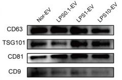
 and scanning electron microscopy (right). The red arrows indicate representative exosomes; C: Representative flow cytometry plots showing the phenotypes of exosome markers, including CD9, CD47, CD63, and CD81; D: The particle size distribution of C2C12-Exos was analyzed by the qNano platform; E: Western blotting showed the presence of exosomal markers, including CD63, HSP60, Alix, and CD9. The four lanes represent different exosomal proteins and deproteinized supernatants extracted from the two independent conditioned media. LPS: Lipopolysaccharide.")
 Morphology of PUMSCs. (B) Immunofluorescence staining results show that the isolated PUMSCs express α-SMA, a smooth muscle cell surface marker. (C) Growth curve of PUMSCs. (D) Flow cytometric analysis of MSC surface markers shows that UCMSCs express high levels = of CD29, CD44, CD73, and CD90 but do not express CD45 and HLA-DR. (E) Transmission electron microscopy shows that the UCMSC-sEV are cup-shaped vesicles. Scale bar = 100 nm. (F) NanoFCM analysis of the particle size of UCMSC-sEV. (G) Western blotting was used to detect the expression of the sEV marker proteins CD9, CD63, CD81, and TSG101.")
 Representative flow cytometry histograms of BMSCs and ADSCs show positive staining for CD29 and CD90 but not for CD34 and CD45. (B) BMSCs and ADSCs were successfully induced into osteoblasts (positively stained with Alizarin Red S) and adipocytes (positively stained with Oil Red O). The magnification is 100×. (C) Representative immunofluorescence results of CCSMCs show positive expression for α-SMA and desmin. Scale bars = 50 μm. (D) Exosomes derived from CCSMCs, BMSCs and ADSCs were observed using transmission electron microscopy, and the particle size distributions of the exosomes were measured by nanoparticle tracking analysis. Scale bars = 100 nm. (E) Representative results of Western blot analysis of exosomes derived from CCSMCs, BMSCs and ADSCs show positive expression for CD9, CD63 and TSG101 but not for calnexin. CCSMC: corpus cavernosum smooth muscle cell; BMSC: bone marrow stem cell; ADSC: adipose-derived stem cell; CCSMC-EXOs: exosomes derived from corpus cavernosum smooth muscle cells; BMSC-EXOs: exosomes derived from bone marrow stem cells; ADSC-EXOs: exosomes derived from adipose-derived stem cells; α-SMA: α-smooth muscle actin; DAPI: 4’,6-diamidino-2-phenylindole")
. (a) Transmission electron microscopy. (Scale bar =200 nm) (b and c) Nanoparticle Tracking analysis. (d) The average levels of TSG101, Alix, CD81, CD9, CD63, HSP70 and C-myc. ∗∗P <0.01 compared to the cell group.")
 Morphological diagram of PUMSLCs: PUMSLCs differentiated into lipoblasts (B), osteoblasts (C), and chondroblasts (D) after in vitro induction. (E) Flow cytometry for MSC surface markers showed that PUMSLCs expressed high levels of CD29, CD73, CD90, and CD105 but did not express CD34, CD45, or HLA-DR. (F) Western blotting for the PUMSLC-sEV exosome marker proteins CD9, CD63, CD81, and TSG101. (G) NanoFCM determined the particle size of PUMSLC-sEVs. (H) Transmission electron microscopy showed that PUMSLC-sEVs were goblet-shaped. (I) PUMSLC-sEVs were endocytosed by HUVECs.")
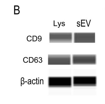
; B. Identification of hucMSC-Ex was done through transmission electron microscopy (TEM); C. Western blot analysis was performed to identify the surface markers of hucMSC-Ex.")
 served as negative control. Representative morphology of B, PCOS-EVs, and C, CTRL-EVs, are shown. Representative nanoparticle tracking analysis images of D, PCOS-EVs, and E, CTRL-EVs, are shown. F, The median size of PCOS-EVs (131.87 ± 3.85 nm) and CTRL-EVs (140.33 ± 12.04 nm) are compared. CTRL-EVs, follicular extracellular vesicles of control women; PCOS-EVs, follicular extracellular vesicles of women with polycystic ovary syndrome.")
 RT-qPCR analysis of miR-21-5p level in A549 cells transfected with miR-21-5p mimics or miR-21-5p inhibitor. (B) Ki67 staining analysis of cell proliferation in A549 cells transfected with miR-21-5p mimics or miR-21-5p inhibitor. (C, D) Exosomes (A549 Exo, A549miR-21-5p Exo) were collected from A549 cells and miR-21-5p mimics-transfected A549 cells respectively. The phenotype of the isolated exosomes was analyzed by TEM and NTA analysis.")
 Flow cytometry was used to identify cells. Flow quadrant diagram of Tregs (quadrant 2). (B) Transmission electron microscope was used to observe morphology of exosomes and the representative images of exosomes under transmission electron microscope were shown. Magnification ×60k, scale bar = 100 and 50 nm. (C) Nanoparticle Tracking Analysis was used for particle size distribution of exosomes. There was a total of 21,714,571 events. n = 5. (D) Western blot was used to detect exosome biomarkers and there were representative band images of exosome biomarkers CD63, CD9, and TSG101. n = 3. The “n” means number of experimental replicates")
 and old (red) reproductive age. The data were obtained using nanoparticle tracking analysis. Shown are the means ± SEM. Detection of extracellular vesicle markers, including CD63, CD9, and CD81, using Western blot analysis.")
. (A) Examination of isolated EVs by TEM. (B) The size distribution of exosomes was determined by DLS. (C) The expressions of EVs biomarkers CD9, CD63 in EVs samples were detected by Western blot.")
产品描述
*The optimal dilutions should be determined by the end user. For optimal experimental results, antibody reuse is not recommended.
*Tips:
WB: 适用于变性蛋白样本的免疫印迹检测. IHC: 适用于组织样本的石蜡(IHC-p)或冰冻(IHC-f)切片样本的免疫组化/荧光检测. IF/ICC: 适用于细胞样本的荧光检测. ELISA(peptide): 适用于抗原肽的ELISA检测.
引用格式: Affinity Biosciences Cat# AF5139, RRID:AB_2837625.
展开/折叠
Tetraspanin 29; 5H9; 5H9 antigen; Antigen defined by monoclonal antibody 602 29; Antigen defined by monoclonal antibody 60229; BA-2/p24 antigen; BA2; BTCC 1; BTCC1; CD9; CD9 antigen; CD9 antigen p24; CD9 molecule; CD9_HUMAN; Cell growth inhibiting gene 2 protein; Cell growth-inhibiting gene 2 protein; DRAP 27; DRAP27; GIG2; Growth inhibiting gene 2 protein; Leukocyte antigen MIC3; MIC3; Motility related protein; Motility-related protein; MRP 1; MRP-1; MRP1; p24; p24 antigen; Tetraspanin-29; Tspan 29; Tspan-29; TSPAN29;
抗原和靶标
A synthesized peptide derived from human CD9, corresponding to a region within the internal amino acids.
Detected in platelets (at protein level) (PubMed:19640571). Expressed by a variety of hematopoietic and epithelial cells (PubMed:19640571).
- P21926 CD9_HUMAN:
- Protein BLAST With
- NCBI/
- ExPASy/
- Uniprot
MPVKGGTKCIKYLLFGFNFIFWLAGIAVLAIGLWLRFDSQTKSIFEQETNNNNSSFYTGVYILIGAGALMMLVGFLGCCGAVQESQCMLGLFFGFLLVIFAIEIAAAIWGYSHKDEVIKEVQEFYKDTYNKLKTKDEPQRETLKAIHYALNCCGLAGGVEQFISDICPKKDVLETFTVKSCPDAIKEVFDNKFHIIGAVGIGIAVVMIFGMIFSMILCCAIRRNREMV
种属预测
score>80的预测可信度较高,可尝试用于WB检测。*预测模型主要基于免疫原序列比对,结果仅作参考,不作为质保凭据。
High(score>80) Medium(80>score>50) Low(score<50) No confidence
研究背景
Integral membrane protein associated with integrins, which regulates different processes, such as sperm-egg fusion, platelet activation and aggregation, and cell adhesion. Present at the cell surface of oocytes and plays a key role in sperm-egg fusion, possibly by organizing multiprotein complexes and the morphology of the membrane required for the fusion (By similarity). In myoblasts, associates with CD81 and PTGFRN and inhibits myotube fusion during muscle regeneration (By similarity). In macrophages, associates with CD81 and beta-1 and beta-2 integrins, and prevents macrophage fusion into multinucleated giant cells specialized in ingesting complement-opsonized large particles. Also prevents the fusion between mononuclear cell progenitors into osteoclasts in charge of bone resorption (By similarity). Acts as a receptor for PSG17 (By similarity). Involved in platelet activation and aggregation. Regulates paranodal junction formation (By similarity). Involved in cell adhesion, cell motility and tumor metastasis.
Palmitoylated at a low, basal level in unstimulated platelets. The level of palmitoylation increases when platelets are activated by thrombin (in vitro). The protein exists in three forms with molecular masses between 22 and 27 kDa, and is known to carry covalently linked fatty acids.
Cell membrane>Multi-pass membrane protein. Membrane>Multi-pass membrane protein. Secreted>Extracellular exosome.
Note: Present at the cell surface of oocytes. Accumulates in the adhesion area between the sperm and egg following interaction between IZUMO1 and its receptor IZUMO1R/JUNO.
Detected in platelets (at protein level). Expressed by a variety of hematopoietic and epithelial cells.
Belongs to the tetraspanin (TM4SF) family.
研究领域
· Organismal Systems > Immune system > Hematopoietic cell lineage. (View pathway)
文献引用

Application: WB Species: Mouse Sample:
Application: WB Species: Human Sample: HEK293T cells

Application: WB Species: Human Sample: HEK293T cells
Application: WB Species: Human Sample: DPSCs-sEV

Application: WB Species: human Sample:
Application: WB Species: Human Sample: SCAPs
限制条款
产品的规格、报价、验证数据请以官网为准,官网链接:www.affbiotech.com | www.affbiotech.cn(简体中文)| www.affbiotech.jp(日本語)产品的数据信息为Affinity所有,未经授权不得收集Affinity官网数据或资料用于商业用途,对抄袭产品数据的行为我们将保留诉诸法律的权利。
产品相关数据会因产品批次、产品检测情况随时调整,如您已订购该产品,请以订购时随货说明书为准,否则请以官网内容为准,官网内容有改动时恕不另行通知。
Affinity保证所销售产品均经过严格质量检测。如您购买的商品在规定时间内出现问题需要售后时,请您在Affinity官方渠道提交售后申请。产品仅供科学研究使用。不用于诊断和治疗。
产品未经授权不得转售。
Affinity Biosciences将不会对在使用我们的产品时可能发生的专利侵权或其他侵权行为负责。Affinity Biosciences, Affinity Biosciences标志和所有其他商标所有权归Affinity Biosciences LTD.