

 and mouse anti-beta tubulin Ab(T0023 1:200) for 1 hour at 37°C. An AlexaFluor594 conjugated goat anti-rabbit IgG(H+L) Ab(Red) and an AlexaFluor488 conjugated goat anti-mouse IgG(H+L) Ab(Green) were used as the secondary antibody.
The nuclear counter stain is DAPI(blue).")
 Photographs of the PASMCs migration through the polycarbonate membrane stained by 0.2% crystal violet in hypoxia and treated with increasing concentrations of quercetin for 24 h. (B) Quantification of the number of cells migrating through the polycarbonate membrane of average of 3 independent experiments. (C) Full-length blots of MMP-2, MMP-9, CXCR4, Integrin α1, β1, and α5 and GAPDH are presented. (D) Results were quantified by densitometry analysis of the bands form (C) and then normalization to GAPDH protein. *Po0.05, **Po0.01 compared with control; #Po0.05, ##Po0.01 compared with hypoxia and quercetin treated PASMCs.")
 Quantitative polymerase chain reaction (qPCR) data showing the levels of CTSS, SDF-1, CXCR4, IL-17, IL-18, MCP-1, ICAM-1, VCAM-1, TNF-α, p22phox, p47phox, p67phox, gp91phox, PGC1-α, and PPAR-γ mRNAs. (B) Representative immunoblotting images and quantitative data (C) for CTSS, SDF-1, CXCR4 in gastrocnemius muscles at Day 14 after stress (n = 4). (D and E) Activities of Na+-K+-ATPase and mitochondrial complex IV of the four groups of mice. Data are mean ± SEM, and p-values were determined by a one-way ANOVA followed by Bonferroni post hoc tests (C). **p")
产品描述
*The optimal dilutions should be determined by the end user.
*Tips:
WB: 适用于变性蛋白样本的免疫印迹检测. IHC: 适用于组织样本的石蜡(IHC-p)或冰冻(IHC-f)切片样本的免疫组化/荧光检测. IF/ICC: 适用于细胞样本的荧光检测. ELISA(peptide): 适用于抗原肽的ELISA检测.
引用格式: Affinity Biosciences Cat# AF5279, RRID:AB_2837765.
展开/折叠
C-X-C chemokine receptor type 4; CD184; CD184 antigen; Chemokine (C X C motif) receptor 4; Chemokine CXC Motif Receptor 4; CXC-R4; CXCR-4; CXCR4; CXCR4_HUMAN; D2S201E; FB22; Fusin; HM89; HSY3RR; LAP 3; LAP3; LCR1; LESTR; Leukocyte derived seven transmembrane domain receptor; Leukocyte-derived seven transmembrane domain receptor; Lipopolysaccharide associated protein 3; Neuropeptide Y receptor Y3; NPY3R; NPYR; NPYRL; NPYY3; NPYY3R; Probable G protein coupled receptor lcr1 homolog; SDF 1 receptor; SDF-1 receptor; SEVEN-TRANSMEMBRANE-SEGMENT RECEPTOR; Stromal cell derived factor 1 receptor; Stromal cell-derived factor 1 receptor; WHIM; WHIMS;
抗原和靶标
Expressed in numerous tissues, such as peripheral blood leukocytes, spleen, thymus, spinal cord, heart, placenta, lung, liver, skeletal muscle, kidney, pancreas, cerebellum, cerebral cortex and medulla (in microglia as well as in astrocytes), brain microvascular, coronary artery and umbilical cord endothelial cells. Isoform 1 is predominant in all tissues tested.
- P61073 CXCR4_HUMAN:
- Protein BLAST With
- NCBI/
- ExPASy/
- Uniprot
MEGISIYTSDNYTEEMGSGDYDSMKEPCFREENANFNKIFLPTIYSIIFLTGIVGNGLVILVMGYQKKLRSMTDKYRLHLSVADLLFVITLPFWAVDAVANWYFGNFLCKAVHVIYTVNLYSSVLILAFISLDRYLAIVHATNSQRPRKLLAEKVVYVGVWIPALLLTIPDFIFANVSEADDRYICDRFYPNDLWVVVFQFQHIMVGLILPGIVILSCYCIIISKLSHSKGHQKRKALKTTVILILAFFACWLPYYIGISIDSFILLEIIKQGCEFENTVHKWISITEALAFFHCCLNPILYAFLGAKFKTSAQHALTSVSRGSSLKILSKGKRGGHSSVSTESESSSFHSS
种属预测
score>80的预测可信度较高,可尝试用于WB检测。*预测模型主要基于免疫原序列比对,结果仅作参考,不作为质保凭据。
High(score>80) Medium(80>score>50) Low(score<50) No confidence
研究背景
Receptor for the C-X-C chemokine CXCL12/SDF-1 that transduces a signal by increasing intracellular calcium ion levels and enhancing MAPK1/MAPK3 activation. Involved in the AKT signaling cascade. Plays a role in regulation of cell migration, e.g. during wound healing. Acts as a receptor for extracellular ubiquitin; leading to enhanced intracellular calcium ions and reduced cellular cAMP levels. Binds bacterial lipopolysaccharide (LPS) et mediates LPS-induced inflammatory response, including TNF secretion by monocytes. Involved in hematopoiesis and in cardiac ventricular septum formation. Also plays an essential role in vascularization of the gastrointestinal tract, probably by regulating vascular branching and/or remodeling processes in endothelial cells. Involved in cerebellar development. In the CNS, could mediate hippocampal-neuron survival (By similarity).
(Microbial infection) Acts as a coreceptor (CD4 being the primary receptor) for human immunodeficiency virus-1/HIV-1 X4 isolates and as a primary receptor for some HIV-2 isolates. Promotes Env-mediated fusion of the virus.
Phosphorylated on agonist stimulation. Rapidly phosphorylated on serine and threonine residues in the C-terminal. Phosphorylation at Ser-324 and Ser-325 leads to recruitment of ITCH, ubiquitination and protein degradation.
Ubiquitinated after ligand binding, leading to its degradation. Ubiquitinated by ITCH at the cell membrane on agonist stimulation. The ubiquitin-dependent mechanism, endosomal sorting complex required for transport (ESCRT), then targets CXCR4 for lysosomal degradation. This process is dependent also on prior Ser-/Thr-phosphorylation in the C-terminal of CXCR4. Also binding of ARRB1 to STAM negatively regulates CXCR4 sorting to lysosomes though modulating ubiquitination of SFR5S.
Sulfation on Tyr-21 is required for efficient binding of CXCL12/SDF-1alpha and promotes its dimerization. Tyr-7 and Tyr-12 are sulfated in a sequential manner after Tyr-21 is almost fully sulfated, with the binding affinity for CXCL12/SDF-1alpha increasing with the number of sulfotyrosines present. Sulfotyrosines Tyr-7 and Tyr-12 occupy clefts on opposing CXCL12 subunits, thus bridging the CXCL12 dimer interface and promoting CXCL12 dimerization.
O- and N-glycosylated. Asn-11 is the principal site of N-glycosylation. There appears to be very little or no glycosylation on Asn-176. N-glycosylation masks coreceptor function in both X4 and R5 laboratory-adapted and primary HIV-1 strains through inhibiting interaction with their Env glycoproteins. The O-glycosylation chondroitin sulfate attachment does not affect interaction with CXCL12/SDF-1alpha nor its coreceptor activity.
Cell membrane>Multi-pass membrane protein. Cell junction. Early endosome. Late endosome. Lysosome.
Note: In unstimulated cells, diffuse pattern on plasma membrane. On agonist stimulation, colocalizes with ITCH at the plasma membrane where it becomes ubiquitinated. In the presence of antigen, distributes to the immunological synapse forming at the T-cell-APC contact area, where it localizes at the peripheral and distal supramolecular activation cluster (SMAC).
Expressed in numerous tissues, such as peripheral blood leukocytes, spleen, thymus, spinal cord, heart, placenta, lung, liver, skeletal muscle, kidney, pancreas, cerebellum, cerebral cortex and medulla (in microglia as well as in astrocytes), brain microvascular, coronary artery and umbilical cord endothelial cells. Isoform 1 is predominant in all tissues tested.
Monomer. Can form homodimers. Interacts with CD164. Interacts with ARRB2; the interaction is dependent on the C-terminal phosphorylation of CXCR4 and allows activation of MAPK1 and MAPK3. Interacts with ARRC; the interaction is dependent on the C-terminal phosphorylation of CXCR4 and modulates calcium mobilization. Interacts with RNF113A; the interaction, enhanced by CXCL12, promotes CXCR4 ubiquitination and subsequent degradation. Interacts (via the cytoplasmic C-terminal) with ITCH (via the WW domains I and II); the interaction, enhanced by CXCL12, promotes CXCR4 ubiquitination and leads to its degradation. Interacts with extracellular ubiquitin. Interacts with DBN1; this interaction is enhanced by antigenic stimulation. Following LPS binding, may form a complex with GDF5, HSP90AA1 and HSPA8.
(Microbial infection) Interacts with HIV-1 surface protein gp120 and Tat.
(Microbial infection) Interacts with HHV-8 protein ORF K4.
(Microbial infection) May interact with human cytomegalovirus/HHV-5 protein UL78.
(Microbial infection) Interacts with Staphylococcus aureus protein SSL10.
The amino-terminus is critical for ligand binding. Residues in all four extracellular regions contribute to HIV-1 coreceptor activity.
Belongs to the G-protein coupled receptor 1 family.
研究领域
· Cellular Processes > Transport and catabolism > Endocytosis. (View pathway)
· Environmental Information Processing > Signaling molecules and interaction > Cytokine-cytokine receptor interaction. (View pathway)
· Human Diseases > Cancers: Overview > Pathways in cancer. (View pathway)
· Organismal Systems > Immune system > Chemokine signaling pathway. (View pathway)
· Organismal Systems > Development > Axon guidance. (View pathway)
· Organismal Systems > Immune system > Leukocyte transendothelial migration. (View pathway)
· Organismal Systems > Immune system > Intestinal immune network for IgA production. (View pathway)
文献引用

Application: IF/ICC Species: human Sample: breast cancers
Application: WB Species: Human Sample: BMSCs

Application: WB Species: Mouse Sample: UCMSCs
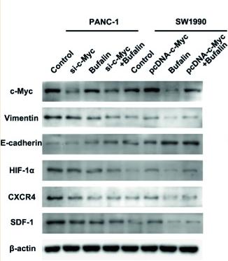
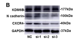
Application: WB Species: Human Sample: pancreatic cancer cells
限制条款
产品的规格、报价、验证数据请以官网为准,官网链接:www.affbiotech.com | www.affbiotech.cn(简体中文)| www.affbiotech.jp(日本語)产品的数据信息为Affinity所有,未经授权不得收集Affinity官网数据或资料用于商业用途,对抄袭产品数据的行为我们将保留诉诸法律的权利。
产品相关数据会因产品批次、产品检测情况随时调整,如您已订购该产品,请以订购时随货说明书为准,否则请以官网内容为准,官网内容有改动时恕不另行通知。
Affinity保证所销售产品均经过严格质量检测。如您购买的商品在规定时间内出现问题需要售后时,请您在Affinity官方渠道提交售后申请。产品仅供科学研究使用。不用于诊断和治疗。
产品未经授权不得转售。
Affinity Biosciences将不会对在使用我们的产品时可能发生的专利侵权或其他侵权行为负责。Affinity Biosciences, Affinity Biosciences标志和所有其他商标所有权归Affinity Biosciences LTD.