产品: TRAF6 抗体
货号: AF5376
描述: Rabbit polyclonal antibody to TRAF6
应用: WB IHC IF/ICC
反应: Human, Mouse, Rat
预测: Pig, Bovine, Sheep, Rabbit, Dog, Chicken
分子量: 58 kDa; 60kD(Calculated).
蛋白号: Q9Y4K3
RRID: AB_2810280

浏览相似产品>>

   规格 价格 库存
 50ul RMB¥ 1250 现货
 100ul RMB¥ 2300 现货
 200ul RMB¥ 3000 现货

货期: 当天发货

联系销售

产品描述

来源:
Rabbit
应用:
WB 1:500-1:2000, IHC 1:50-1:200, IF/ICC 1:100-1:500
*The optimal dilutions should be determined by the end user.
*Tips:

WB: 适用于变性蛋白样本的免疫印迹检测. IHC: 适用于组织样本的石蜡(IHC-p)或冰冻(IHC-f)切片样本的免疫组化/荧光检测. IF/ICC: 适用于细胞样本的荧光检测. ELISA(peptide): 适用于抗原肽的ELISA检测.

反应:
Human,Mouse,Rat
预测:
Pig(100%), Bovine(90%), Sheep(90%), Rabbit(100%), Dog(100%), Chicken(90%)
克隆:
Polyclonal
特异性:
TRAF6 Antibody detects endogenous levels of total TRAF6.
RRID:
AB_2810280
引用格式: Affinity Biosciences Cat# AF5376, RRID:AB_2810280.
偶联:
Unconjugated.
纯化:
The antiserum was purified by peptide affinity chromatography using SulfoLink™ Coupling Resin (Thermo Fisher Scientific).
保存:
Rabbit IgG in phosphate buffered saline , pH 7.4, 150mM NaCl, 0.02% sodium azide and 50% glycerol. Store at -20 °C. Stable for 12 months from date of receipt.
别名:

展开/折叠

E3 ubiquitin-protein ligase TRAF6; Interleukin 1 signal transducer; Interleukin-1 signal transducer; MGC 3310; MGC:3310; MGC3310; OTTHUMP00000232772; OTTHUMP00000232773; RING finger protein 85; RNF 85; RNF85; TNF receptor associated factor 6; TNF receptor-associated factor 6; TNF receptor-associated factor 6, E3 ubiquitin protein ligase; TRAF 6; Traf6; TRAF6_HUMAN;

抗原和靶标

免疫原:
Uniprot:
基因/基因ID:
表达:
Q9Y4K3 TRAF6_HUMAN:

Expressed in heart, brain, placenta, lung, liver, skeletal muscle, kidney and pancreas.

描述:
Tumor necrosis factor receptor-associated factor 6 (TRAF6) regulates adaptive immunity, innate immunity and bone metabolism. TRAF6 is a ubiquitin (Ub) ligase that mediates the activation of protein kinases, such as transforming growth factor beta-activated kinase (TAK1) and IκB kinase (IKK)
序列:
MSLLNCENSCGSSQSESDCCVAMASSCSAVTKDDSVGGTASTGNLSSSFMEEIQGYDVEFDPPLESKYECPICLMALREAVQTPCGHRFCKACIIKSIRDAGHKCPVDNEILLENQLFPDNFAKREILSLMVKCPNEGCLHKMELRHLEDHQAHCEFALMDCPQCQRPFQKFHINIHILKDCPRRQVSCDNCAASMAFEDKEIHDQNCPLANVICEYCNTILIREQMPNHYDLDCPTAPIPCTFSTFGCHEKMQRNHLARHLQENTQSHMRMLAQAVHSLSVIPDSGYISEVRNFQETIHQLEGRLVRQDHQIRELTAKMETQSMYVSELKRTIRTLEDKVAEIEAQQCNGIYIWKIGNFGMHLKCQEEEKPVVIHSPGFYTGKPGYKLCMRLHLQLPTAQRCANYISLFVHTMQGEYDSHLPWPFQGTIRLTILDQSEAPVRQNHEEIMDAKPELLAFQRPTIPRNPKGFGYVTFMHLEALRQRTFIKDDTLLVRCEVSTRFDMGSLRREGFQPRSTDAGV

种属预测

种属预测:

score>80的预测可信度较高,可尝试用于WB检测。*预测模型主要基于免疫原序列比对,结果仅作参考,不作为质保凭据。

Species
Results
Score
Pig
100
Dog
100
Rabbit
100
Bovine
90
Sheep
90
Chicken
90
Zebrafish
78
Horse
0
Xenopus
0
Model Confidence:
High(score>80) Medium(80>score>50) Low(score<50) No confidence

翻译修饰 - Q9Y4K3 作为底物

Site PTM Type Enzyme
K104 Ubiquitination
K124 Ubiquitination
S188 Phosphorylation
Y326 Phosphorylation
K331 Ubiquitination
K365 Ubiquitination
T486 Phosphorylation
K489 Ubiquitination
T492 Phosphorylation
S507 Phosphorylation

研究背景

功能:

E3 ubiquitin ligase that, together with UBE2N and UBE2V1, mediates the synthesis of 'Lys-63'-linked-polyubiquitin chains conjugated to proteins, such as IKBKG, IRAK1, AKT1 and AKT2. Also mediates ubiquitination of free/unanchored polyubiquitin chain that leads to MAP3K7 activation. Leads to the activation of NF-kappa-B and JUN. May be essential for the formation of functional osteoclasts. Seems to also play a role in dendritic cells (DCs) maturation and/or activation. Represses c-Myb-mediated transactivation, in B-lymphocytes. Adapter protein that seems to play a role in signal transduction initiated via TNF receptor, IL-1 receptor and IL-17 receptor. Regulates osteoclast differentiation by mediating the activation of adapter protein complex 1 (AP-1) and NF-kappa-B, in response to RANK-L stimulation. Together with MAP3K8, mediates CD40 signals that activate ERK in B-cells and macrophages, and thus may play a role in the regulation of immunoglobulin production.

翻译修饰:

Sumoylated on Lys-124, Lys-142 and Lys-453 with SUMO1.

Polyubiquitinated on Lys-124; after cell stimulation with IL-1-beta or TGF-beta. This ligand-induced cell stimulation leads to dimerization/oligomerization of TRAF6 molecules, followed by auto-ubiquitination which involves UBE2N and UBE2V1 and leads to TRAF6 activation. This 'Lys-63' site-specific poly-ubiquitination appears to be associated with the activation of signaling molecules. Endogenous autoubiquitination occurs only for the cytoplasmic form. Deubiquitinated by USP10 in a TANK-dependent manner, leading to the negative regulation of NF-kappaB signaling upon DNA damage.

细胞定位:

Cytoplasm. Cytoplasm>Cell cortex. Nucleus. Lipid droplet.
Note: Found in the nuclei of some aggressive B-cell lymphoma cell lines as well as in the nuclei of both resting and activated T- and B-lymphocytes. Found in punctate nuclear body protein complexes. Ubiquitination may occur in the cytoplasm and sumoylation in the nucleus. RSAD2/viperin recruits it to the lipid droplet (By similarity).

Extracellular region or secreted Cytosol Plasma membrane Cytoskeleton Lysosome Endosome Peroxisome ER Golgi apparatus Nucleus Mitochondrion Manual annotation Automatic computational assertionSubcellular location
组织特异性:

Expressed in heart, brain, placenta, lung, liver, skeletal muscle, kidney and pancreas.

亚基结构:

Homotrimer. Homooligomer. N-terminal region is dimeric while C-terminal region is trimeric; maybe providing a mode of oligomerization. Upon IL1B treatment, forms a complex with PELI1, IRAK1, IRAK4 and MYD88; this complex recruits MAP3K7/TAK1, TAB1 and TAB2 to mediate NF-kappa-B activation. Direct binding of SMAD6 to PELI1 prevents the complex formation and hence negatively regulates IL1R-TLR signaling and eventually NF-kappa-B-mediated gene expression. Binds to TNFRSF5/CD40 and TNFRSF11A/RANK. Associates with NGFR, TNFRSF17, IRAK2, IRAK3, RIPK2, MAP3K1, MAP3K5, MAP3K14, CSK, TRAF, TRAF-interacting protein TRIP and TNF receptor associated protein TDP2. Interacts with IL17R. Interacts with SQSTM1 bridging NTRK1 and NGFR. Forms a ternary complex with SQSTM1 and PRKCZ (By similarity). Interacts with PELI2 and PELI3. Binds UBE2V1. Interacts with TAX1BP1. Interacts with ZNF675. Interacts with ARRB1 and ARRB2. Interacts with MAP3K7 and TAB1/MAP3K7IP1; during IL-1 signaling. Interacts with UBE2N. Interacts with TGFBR1, HDAC1 and RANGAP1. Interacts with AKT1, AKT2 and AKT3. Interacts (via TRAF domains) with NUMBL (via C-terminal). Interacts with RBCK1. Interacts with TRAF3IP2. Interacts with LIMD1 (via LIM domains) (By similarity). Interacts with RSAD2/viperin (By similarity). Interacts (via C-terminus) with EIF2AK2/PKR (via the kinase catalytic domain) (By similarity). Interacts with ZFAND5. Interacts with IL1RL1. Interacts with TRAFD1. Interacts with AJUBA. Interacts with MAVS/IPS1. Interacts (via TRAF domains) with WDR34 (via WD domains). Interacts with IFIT3 (via N-terminus). Interacts with TICAM2. Interacts with CARD14. Interacts with CD40 and MAP3K8; the interaction is required for ERK activation (By similarity). Interacts with TICAM1 and this interaction is enhanced in the presence of WDFY1. Interacts with TANK; this interaction increases in response to DNA damage. Interacts with USP10; this interaction increases in response to DNA damage. Interacts with ZC3H12A; this interaction increases in response to DNA damage and is stimulated by TANK. Interacts with WDFY3 (By similarity). Interacts with TRIM13. Interacts with GPS2 (By similarity). Interacts (via C-terminus) with SASH1.

蛋白家族:

The coiled coil domain mediates homo- and hetero-oligomerization.

The MATH/TRAF domain binds to receptor cytoplasmic domains.

Belongs to the TNF receptor-associated factor family. A subfamily.

研究领域

· Cellular Processes > Transport and catabolism > Autophagy - animal.   (View pathway)

· Cellular Processes > Transport and catabolism > Endocytosis.   (View pathway)

· Environmental Information Processing > Signal transduction > MAPK signaling pathway.   (View pathway)

· Environmental Information Processing > Signal transduction > NF-kappa B signaling pathway.   (View pathway)

· Genetic Information Processing > Folding, sorting and degradation > Ubiquitin mediated proteolysis.   (View pathway)

· Human Diseases > Infectious diseases: Bacterial > Pertussis.

· Human Diseases > Infectious diseases: Parasitic > Leishmaniasis.

· Human Diseases > Infectious diseases: Parasitic > Chagas disease (American trypanosomiasis).

· Human Diseases > Infectious diseases: Parasitic > Toxoplasmosis.

· Human Diseases > Infectious diseases: Bacterial > Tuberculosis.

· Human Diseases > Infectious diseases: Viral > Hepatitis C.

· Human Diseases > Infectious diseases: Viral > Measles.

· Human Diseases > Infectious diseases: Viral > Herpes simplex infection.

· Human Diseases > Infectious diseases: Viral > Epstein-Barr virus infection.

· Human Diseases > Cancers: Overview > Pathways in cancer.   (View pathway)

· Human Diseases > Cancers: Specific types > Small cell lung cancer.   (View pathway)

· Organismal Systems > Development > Osteoclast differentiation.   (View pathway)

· Organismal Systems > Immune system > Toll-like receptor signaling pathway.   (View pathway)

· Organismal Systems > Immune system > NOD-like receptor signaling pathway.   (View pathway)

· Organismal Systems > Immune system > RIG-I-like receptor signaling pathway.   (View pathway)

· Organismal Systems > Immune system > IL-17 signaling pathway.   (View pathway)

· Organismal Systems > Nervous system > Neurotrophin signaling pathway.   (View pathway)

文献引用

1). Breaking the vicious loop between inflammation, oxidative stress and coagulation, a novel anti-thrombus insight of nattokinase by inhibiting LPS-induced inflammation and oxidative stress. Redox Biology, 2020 (PubMed: 32193146) [IF=11.4]

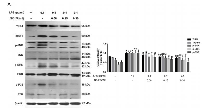
Application: WB    Species: Mice    Sample: RAW264.7 cells

Fig. 5. NK diminished LPS-induced TLR4 activation likely due to promoting TRL4 proteolysis in RAW264.7 cells. (A) Effect of NK on LPS-induced TLR4 signaling pathways. Cells were pretreated with indicated concentrations of NK for 1 h and then exposed to LPS (0.1 μg/mL) for 12 h. Equal amounts of total cell lysates were loaded and subjected to immunoblot analysis. Data represent the mean ± SD from three independent experiments. (B) NK induced TRL4 degradation via its serine protease activity in RAW264.7 cells. Cells were treated with NK (0.3 FU/mL) for indicated time points with or without PMSF pretreatment for 30 min. Equal amounts of total cell lysates were loaded and subjected to immunoblot analysis. Data represent mean ± SD from three independent experiments. *P < 0.05, **P < 0.01 compared to control group; #P < 0.05, ##P < 0.05 compared to NK 12h group; $ P < 0.05, $$P < 0.05 compared to NK 24h group.

2). The YTHDF1–TRAF6 pathway regulates the neuroinflammatory response and contributes to morphine tolerance and hyperalgesia in the periaqueductal gray. Journal of Neuroinflammation, 2022 (PubMed: 36550542) [IF=9.3]

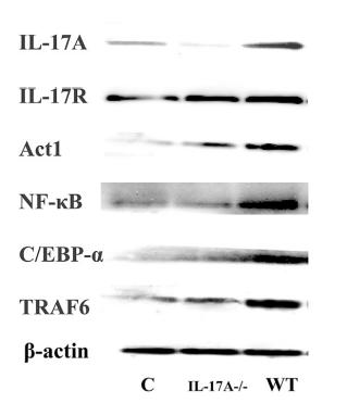
3). Interleukin 17A deficiency alleviates fluoride-induced testicular injury by inhibiting the immune response and apoptosis. Chemosphere, 2021 (PubMed: 33297146) [IF=8.8]

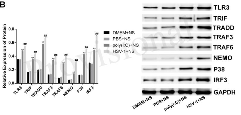
Application: WB    Species: Mice    Sample: testes

Fig. 6. F can stimulate downstream proteins in IL-17A signal pathway in the testes (n ¼ 13. Three times repetition). Statistical analysis was carried out using one-way ANOVA with Dunnett’s test. Error bars denote the mean ± s.e.m. *P < 0.05, **P < 0.01.

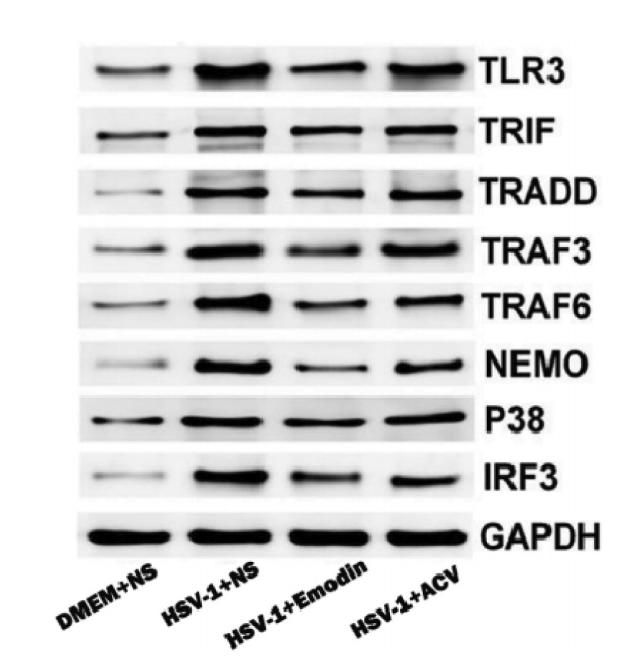
4). The matrix protein of Newcastle disease virus inhibits inflammatory response through IRAK4/TRAF6/TAK1/NF-κB signaling pathway. International Journal of Biological Macromolecules, 2022 (PubMed: 35872314) [IF=8.2]

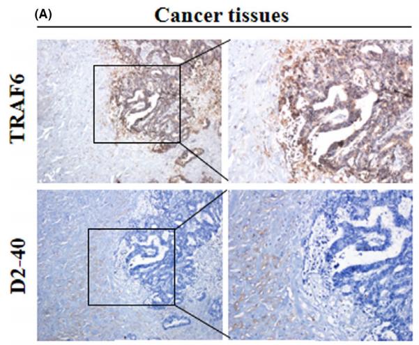
5). TRAF6 promotes the progression and growth of colorectal cancer through nuclear shuttle regulation NF-kB/c-jun signaling pathway. LIFE SCIENCES, 2019 (PubMed: 31487530) [IF=6.1]

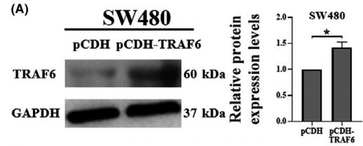
6). TRAF6 regulates the signaling pathway influencing colorectal cancer function through ubiquitination mechanisms. Cancer Science, 2022 (PubMed: 35179811) [IF=5.7]

Application: WB    Species: human    Sample: SW480 and HCT116 cells

FIGURE 2|The role of TRAF6 in HLEC tube formation. (A, C) Western blotting analysis of TRAF6 protein expression in SW480 and HCT116 cells transfected with pCDH and pCDH-TRAF6, respectively. GAPDH served as a loading control. The relative protein expression levels of TRAF6 in SW480 and HCT116 cells were quantified.

Application: IHC    Species: human    Sample: colorectal cancer tissues

FIGURE 1|Expression of TRAF6 and lymphatic vessels in colorectal cancer clinical samples. (A, B) Representative immunohistochemical staining images for TRAF6 protein and D2-40 in colorectal cancer (CRC) and adjacent normal tissues from a tissue array are shown.Magnification, ×40 and ×100.

7). Chang-Kang-Fang alleviates diarrhea predominant irritable bowel syndrome (IBS-D) through inhibiting TLR4/NF-κB/NLRP3 pathway. Journal of ethnopharmacology, 2024 (PubMed: 38670405) [IF=5.4]

8). Adamantane-linked isothiourea derivatives suppress the growth of experimental hepatocellular carcinoma via inhibition of TLR4-MyD88-NF-κB signaling. American Journal of Cancer Research, 2023 (PubMed: 33575076) [IF=5.3]

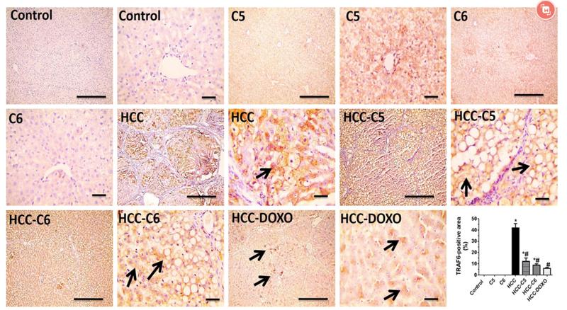
Application: IHC    Species: rat    Sample: hepatic tissues

Figure 10.| Effect of compounds 5 and 6 on hepatic on hepatic TRAF-6 protein expression in TAA-administered rats. Representative microimages of immunostaining for TRAF-6 protein (IHC counterstained with Mayer’s hematoxylin) in hepatic tissues and statistical analysis of positive area of immunolabelling (%) are shown. Black arrows denote positive immunoexpression. Images were captured at magnification of 100× (scale bar, 100 µm) or 400× (scale bar, 50 µm). *P < 0.05 vs. control group, #P < 0.05 vs. HCC group. DOXO: doxorubicin, HCC: hepatocellular carcinoma, IHC: immunohistochemistry, TAA: thioacetamide, TRAF-6: tumor necrosis factor receptor-associated factor-6.

9). Cornuside protects against ischemic stroke in rats by suppressing the IL-17F/TRAF6/NF-κB pathway via the brain-gut axis. Experimental neurology, 2024 (PubMed: 38169196) [IF=5.3]

10). TMT-based quantitative proteomics analysis reveals the attenuated replication mechanism of Newcastle disease virus caused by nuclear localization signal mutation in viral matrix protein. Virulence, 2020 (PubMed: 32420802) [IF=5.2]

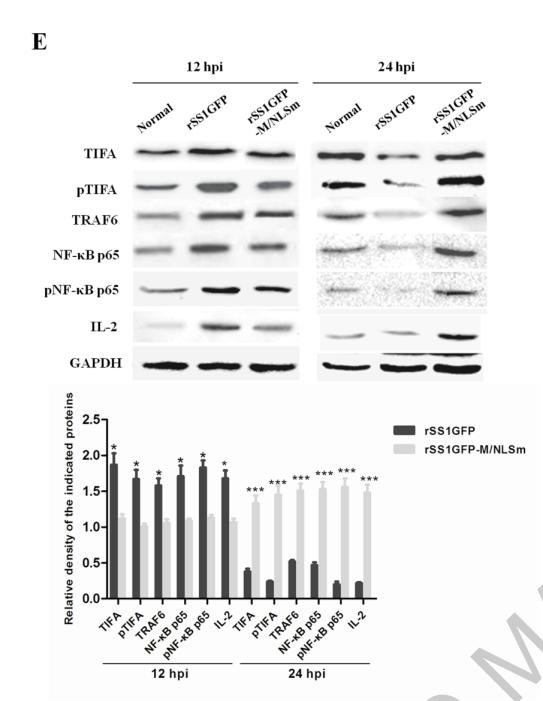
Application: WB    Species: Mouse    Sample: BSR-T7/5 cells

Figure 9. rSS1GFP replication is enhanced by inhibiting TIFA/TRAF6/NF-κB signaling pathway later in infection. (A) The mRNA expression levels of TIFA gene in BSR-T7/5 cells infected with rSS1GFP and rSS1GFP-M/NLSm were verified by qRT-PCR. (B) The protein expression levels of TIFA in BSR-T7/5 cells infected with rSS1GFP and rSS1GFP-M/NLSm were examined by Western blotting. The relative expression levels of TIFA were compared with the control GAPDH expression. (C) The subcellular localization of EGFP-M or EGFP-M/NLSm and HA-TIFA in plasmids co-transfected BSR-T7/5 cells. Original magnification was 1 × 200. (D) The effect of different dosage EGFP-M or EGFP-M/NLSm on the expression level of endogenous TIFA in plasmid transfected BSR-T7/5 cells. The relative expression levels of TIFA were compared with the control GAPDH expression. (E) The expression patterns of TIFA, pTIFA, TRAF6, NF-κB p65, pNF-κB p65, and IL-2 in BSR-T7/5 cells infected with rSS1GFP and rSS1GFP-M/NLSm at 12 and 24 hpi. The relative expression levels of these proteins were compared with the control GAPDH expression. (F) The effect of TIFA overexpression on the expression of IL-2 and virus titers of rSS1GFP and rSS1GFP-M/NLSm at 12 and 24 hpi. (G) The effect of siRNA-mediated knockdown of TIFA on the expression of IL-2 and virus titers of rSS1GFP and rSS1GFP-M/NLSm at 12 and 24 hpi. (H) The schematic diagram illustrated that the M protein in the cytoplasm inhibited host cell immune response by down-regulating TIFA/TRAF6/NF-κB signaling pathway

加载更多

限制条款

产品的规格、报价、验证数据请以官网为准,官网链接:www.affbiotech.com | www.affbiotech.cn(简体中文)| www.affbiotech.jp(日本語)

产品的数据信息为Affinity所有,未经授权不得收集Affinity官网数据或资料用于商业用途,对抄袭产品数据的行为我们将保留诉诸法律的权利。

产品相关数据会因产品批次、产品检测情况随时调整,如您已订购该产品,请以订购时随货说明书为准,否则请以官网内容为准,官网内容有改动时恕不另行通知。

Affinity保证所销售产品均经过严格质量检测。如您购买的商品在规定时间内出现问题需要售后时,请您在Affinity官方渠道提交售后申请。

产品仅供科学研究使用。不用于诊断和治疗。 

产品未经授权不得转售。

Affinity Biosciences将不会对在使用我们的产品时可能发生的专利侵权或其他侵权行为负责。Affinity Biosciences, Affinity Biosciences标志和所有其他商标所有权归Affinity Biosciences LTD.