, using SQSTM1/p62 Antibody. The lane on the left was treated with blocking peptide.")


.")


 and mouse anti-beta tubulin Ab(#T0023) for 1 hour at 37°C. An AlexaFluor594 conjugated goat anti-rabbit IgG Ab(Red) and an AlexaFluor488 conjugated goat anti-mouse IgG Ab(Green) were used as the secondary antibody.
The nuclear counter stain is DAPI (blue).")
 and mouse anti-beta tubulin Ab(T0023 1:200) for 1 hour at 37°C. An AlexaFluor594 conjugated goat anti-rabbit IgG(H+L) Ab(Red) and an AlexaFluor488 conjugated goat anti-mouse IgG(H+L) Ab(Green) were used as the secondary antibody.
The nuclear counter stain is DAPI(blue).")
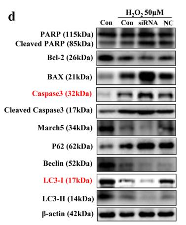
 were used as negative control.(D) Sertoli cells were pretreated with NAC (10 mM) for 1 h, followed by 400 μg/ml OLA treatment for 24 h prior to immunblot analysis for the expression of each protein. Semiquantitative analysis of protein expression in following histogram (mean ± S.E.M., three independent replicates per groups). * p < 0.05; ** p < 0.01.")

 Western blots were conducted to determine LC3‑II and p62 expression, β‑actin expression was detected as loading control. Quantified data of blots are presented as the mean ± standard deviation of three separate experiments. *P<0.05 vs. the control group. #P<0.05 vs. the hIAPP group. hIAPP, human islet amyloid polypeptide; rIAPP, rodent islet amyloid polypeptide; LC3, microtubule associated protein 1 light chain 3; ctrl, control group.")
 The expression and quantification of HMGB1, RAGE, LC3, and Beclin1 in the A498 cell line were analyzed by Western blot analysis.")
Cells were transfected either with Nrf2 siRNA (10 or 20 µM) or scrambled siRNA, and then treated with Andro (20 µM) for 24 h. Then, Nrf2, p62, and p21 protein levels were evaluated by Western blot analysis")
 for 48 hours. Cell apoptosis was detected via flow cytometry analysis. The apoptotic ratio was calculated as the percentage of early apoptotic (Annexin V+/PI-) cells plus late apoptotic (Annexin V+/PI+) cells (A,B).The expression of apoptosis-related proteins and autophagy markers was measured via Western blot analysis (C,D).")
 The degree of pulmonary artery remodeling in MCT-treated mice increased significantly compared with control mice. (B) Analysis of pulmonary arteries. (C) Western blot analysis using lung tissue samples showing significantly increased expression levels of SUMO1, HIF-1α and autophagy marker LC3b, and a significantly decreased expression level of autophagy marker p62 in MCT-treated mice than in control mice. (D) Western blot data in (C) were quantified and plotted.")

 was used to inhibit cell autophagy in these experiments. (A‑C) Western blot analysis results for the autophagy‑associated protein levels of LC3‑I/II and SQSTM1/p62. Tubulin was used as an internal control. The results are expressed as the means ± SD (n=3 for each group). *P<0.05; **P<0.001; and ****P<0.0001 compared with the control group.")

 TUNEL assay of brain tissues. The black arrow points to cells with positive staining. Scale bar = 30 μm (n = 4). Data are presented as mean ± SD. P values were determined by ANOVA followed by the Tukey post-hoc test,****p < 0.0001. (B–C) Relative protein levels of caspase3, Bax, Bcl-2. Beclin1, LC3II/I, p62 in the sham group, the AAV-mCherry group, and the AAVLRG1 group. Quantification of protein levels was normalized to GAPDH (n = 3). Data are presented as mean ± SD. P values were determined by ANOVA followed by the Tukey post-hoc test, * p < 0.05.")
 Autophagy related proteins LC3-II/LC3-I, Beclin-1, and P62 were detected and quantified by Western blot in THP-1, THP-1/ADM, K562 and K562/ADM cells.")
 The effect of PA on LC3-II and p62 protein levels in hippocampus were in-vestigated by western blot analysis.")
 LC3-II/LC3-I ratios and (E) P62 levels were analyzed in macrophages by western blot.")
 Expression levels of beclin 1, LC3 II, and p62 proteins in the LV of mice. n = 6, ∗P < 0.05, ∗∗P < 0.01 vs. control group; #P < 0.05, ##P < 0.01 vs. Ang II group. (b) Representative images of the ultrastructure of H9c2 cells showing the changes of autophagy under a transmission electron microscope. Scale bar in (i), 2 μm and magnification, ×10000; in (ii), 1 μm, ×20000. Yellow arrow refers to the autophagosome, which contains dense electronic content such as autolysosomes or mitochondria. (c) Expression of beclin 1 and LC3 mRNA in H9c2 cells. (d, e) Expression of beclin 1, LC3II, and P62 proteins in H9c2 cells. In cell experiments, (1) control, (2) Ang II, (3) NC, (4) NC + Ang II, (5) miR-128 antag, (6) miR-128 antag + Ang II, (7) Ang II + CQ (chloroquine), and (8) miR-128 antag + Ang II + CQ. n = 3 independent batches, repeated 2 ~ 3 wells/each batch. Data are expressed as mean ± SD. Statistical significance was assessed by using one-way ANOVA followed by LSD. ∗P < 0.05, ∗∗P < 0.01 vs. control group; #P < 0.05, ##P < 0.01 vs. NC + Ang II group; ∆P < 0.05, P < 0.01 vs. Ang II group; ▴P < 0.05 vs. Ang II + CQ group.")
 Representative images of Beclin1, LC3 and P62 (immunohistochemical staining; magnification, ×400). Quantitative analysis of (B) Beclin1, (C) LC3 and (D) P62 were normalized to the normal control group. (E) Protein expression levels of Beclin1, LC3 and P62 in the liver tissues were detected via western blotting. Semi-quantitative analysis of (F) Beclin1, (G) LC3 and (H) P62 was normalised to β-actin. Data are presented as the mean ± SD, n=3. *P<0.05, ***P<0.001 vs. control group; #P<0.05, ##P<0.01, ###P<0.001 vs. model group. Con, Control; Mod, Model; AS-IV, Astragaloside IV (80 mg/kg); Met, Metformin (200 mg/kg).")
 Representative images of Beclin1, LC3 and P62 (immunohistochemical staining; magnification, ×400). Quantitative analysis of (B) Beclin1, (C) LC3 and (D) P62 were normalized to the normal control group. (E) Protein expression levels of Beclin1, LC3 and P62 in the liver tissues were detected via western blotting. Semi-quantitative analysis of (F) Beclin1, (G) LC3 and (H) P62 was normalised to β-actin. Data are presented as the mean ± SD, n=3. *P<0.05, ***P<0.001 vs. control group; #P<0.05, ##P<0.01, ###P<0.001 vs. model group. Con, Control; Mod, Model; AS-IV, Astragaloside IV (80 mg/kg); Met, Metformin (200 mg/kg).")

 activity and autophagic fux in CD4+T cells. (A–E) Protein levels of phospho (p)-mTOR,p-P70 S6 kinase (S6K), LC3II/I and p62/sequestome 1 (SQSTM1) in CD4+T cells were quantifed by western blotting in wild-type (WT) mice, mTOR knockout via Cre expression under the control of lymphocyte-specifc protein tyrosine kinase (Lck) (Lck-mTOR) cecal ligation and puncture (CLP) mice, and tuberous sclerosis complex 1(TSC1) knockout (Lck-TSC1)+CLP mice. The amount of each protein level was normalized to that of β-actin. Means±standard deviations (SDs) of four mice per group are shown. **P<0.01, ***P<0.001,****P<0.0001.")
. B, Detection of the autophagy-related proteins LC3II/I, Beclin1, AKT, PTEN, mTOR and STX3 by Western blotting.")
 Expression levels of beclin 1, LC3 II, and p62 proteins in the LV of mice. n = 6, ∗P < 0:05, ∗∗P < 0:01 vs. control group; #P < 0:05, ##P < 0:01 vs. Ang II group.")
 To confrm that the efect of baicalin is mediated by regulating autophagy, 3-MA was used with baicalin treatment, and the levels of the autophagy-related proteins LC3II/I, Beclin1, P62, p-AMPK, AMPK, and mTOR were measured by Western blotting.")

 By IHC staining, the number of Beclin 1+ and LC3II+ cells was significantly reduced in patients with eCRSwNP or noeCRSwNP, compared with control tissues. p62 staining revealed no significant differences among the groups. (c, d) Western blot analysis showed that the levels of Beclin 1 protein in nasal polyps were downregulated in patients with eCRSwNP or noeCRSwNP, compared with control tissues. p62 protein levels increased, whereas LC3II/LC3I levels were significantly reduced, only in the nasal polyps of patients with eCRSwNP, compared with control tissues. (e) TEM showed that autophagosomes formed less frequently in the nasal polyps of patients with eCRSwNP or noeCRSwNP, compared with control tissues. Black arrow: autophagosome. ∗P < 0.05, ∗∗P < 0.001.")

 Representative protein blots of p62, LC3, and Beclin 1. (b) Quantitative analysis of p62, LC3, and Beclin 1 proteins. (c) Representative protein blots of PI3K/AKT pathway-related proteins. (d) Quantitative analysis of PI3K pathway-related targets in each group. ∗P < 0.05 and ∗∗P < 0.01 vs. sham group. ▲P < 0.05 and ▲▲P < 0.01 vs. IR group.")
 p62 expression was detected by immunofluorescence analysis in LBAG-treated H/R-injured H9c2 cells. Scale bar: 50 μm. (b, c) p62, LC3, Beclin 1, and PI3K/AKT pathway-related protein levels were estimated with western blotting in LBAG-treated H/R-injured H9c2 cells. ∗P < 0.05 and ∗∗P < 0.01 vs. control group. ▲P < 0.05 and ▲▲P < 0.01 vs. H/R group. #P < 0.05 and ##P < 0.01 vs. 10 μmol/L LBAG group.")
 and model groups (0.1 N/0.3/0.5 N group); b–c Graphic display of LC3 II (b) and P62 (c) protein level in each group (n = 3); Columns, mean ± SE; #p < 0.05 or ##p < 0.01, compared with the SH group; *p < 0.05 or **p < 0.01, comparison between all model groups")

 Protein levels of beclin-1, p62, and LC3II as measured by western blotting. (B–D) Relative density values showing P62, beclin1, LC3- II/LC3- I expression. (E,F) Representative images of TEM and immunohistochemical staining for P62. The white arrows indicate autophagosomes and autolysosomes, the yellow arrowheads indicate abnormal mitochondria. Data are expressed as *p < 0.05, **p < 0.01, compared with sham group; #p < 0.05, ##p < 0.01, compared with model group.")
 post‐oxygen–glucose deprivation (OGD). (A, B) Apoptosis of BMSCs in different groups was stained by Annexin V‐FITC/PI, and then assessed by flow cytometry (FCM). (C–E) Western blot images and relative quantification of apoptosis‐related proteins (Bcl‐2, Bax) and Gapdh (n = 4). (F–I) Western blot analysis of P62, Beclin‐1, LC3, and Gapdh and quantitative analysis (n = 4). * means p value <0.05, ** means p value <0.01, and *** means p value <0.001, p values were calculated with one‐way ANOVA, followed by Bonferroni's multiple comparison test")
 Representative Western blot of Parkin, PINK1, SQSTM1/p62, LC3A/B-I:II, BNIP3L, C1-C5-CR, T1-T5-HR. (B–F) Relative levels of appropriate proteins with respect to the loading control (GAPDH). CR, control; HR, hyperthyroidism. **—p < 0.02, ***—p < 0.001 as compared with the control data (n = 5–6).")
 Immunofluorescence staining of LC3 and F4/80 in the lung tissues of WT and Nrf2−/− mice before or after CLP treatment. (C,D) Western blot analyses of autophagy-associated proteins including LC3 I, LC3 II, Beclin1, and p62 in lung tissues of each group. ** p < 0.01, *** p < 0.001. (E) Transmission electron microscopy assay detected the numbers of double-membrane autophagosomes and autolysosomes (red arrows in the pictures) in lung tissues of each group.")
 treated VSMCs. The relative protein expressions in Ang II-treated VSMCs with presence of C/EBPα overexpression or knockdown were analyzed by Western blot. GAPDH was used as an internal control. Data are presented as means ± SEM. (a, b) Evaluation of the influence of C/EBPα on the expression of α-SMA, SM-MHC, and OPN proteins. (c, d) Evaluation of the influence of C/EBPα on the expression of PIK3C2A protein. (e, f) Evaluation of the influence of C/EBPα on the expression of Beclin-1, p62, LC3II, and LC3I proteins. (g, h) Evaluation of the influence of C/EBPα on the expression of MMP-2 and MMP-9 proteins. (i) Intracellular ROS was measured by flow cytometry using DCFH-DA probe with presence of C/EBPα overexpression or knockdown. Control: control group; model: treated with 1 μM Ang II; vector: treated with 1 μM Ang II in vector transfected VSMCs; pcC/EBPα: treated with 1 μM Ang II in C/EBPα-overexpressed VSMCs; siNC: treated with 1 μM Ang II in siRNA NC-transfected VSMCs; siC/EBPα: treated with 1 μM Ang II in C/EBPα-knockdown VSMCs. ∗P < 0.05 vs. control,")
 of P62 (A, B), LC3-II (A, C), and Beclin-1 (A, D) were analyzed. The proteins expression of P62 (E, F), LC3-II (E, G), and Beclin-1 (E, H) in DEX-treated MC3T3-E1 cells were determined by western blot. * *P˂0.01. NC, negative control group; DEX, model rat group treated with DEX (5 mg/kg).")
 of P62 (A, B), LC3-II (A, C), and Beclin-1 (A, D) were analyzed. The proteins expression of P62 (E, F), LC3-II (E, G), and Beclin-1 (E, H) in DEX-treated MC3T3-E1 cells were determined by western blot. * *P˂0.01. NC, negative control group; DEX, model rat group treated with DEX (5 mg/kg).")
 Protein levels of PINK1, Beclin1, p62, and LC3II as measured by western blotting. (b–f) Relative density values showing PINK1, Beclin1, LC3-II, LC3-II/LC3-I, and p62 expression. (g) Representative images of immunohistochemical staining (400×) for p62. ∗P < 0.05, ∗∗P < 0.01, compared with the NC group; #P < 0.05, ##P < 0.01, compared with BLM group.")
, p62 (B), and LC3(C) were captured under fluorescence microscopy, and the relative fluorescence intensity was calculated. All images were obtained at identical magnification, ×200, scale bar = 50 μm. Data are represented as mean ± SEM (n = 3). **P")

 HEK−293T cells were transfected with PPRV−N expression plasmid or treated with rapamycin for 48 h. The LC3−II levels in the cells treated with or not treated with chloroquine were compared between control cells and either PPRV−N−expressing cells or rapamycin-treated cells. Corresponding graphs depict the levels of SQSTM1/p62 (B,C) and LC3B−II (D,E). (F): The line graph showing the comparison between SQSTM1/p62 and LC3B−II, in the absence and presence of chloroquine. (G): HEK−293T cells were pretreated with wortmannin for 6 h and transfected with PPRV−N expression plasmid for 48 h. The LC3B conversion and SQSTM1/p62 levels were compared both in the presence and absence of chloroquine between mock transfected and PPRV−N−transfected cells. Corresponding graphs depict the levels of SQSTM1/p62 (H,I) and LC3B−II (J,K). (L): The line graph showing the comparison between SQSTM1/p62 and LC3B−II in the absence and presence of wortmannin. GAPDH was used as the internal loading control. Antibodies against SQSTM1/p62 and LC3B were used in immunoblotting analysis. The data represents the mean ± SD of three independent experiments. Paired Student’s t-test was used to determine the significance. *** p < 0.0005, ** p < 0.005, * p < 0.05; ns: non-significant.")
. Western blot detection of proteins in the NLRP3 inflammatory body: NLRP3, cleaved-CASP1, and cleaved-IL-1β (B). Western blot detection of proteins in the autophagy pathway: Beclin1, p-VPS34, and p62 (C). The expression of NLRP3 inflammatory bodies and autophagic vesicles in uterine tissues was observed by IF: NLRP3 was labeled with red fluorescence, VPS34 was labeled with green fluorescence, and nuclei were labeled with DAPI with blue fluorescence (D). Western blot detection of proteins in the lysosomal pathway: LAMP2, Ubiquitin and LC3 Ⅱ. (E). Results are presented as the mean ± S.E.M.")
 B16 cells were treated with 0, 1 and 10 nM RAPA for 48 h. The formation of autophagic vesicles was observed by transmission electron microscopy. M indicates mitochondrial structure, N the nucleus, C the cytoplasm, and red triangles indicate autophagic lysosomes. Magnification, ×8,000; scale bar, 2.0 nm. (B) Expression levels of autophagy-related LC3, p62 and Beclin-1 in B16 cells were detected by western blotting. Relative expression of (C) LC3II, (D) Beclin-1 and (E) p62 proteins. Relative mRNA expression levels of (F) LC3 II, (G) Beclin-1 and (H) p62. (I) B16 cells were treated with rapamycin (100 nM) and/or CQ (10 µM) for 48 h. The protein expression levels of LC3 and p62 were detected by western blotting. (J) Relative expression of LC3II protein. (K) Relative expression of LC3II protein. Data are presented as the mean ± SD (n=3). *P")
 Phalloidin staining for VSMC morphology. Scale bar: 100 μm. (b) Flowchart, (c) protein-protein interaction network, (d) gene ontology, and (e) KEGG analysis on network pharmacology for HLJDD treating AS. Western blotting presented the effect of HLJDD on the PI3K/AKT pathway (f–i, n = 4 per group) in vivo and (j–m, n = 4–5 per group) in vitro. Data were presented as mean ± standard deviation in one-way ANOVA, with Dunnett post hoc tests. ns: not significant")
 Western blot analysis of apoptosis-related protein expression levels. (B–D) Quantification of cleaved-caspase-3 (B), Bax (C), and Bcl-2 (D) protein levels from the western blot data. (E) Western blot analysis of autophagy-related protein expression levels. (F–H) Quantification of LC3II/LC3I (F), Beclin-1 (G), and P62 (H) protein levels from the western blot data. (I) Western blot analysis of oxidative stress-related protein expression levels. β-Actin was used as an internal reference for cleaved-caspase-3, Bax, and Bcl-2. GAPDH was used as an internal reference for LC3I, LC3II, Beclin-1, P62, Nrf2, and HO-1. (J, K) Western blot analysis of Nrf2 (J) and HO-1 (K) protein expression levels. All western blot data were normalized to the sham group. Data are expressed as mean ± SD (n = 3). **P < 0.01, vs. sham group; #P < 0.05, ##P < 0.01, vs. model group (one-way analysis of variance followed by Tukey's post hoc test). BA: Biochanin A; HO-1: heme oxygenase-1; LC3: microtube-associated protein 1 light-chain 3; Nrf2: nuclear factor erythroid2-related factor 2; P62: sequestosome-1; PC: positive control; SCI: spinal cord injury.")
 The number of autophagosomes in gastric mucosal epithelial cells was shown by transmission electron microscopy (indicated by red arrows), Magnification ×12,000. (B) The expression of LC-3 was detected by immunofluorescence assay, magnification ×400; scale bar: 20 µm (n = 6). (C) Using ImageJ software to analyse the fluorescence intensity of LC-3. (D) The expression of Beclin1 was detected by immunofluorescence assay, magnification ×400; scale bar: 20 µm (n = 6). (E) Using ImageJ software to analyse the fluorescence intensity of Beclin1. (F) Western blotting bands of autophagy-related proteins. (G,H) Grey intensity analysis of P62/β‐actin and LC-3II/LC-3I ratio (n = 3). These data are means ± SD, ###p < 0.001 when compared with the control group. *p < 0.05, **p < 0.01 and ***p < 0.001 when compared with the model group. ns: no significant difference.")
 Expression of autophagy-related genes. (B, C) Expression of autophagy-related proteins in the whole cell lysate. (D, E) Expression of mitophagy-related proteins in the mitochondria protein extract. n=6 per group and the values are the mean ± SEM. ** P")
产品描述
*The optimal dilutions should be determined by the end user.
*Tips:
WB: 适用于变性蛋白样本的免疫印迹检测. IHC: 适用于组织样本的石蜡(IHC-p)或冰冻(IHC-f)切片样本的免疫组化/荧光检测. IF/ICC: 适用于细胞样本的荧光检测. ELISA(peptide): 适用于抗原肽的ELISA检测.
引用格式: Affinity Biosciences Cat# AF5384, RRID:AB_2837869.
展开/折叠
A170; DMRV; EBI 3 associated protein of 60 kDa; EBI 3 associated protein p60; EBI3 associated protein of 60 kDa; EBI3 associated protein p60; EBI3-associated protein of 60 kDa; EBIAP; FTDALS3; MGC127197; ORCA; OSF-6; Osi; OSIL; Oxidative stress induced like; p60; p62; p62B; Paget disease of bone 3; PDB 3; PDB3; Phosphotyrosine independent ligand for the Lck SH2 domain of 62 kDa; Phosphotyrosine independent ligand for the Lck SH2 domain p62; Phosphotyrosine-independent ligand for the Lck SH2 domain of 62 kDa; PKC-zeta-interacting protein; Protein kinase C-zeta-interacting protein; Sequestosome 1; Sequestosome-1; SQSTM 1; SQSTM_HUMAN; Sqstm1; STAP; STONE14; Ubiquitin binding protein p62; Ubiquitin-binding protein p62; ZIP 3; ZIP; ZIP3;
抗原和靶标
- Q13501 SQSTM_HUMAN:
- Protein BLAST With
- NCBI/
- ExPASy/
- Uniprot
MASLTVKAYLLGKEDAAREIRRFSFCCSPEPEAEAEAAAGPGPCERLLSRVAALFPALRPGGFQAHYRDEDGDLVAFSSDEELTMAMSYVKDDIFRIYIKEKKECRRDHRPPCAQEAPRNMVHPNVICDGCNGPVVGTRYKCSVCPDYDLCSVCEGKGLHRGHTKLAFPSPFGHLSEGFSHSRWLRKVKHGHFGWPGWEMGPPGNWSPRPPRAGEARPGPTAESASGPSEDPSVNFLKNVGESVAAALSPLGIEVDIDVEHGGKRSRLTPVSPESSSTEEKSSSQPSSCCSDPSKPGGNVEGATQSLAEQMRKIALESEGRPEEQMESDNCSGGDDDWTHLSSKEVDPSTGELQSLQMPESEGPSSLDPSQEGPTGLKEAALYPHLPPEADPRLIESLSQMLSMGFSDEGGWLTRLLQTKNYDIGAALDTIQYSKHPPPL
种属预测
score>80的预测可信度较高,可尝试用于WB检测。*预测模型主要基于免疫原序列比对,结果仅作参考,不作为质保凭据。
High(score>80) Medium(80>score>50) Low(score<50) No confidence
研究背景
Autophagy receptor required for selective macroautophagy (aggrephagy). Functions as a bridge between polyubiquitinated cargo and autophagosomes. Interacts directly with both the cargo to become degraded and an autophagy modifier of the MAP1 LC3 family. Along with WDFY3, involved in the formation and autophagic degradation of cytoplasmic ubiquitin-containing inclusions (p62 bodies, ALIS/aggresome-like induced structures). Along with WDFY3, required to recruit ubiquitinated proteins to PML bodies in the nucleus. May regulate the activation of NFKB1 by TNF-alpha, nerve growth factor (NGF) and interleukin-1. May play a role in titin/TTN downstream signaling in muscle cells. May regulate signaling cascades through ubiquitination. Adapter that mediates the interaction between TRAF6 and CYLD (By similarity). May be involved in cell differentiation, apoptosis, immune response and regulation of K(+) channels. Involved in endosome organization by retaining vesicles in the perinuclear cloud: following ubiquitination by RNF26, attracts specific vesicle-associated adapters, forming a molecular bridge that restrains cognate vesicles in the perinuclear region and organizes the endosomal pathway for efficient cargo transport. Promotes relocalization of 'Lys-63'-linked ubiquitinated STING1 to autophagosomes. Acts as an activator of the NFE2L2/NRF2 pathway via interaction with KEAP1: interaction inactivates the BCR(KEAP1) complex, promoting nuclear accumulation of NFE2L2/NRF2 and subsequent expression of cytoprotective genes.
Phosphorylated. May be phosphorylated by PRKCZ (By similarity). Phosphorylated in vitro by TTN. Phosphorylation at Ser-403 by ULK1 is stimulated by SESN2. Phosphorylated at Ser-403 by TBK1, leading to promote relocalization of 'Lys-63'-linked ubiquitinated STING1 to autophagosomes. Phosphorylation at Ser-349 by MTOR promotes interaction with KEAP1 and inactivation of the BCR(KEAP1) complex, promoting NFE2L2/NRF2 nuclear accumulation and expression of phase II detoxifying enzymes (By similarity).
Ubiquitinated by RNF26: ubiquitinated SQSTM1 attracts specific vesicle-associated adapters, forming a molecular bridge that restrains cognate vesicles in the perinuclear region and organizes the endosomal pathway for efficient cargo transport. Deubiquitination by USP15 releases target vesicles for fast transport into the cell periphery. Ubiquitinated by the BCR(KEAP1) complex at Lys-420, increasing SQSTM1 sequestering activity and promoting its degradation. Ubiquitinated via 'Lys-29' and 'Lys-33'-linked polyubiquitination leading to xenophagic targeting of bacteria and inhibition of their replication.
Cytoplasm>Cytosol. Late endosome. Lysosome. Cytoplasmic vesicle>Autophagosome. Nucleus. Endoplasmic reticulum. Nucleus>PML body. Cytoplasm>Myofibril>Sarcomere.
Note: In cardiac muscle, localizes to the sarcomeric band (By similarity). Commonly found in inclusion bodies containing polyubiquitinated protein aggregates. In neurodegenerative diseases, detected in Lewy bodies in Parkinson disease, neurofibrillary tangles in Alzheimer disease, and HTT aggregates in Huntington disease. In protein aggregate diseases of the liver, found in large amounts in Mallory bodies of alcoholic and nonalcoholic steatohepatitis, hyaline bodies in hepatocellular carcinoma, and in SERPINA1 aggregates. Enriched in Rosenthal fibers of pilocytic astrocytoma. In the cytoplasm, observed in both membrane-free ubiquitin-containing protein aggregates (sequestosomes) and membrane-surrounded autophagosomes. Colocalizes with TRIM13 in the perinuclear endoplasmic reticulum. Co-localizes with TRIM5 in cytoplasmic bodies. When nuclear export is blocked by treatment with leptomycin B, accumulates in PML bodies.
Ubiquitously expressed.
Homooligomer or heterooligomer; may form homotypic arrays. Dimerization interferes with ubiquitin binding. Interacts directly with PRKCI and PRKCZ (Probable). Forms ternary complexes with PRKCZ and KCNAB2 or PRKCZ and GABBR3. Also interacts with KCNAB1, GABRR1, GABRR2 and GABRR3. Forms an NGF-induced complex with IKBKB, PRKCI and TRAF6 (By similarity). Interacts with EBI3, LCK, RASA1, PRKCZ, PRKCI, NR2F2, NTRK1, NTRK2, NTRK3, NBR1, MAP2K5, TRIM13, TRIM55 and MAPKAPK5. Interacts with the proteasome subunits PSMD4 and PSMC2. Interacts with K63-polyubiquitinated MAPT/TAU. Interacts with IKBKB through PRKCZ and PRKCI. Interacts with NGFR through TRAF6 and bridges that complex to NTRK1. Forms a complex with MAP2K5 and PRKCZ or PRKCI. Component of a ternary complex with PAWR and PRKCZ. Upon TNF-alpha stimulation, interacts with RIPK1 probably bridging IKBKB to the TNF-R1 complex composed of TNF-R1/TNFRSF1A, TRADD and RIPK1. Forms a complex with JUB/Ajuba, PRKCZ and TRAF6. Interacts with TRAF6 and CYLD. Identified in a complex with TRAF6 and CYLD (By similarity). Identified in a heterotrimeric complex with ubiquitin and ZFAND5, where ZFAND5 and SQSTM1 both interact with the same ubiquitin molecule. Directly interacts with MAP1LC3A and MAP1LC3B, as well as with other MAP1 LC3 family members, including GABARAP, GABARAPL1 and GABARAPL2; these interactions are necessary for the recruitment MAP1 LC3 family members to inclusion bodies containing polyubiquitinated protein aggregates and for their degradation by autophagy. Interacts with FHOD3. Interacts with TRMI5. Interacts with SESN1. Interacts with SESN2. Interacts with ULK1. Interacts with UBD. Interacts with WDR81; the interaction is direct and regulates the interaction of SQSTM1 with ubiquitinated proteins. Interacts with WDFY3; this interaction is required to recruit WDFY3 to cytoplasmic bodies and to PML bodies. Interacts with TRIM23. Interacts with LRRC25. Interacts with TRIM50. Interacts with TRIM16. Interacts with STING1; leading to relocalization of STING1 to autophagosomes. Interacts (when phosphorylated at Ser-349) with KEAP1; the interaction is direct and inactivates the BCR(KEAP1) complex by sequestering KEAP1 in inclusion bodies, promoting its degradation.
The UBA domain binds specifically 'Lys-63'-linked polyubiquitin chains of polyubiquitinated substrates. Mediates the interaction with TRIM55. Both the UBA and PB1 domains are necessary and sufficient for the localization into the ubiquitin-containing inclusion bodies.
The PB1 domain mediates homooligomerization and interactions with FHOD3, MAP2K5, NBR1, PRKCI, PRKCZ and WDR81. Both the PB1 and UBA domains are necessary and sufficient for the localization into the ubiquitin-containing inclusion bodies.
The ZZ-type zinc finger mediates the interaction with RIPK1.
The LIR (LC3-interacting region) motif mediates the interaction with ATG8 family proteins.
研究领域
· Cellular Processes > Cell growth and death > Necroptosis. (View pathway)
· Cellular Processes > Cell growth and death > Cellular senescence. (View pathway)
· Organismal Systems > Development > Osteoclast differentiation. (View pathway)
文献引用

Application: WB Species: human Sample: A549 cells
Application: WB Species: mice Sample: bone marrow mesenchymal stem (BMSCs)
Application: WB Species: human Sample: MDA-MB-231 cells

Application: WB Species: Human Sample: MDA-MB-231 cells

Application: WB Species: rat Sample: PC12 cells
限制条款
产品的规格、报价、验证数据请以官网为准,官网链接:www.affbiotech.com | www.affbiotech.cn(简体中文)| www.affbiotech.jp(日本語)产品的数据信息为Affinity所有,未经授权不得收集Affinity官网数据或资料用于商业用途,对抄袭产品数据的行为我们将保留诉诸法律的权利。
产品相关数据会因产品批次、产品检测情况随时调整,如您已订购该产品,请以订购时随货说明书为准,否则请以官网内容为准,官网内容有改动时恕不另行通知。
Affinity保证所销售产品均经过严格质量检测。如您购买的商品在规定时间内出现问题需要售后时,请您在Affinity官方渠道提交售后申请。产品仅供科学研究使用。不用于诊断和治疗。
产品未经授权不得转售。
Affinity Biosciences将不会对在使用我们的产品时可能发生的专利侵权或其他侵权行为负责。Affinity Biosciences, Affinity Biosciences标志和所有其他商标所有权归Affinity Biosciences LTD.