产品: CD44 抗体
货号: DF6392
描述: Rabbit polyclonal antibody to CD44
应用: WB IHC IF/ICC
反应: Human, Mouse, Rat
预测: Pig, Bovine, Horse, Sheep, Rabbit, Dog
分子量: 82kDa; 82kD(Calculated).
蛋白号: P16070
RRID: AB_2838355

浏览相似产品>>

   规格 价格 库存
 50ul RMB¥ 1250 现货
 100ul RMB¥ 2300 现货
 200ul RMB¥ 3000 现货

货期: 当天发货

联系销售

产品描述

来源:
Rabbit
应用:
WB 1:500-1:2000, IHC 1:50-1:200, IF/ICC 1:100-1:500
*The optimal dilutions should be determined by the end user.
*Tips:

WB: 适用于变性蛋白样本的免疫印迹检测. IHC: 适用于组织样本的石蜡(IHC-p)或冰冻(IHC-f)切片样本的免疫组化/荧光检测. IF/ICC: 适用于细胞样本的荧光检测. ELISA(peptide): 适用于抗原肽的ELISA检测.

反应:
Human,Mouse,Rat
预测:
Pig(82%), Bovine(82%), Horse(82%), Sheep(82%), Rabbit(82%), Dog(91%)
克隆:
Polyclonal
特异性:
CD44 Antibody detects endogenous levels of total CD44.
RRID:
AB_2838355
引用格式: Affinity Biosciences Cat# DF6392, RRID:AB_2838355.
偶联:
Unconjugated.
纯化:
The antiserum was purified by peptide affinity chromatography using SulfoLink™ Coupling Resin (Thermo Fisher Scientific).
保存:
Rabbit IgG in phosphate buffered saline , pH 7.4, 150mM NaCl, 0.02% sodium azide and 50% glycerol. Store at -20 °C. Stable for 12 months from date of receipt.
别名:

展开/折叠

LHR; BA-1; CD 44; CD44; CD44 antigen; CD44 molecule (Indian blood group); CD44 molecule; CD44_HUMAN; CDw44; Cell surface glycoprotein CD44; chondroitin sulfate proteoglycan 8; CSPG8; ECMR-III; Epican; Extracellular matrix receptor III; GP90 lymphocyte homing/adhesion receptor; HCELL; hematopoietic cell E- and L-selectin ligand; Heparan sulfate proteoglycan; Hermes antigen; homing function and Indian blood group system; HSA; HUTCH-I; HUTCH1; Hyaluronate receptor; IN; INLU-related p80 Glycoprotein; MC56; MDU2; MDU3; MGC10468; MIC4; MUTCH1; PGP-1; PGP-I; PGP1; Phagocytic glycoprotein 1; Phagocytic glycoprotein I; Soluble CD44;

抗原和靶标

免疫原:
Uniprot:
基因/基因ID:
表达:
P16070 CD44_HUMAN:

Isoform 10 (epithelial isoform) is expressed by cells of epithelium and highly expressed by carcinomas. Expression is repressed in neuroblastoma cells.

描述:
CD44 is a type I transmembrane glycoprotein that mediates cell-cell and cell-matrix interaction through its affinity for hyaluronic acid (HA) and possibly through other parts of the extracellular matrix (ECM). CD44 is highly polymorphic, possesses a number of alternative splice variants and undergoes extensive post-translational modifications (1,2). Increased surface levels of CD44 are characteristic of T cell activation, and expression of the protein is upregulated during the inflammatory response. Interactions between CD44 and HER2 have been linked to an increase in ovarian carcinoma cell growth (1-3). CD44 interacts with ezrin, radixin and moesin (ERM), linking the actin cytoskeleton to the plasma membrane and the ECM (4-6). CD44 is constitutively phosphorylated at Ser325 in resting cells. Activation of PKC results in phosphorylation of Ser291, dephosphorylation of Ser325, disassociation of ezrin from CD44, and directional motility (4).
序列:
MDKFWWHAAWGLCLVPLSLAQIDLNITCRFAGVFHVEKNGRYSISRTEAADLCKAFNSTLPTMAQMEKALSIGFETCRYGFIEGHVVIPRIHPNSICAANNTGVYILTSNTSQYDTYCFNASAPPEEDCTSVTDLPNAFDGPITITIVNRDGTRYVQKGEYRTNPEDIYPSNPTDDDVSSGSSSERSSTSGGYIFYTFSTVHPIPDEDSPWITDSTDRIPATTLMSTSATATETATKRQETWDWFSWLFLPSESKNHLHTTTQMAGTSSNTISAGWEPNEENEDERDRHLSFSGSGIDDDEDFISSTISTTPRAFDHTKQNQDWTQWNPSHSNPEVLLQTTTRMTDVDRNGTTAYEGNWNPEAHPPLIHHEHHEEEETPHSTSTIQATPSSTTEETATQKEQWFGNRWHEGYRQTPKEDSHSTTGTAAASAHTSHPMQGRTTPSPEDSSWTDFFNPISHPMGRGHQAGRRMDMDSSHSITLQPTANPNTGLVEDLDRTGPLSMTTQQSNSQSFSTSHEGLEEDKDHPTTSTLTSSNRNDVTGGRRDPNHSEGSTTLLEGYTSHYPHTKESRTFIPVTSAKTGSFGVTAVTVGDSNSNVNRSLSGDQDTFHPSGGSHTTHGSESDGHSHGSQEGGANTTSGPIRTPQIPEWLIILASLLALALILAVCIAVNSRRRCGQKKKLVINSGNGAVEDRKPSGLNGEASKSQEMVHLVNKESSETPDQFMTADETRNLQNVDMKIGV

种属预测

种属预测:

score>80的预测可信度较高,可尝试用于WB检测。*预测模型主要基于免疫原序列比对,结果仅作参考,不作为质保凭据。

Species
Results
Score
Dog
91
Pig
82
Horse
82
Bovine
82
Sheep
82
Rabbit
82
Xenopus
0
Zebrafish
0
Chicken
0
Model Confidence:
High(score>80) Medium(80>score>50) Low(score<50) No confidence

翻译修饰 - P16070 作为底物

Site PTM Type Enzyme
S45 Phosphorylation
K54 Ubiquitination
N57 N-Glycosylation
S58 Phosphorylation
T62 Phosphorylation
N110 N-Glycosylation
K158 Ubiquitination
T163 Phosphorylation
S171 Phosphorylation
T174 O-Glycosylation
S179 Phosphorylation
S182 Phosphorylation
S183 Phosphorylation
S184 Phosphorylation
T197 O-Glycosylation
S199 O-Glycosylation
T200 O-Glycosylation
T222 Phosphorylation
S291 Phosphorylation
T311 Phosphorylation
T318 O-Glycosylation
T325 O-Glycosylation
S330 O-Glycosylation
S332 O-Glycosylation
Y412 Phosphorylation
S420 Phosphorylation
S422 O-Glycosylation
T423 O-Glycosylation
T424 O-Glycosylation
T426 O-Glycosylation
S430 O-Glycosylation
T433 O-Glycosylation
S434 O-Glycosylation
S458 O-Glycosylation
S514 O-Glycosylation
T515 O-Glycosylation
T528 O-Glycosylation
T529 O-Glycosylation
T541 O-Glycosylation
S550 O-Glycosylation
S553 O-Glycosylation
T554 O-Glycosylation
T555 O-Glycosylation
T561 O-Glycosylation
S562 O-Glycosylation
T567 O-Glycosylation
S570 O-Glycosylation
T572 O-Glycosylation
T572 Phosphorylation
T577 O-Glycosylation
S578 O-Glycosylation
T581 O-Glycosylation
S583 O-Glycosylation
T587 Phosphorylation
S672 Phosphorylation P17252 (PRKCA)
K681 Ubiquitination
S686 Phosphorylation
K695 Ubiquitination
S697 Phosphorylation P17612 (PRKACA)
S704 Phosphorylation
K705 Ubiquitination
S706 Phosphorylation Q9UQM7 (CAMK2A)
K715 Ubiquitination
S717 Phosphorylation
S718 Phosphorylation
T720 Phosphorylation
T726 Phosphorylation
K739 Ubiquitination

研究背景

功能:

Cell-surface receptor that plays a role in cell-cell interactions, cell adhesion and migration, helping them to sense and respond to changes in the tissue microenvironment. Participates thereby in a wide variety of cellular functions including the activation, recirculation and homing of T-lymphocytes, hematopoiesis, inflammation and response to bacterial infection. Engages, through its ectodomain, extracellular matrix components such as hyaluronan/HA, collagen, growth factors, cytokines or proteases and serves as a platform for signal transduction by assembling, via its cytoplasmic domain, protein complexes containing receptor kinases and membrane proteases. Such effectors include PKN2, the RhoGTPases RAC1 and RHOA, Rho-kinases and phospholipase C that coordinate signaling pathways promoting calcium mobilization and actin-mediated cytoskeleton reorganization essential for cell migration and adhesion.

翻译修饰:

Proteolytically cleaved in the extracellular matrix by specific proteinases (possibly MMPs) in several cell lines and tumors.

N-glycosylated.

O-glycosylated. O-glycosylation contains more-or-less-sulfated chondroitin sulfate glycans, whose number may affect the accessibility of specific proteinases to their cleavage site(s). It is uncertain if O-glycosylation occurs on Thr-637 or Thr-638.

Phosphorylated; activation of PKC results in the dephosphorylation of Ser-706 (constitutive phosphorylation site), and the phosphorylation of Ser-672.

细胞定位:

Cell membrane>Single-pass type I membrane protein. Cell projection>Microvillus.
Note: Colocalizes with actin in membrane protrusions at wounding edges. Co-localizes with RDX, EZR and MSN in microvilli. Localizes to cholesterol-rich membrane-bound lipid raft domains.

Extracellular region or secreted Cytosol Plasma membrane Cytoskeleton Lysosome Endosome Peroxisome ER Golgi apparatus Nucleus Mitochondrion Manual annotation Automatic computational assertionSubcellular location
组织特异性:

Isoform 10 (epithelial isoform) is expressed by cells of epithelium and highly expressed by carcinomas. Expression is repressed in neuroblastoma cells.

亚基结构:

Interacts with PKN2. Interacts with TIAM1 and TIAM2 (By similarity). Interacts with HA, as well as other glycosaminoglycans, collagen, laminin, and fibronectin via its N-terminal segment. Interacts with UNC119. Interacts with PDPN (via extracellular domain); this interaction is required for PDPN-mediated directional migration and regulation of lamellipodia extension/stabilization during cell spreading and migration. Interacts with RDX, EZR and MSN (By similarity). Interacts with EGFR. Interacts with CD74; this complex is essential for the MIF-induced signaling cascade that results in B cell survival (By similarity).

蛋白家族:

The lectin-like LINK domain is responsible for hyaluronan binding.

研究领域

· Environmental Information Processing > Signaling molecules and interaction > ECM-receptor interaction.   (View pathway)

· Human Diseases > Infectious diseases: Bacterial > Shigellosis.

· Human Diseases > Infectious diseases: Viral > Epstein-Barr virus infection.

· Human Diseases > Cancers: Overview > Proteoglycans in cancer.

· Human Diseases > Cancers: Overview > MicroRNAs in cancer.

· Organismal Systems > Immune system > Hematopoietic cell lineage.   (View pathway)

文献引用

1). Metabolic reprogramming of proinflammatory macrophages by target delivered roburic acid effectively ameliorates rheumatoid arthritis symptoms. Signal Transduction and Targeted Therapy (PubMed: 37500654) [IF=39.3]

Application: IF/ICC    Species: Mouse    Sample: RAW264.7 cells

Fig. 2 RBA-NPs display targeting capability in vitro. a, b Confocal micrographs showed that DiD-NPs (red) enjoyed significantly improved cell uptake compared with free DiD in LPS + IFN-γ activated RAW264.7 cells. Scale bar = 10 μm. c Flow cytometry analysis showed that the fluorescence intensity of DiD-NPs in LPS + IFN-γ activated RAW264.7 cells was higher than that of free DiD in RAW264.7. d The expression of CD44 and folate receptors increased significantly on the surface of LPS + IFN-γ activated RAW264.7 cells (d). Scale bar = 10 μm. e LPS + IFN-γ activated macrophages were pretreated with HA or FA or both HA and FA to compete for the overexpressed CD44 or folate receptors, and cellular uptake of DiD-NPs was reduced. f Confocal micrographs showed co-localization of DiD-NPs (orange) with CD44 receptor (red) and folate receptor (green). Scale bar = 20 μm. Cell nuclei was stained with DAPI (blue). All results are shown as mean ± SD. *P 

2). HYD-PEP06 suppresses hepatocellular carcinoma metastasis, epithelial–mesenchymal transition and cancer stem cell-like properties by inhibiting PI3K/AKT and WNT/β-catenin signaling activation. Acta Pharmaceutica Sinica B (PubMed: 34221870) [IF=14.5]

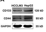
Application: WB    Species: Human    Sample: HepG2 cells

Figure 5 Tumor spheres isolated from hepatocellular carcinoma HepG2 cells obtained the characteristics of cancer stem cells. (A) Comparison of the protein levels of CD133 and CD44 in HCCLM3 and HepG2 cells. The protein expression of (B) CD133 and (C) CD44 was dramatically increased in HepG2 cells compared with those in HCCLM3 cells. ∗P < 0.05, compared with the HCCLM3 cells. (D) The images of 3D tumor spheroids were captured using the fluorescence microscope on Day 0 in attachment surface plates and Days 0, 3, 5, 7, and 10 in ultra-low attachment surface plates with CSC medium (magnification 200 ×); scale bar: 20 μm. (E) The protein expression of CD133 and CD44 was analyzed by Western blot in HepG2 and CSC-like cells. The protein expression of (F) CD133 and (G) CD44 was dramatically increased in CSC-like cells compared with HepG2 cells. Data are presented as mean ± SEM; ∗P < 0.05, ∗∗P < 0.01. (H) The expression of CD133 and CD44 were assessed by immunofluorescence staining in HepG2 and tumor sphere cells. Blue represents DAPI; green represents CD44; red represents CD133. Magnification: 400 ×. (I) CSC augments tumor-initiating in vivo. Representative xenograft tumors derived from CSC-like cells compared to HepG2 cells after two weeks subcutaneous injection in nude mice (n = 3). Left panel: HepG2 cells; right: CSCs. (J) The protein levels of CD133 and CD44 were analyzed by Western blot in the tumor tissues isolated from xenograft models. GAPDH served as a sample loading control. The protein levels of CSC markers (K) CD133 and (L) CD44 were dramatically upregulated in right tissues compared with left tissues. Data are presented as mean ± SEM; ∗P < 0.05, ∗∗∗P < 0.001.

3). CD44‐targeting Drug Delivery System of Exosomes Loading Forsythiaside A Combats Liver Fibrosis via Regulating NLRP3‐mediated Pyroptosis. Advanced Healthcare Materials (PubMed: 36603210) [IF=10.0]

4). ZIF-8 Modified Multifunctional Bone-Adhesive Hydrogels Promoting Angiogenesis and Osteogenesis for Bone Regeneration. ACS Applied Materials & Interfaces (PubMed: 32814397) [IF=9.5]

5). Kaempferol alleviates calcium oxalate crystal-induced renal injury and crystal deposition via regulation of the AR/NOX2 signaling pathway. PHYTOMEDICINE (PubMed: 33852977) [IF=7.9]

Application: IHC    Species: Human    Sample: HK-2 cells

Fig. 1. Effect of kaempferol on renal CaOx crystal deposition and tubular injury in vivo. Eligible mice were divided into five groups (n = 8). After a period of 10-day drug treatments, all mice were sacrificed on the 11 th day for the following tests. Pathological results were shown by the (a) images as well as (b-g) corresponding quantitative analysis through HE staining, Pizzolato staining, PAS staining, TUNEL staining, OPN and CD44 immunohistochemistry staining of paraffin embedded kidney sections. (h-j) Renal dysfunction was determined by serum levels of BUN, creatinine and NAGL among the five groups. (k) Group annotations were applied for all histograms. Data were expressed as the fold changes of experimental group to NC group or GA group, and were represented as means ± SD. * p < 0.05 vs. NC group; ** p < 0.05 as GA+Kae (25mg/kg) vs. GA group, GA + Kae (50 mg/kg) vs. GA group, and GA + Kae (25 mg/kg) vs. GA + Kae (50 mg/kg) group, respectively.

6). Ginsenoside Rg1 prevents bone marrow mesenchymal stem cell senescence via NRF2 and PI3K/Akt signaling. Free radical biology & medicine (PubMed: 34364981) [IF=7.4]

7). 4T1 cell membrane fragments reunited PAMAM polymer disguising as tumor cell clusters for tumor homotypic targeting and anti-metastasis treatment. Biomaterials Science (PubMed: 33355563) [IF=6.6]

Application: WB    Species:    Sample: CCNCs

Fig. 1 |(a) Particle size of PAMAM@DOX, CCM, and CCNCs. (b) Zeta potential of PAMAM@DOX, CCM, and CCNCs with different core-tomembrane mass ratios. (c) TEM images of: (I) PAMAM@DOX, (II) CCM vesicles, and (III, IV) CCNCs. (d) SDS-PAGE analysis of (I) protein marker,(II) 4T1 cell lysate, (III) CCM, and (IV) CCNCs. (e) Membrane protein characterization by the western blotting analysis of (I) 4T1 cell lysate, (II)CCM, and (III) CCNCs. Antigens tested included CD44, CD47, Na+K+-ATPase, and GAPDH

8). CX43 down-regulation promotes cell aggressiveness and 5-fluorouracil-resistance by attenuating cell stiffness in colorectal carcinoma. Cancer Biology & Therapy (PubMed: 37342072) [IF=3.6]

Application: WB    Species: Human    Sample: CRC cells

Figure 5. CX43 increased cell stiffness by polymerizing CRC cytoskeleton. (a and b) Confocal microscopy analysis of α-tubulin polymerization and F-actin cytoskeletal remodeling in CRC cells with CX43 overexpression and knock-down. (c) The young’s modulus of CX43 overexpressed DLD1 cells was measured by atomic mechanics microscope. Mechanical curve (left) and quantitative analysis (right) are displayed. (d) Western blot analyses expression of stem cell characteristic related proteins in CRC cells with CX43 overexpression. (e) The tumor cell spheroidizing ability of CX43 overexpression cells was detected by tumor sphere formation assays. (f) Western blot was used to survey the relative expression of apoptosis pathway related proteins in CX43 overexpression cells. The downregulation of CX43 could rescue the above phenotypes (d, e and f).

9). The capsaicinoid nonivamide suppresses the inflammatory response and attenuates the progression of steatosis in a NAFLD‐rat model. Journal of Biochemical and Molecular Toxicology (PubMed: 36541345) [IF=3.6]

10). ONECUT2 which is targeted by hsa-miR-15a-5p enhances stemness maintenance of gastric cancer stem cells. Experimental Biology and Medicine (PubMed: 34365839) [IF=3.2]

加载更多

限制条款

产品的规格、报价、验证数据请以官网为准,官网链接:www.affbiotech.com | www.affbiotech.cn(简体中文)| www.affbiotech.jp(日本語)

产品的数据信息为Affinity所有,未经授权不得收集Affinity官网数据或资料用于商业用途,对抄袭产品数据的行为我们将保留诉诸法律的权利。

产品相关数据会因产品批次、产品检测情况随时调整,如您已订购该产品,请以订购时随货说明书为准,否则请以官网内容为准,官网内容有改动时恕不另行通知。

Affinity保证所销售产品均经过严格质量检测。如您购买的商品在规定时间内出现问题需要售后时,请您在Affinity官方渠道提交售后申请。

产品仅供科学研究使用。不用于诊断和治疗。 

产品未经授权不得转售。

Affinity Biosciences将不会对在使用我们的产品时可能发生的专利侵权或其他侵权行为负责。Affinity Biosciences, Affinity Biosciences标志和所有其他商标所有权归Affinity Biosciences LTD.