产品: AIF1/IBA1 抗体
货号: DF6442
描述: Rabbit polyclonal antibody to AIF1/IBA1
应用: WB IHC IF/ICC
反应: Human, Mouse, Rat
预测: Pig, Bovine, Horse, Sheep, Rabbit, Dog
分子量: 17kDa; 17kD(Calculated).
蛋白号: P55008
RRID: AB_2838405

浏览相似产品>>

   规格 价格 库存
 50ul RMB¥ 1250 现货
 100ul RMB¥ 2300 现货
 200ul RMB¥ 3000 现货

货期: 当天发货

联系销售

产品描述

来源:
Rabbit
应用:
WB 1:500-1:2000, IHC 1:50-1:200, IF/ICC 1:100-1:500
*The optimal dilutions should be determined by the end user.
*Tips:

WB: 适用于变性蛋白样本的免疫印迹检测. IHC: 适用于组织样本的石蜡(IHC-p)或冰冻(IHC-f)切片样本的免疫组化/荧光检测. IF/ICC: 适用于细胞样本的荧光检测. ELISA(peptide): 适用于抗原肽的ELISA检测.

反应:
Human,Mouse,Rat
预测:
Pig(100%), Bovine(91%), Horse(91%), Sheep(91%), Rabbit(91%), Dog(90%)
克隆:
Polyclonal
特异性:
AIF1/IBA1 Antibody detects endogenous levels of total AIF1/IBA1.
RRID:
AB_2838405
引用格式: Affinity Biosciences Cat# DF6442, RRID:AB_2838405.
偶联:
Unconjugated.
纯化:
The antiserum was purified by peptide affinity chromatography using SulfoLink™ Coupling Resin (Thermo Fisher Scientific).
保存:
Rabbit IgG in phosphate buffered saline , pH 7.4, 150mM NaCl, 0.02% sodium azide and 50% glycerol. Store at -20 °C. Stable for 12 months from date of receipt.
别名:

展开/折叠

AIF 1; AIF-1; Aif1; AIF1 protein; AIF1_HUMAN; Allograft inflammatory factor 1; Allograft inflammatory factor 1 splice variant G; allograft inflammatory factor-1 splice variant Hara-1; balloon angioplasty responsive transcription; BART 1; G1; G1 putative splice variant of allograft inflamatory factor 1; IBA 1; IBA1; interferon gamma responsive transcript; Interferon responsive transcript 1; interferon responsive transcript factor 1; Ionized calcium binding adapter molecule 1; Ionized calcium-binding adapter molecule 1; ionized calcium-binding adapter molecule; IRT 1; IRT1; Microglia response factor; MRF1; Protein g1;

抗原和靶标

免疫原:
Uniprot:
基因/基因ID:
表达:
P55008 AIF1_HUMAN:

Detected in T-lymphocytes and peripheral blood mononuclear cells.

描述:
This gene is induced by cytokines and interferon. Its protein product is thought to be involved in negative regulation of growth of vascular smooth muscle cells, which contributes to the anti-inflammatory response to vessel wall trauma. Three transcript variants encoding different isoforms have been found for this gene. [provided by RefSeq, Sep 2008]
序列:
MSQTRDLQGGKAFGLLKAQQEERLDEINKQFLDDPKYSSDEDLPSKLEGFKEKYMEFDLNGNGDIDIMSLKRMLEKLGVPKTHLELKKLIGEVSSGSGETFSYPDFLRMMLGKRSAILKMILMYEEKAREKEKPTGPPAKKAISELP

种属预测

种属预测:

score>80的预测可信度较高,可尝试用于WB检测。*预测模型主要基于免疫原序列比对,结果仅作参考,不作为质保凭据。

Species
Results
Score
Pig
100
Horse
91
Bovine
91
Sheep
91
Rabbit
91
Dog
90
Xenopus
0
Zebrafish
0
Chicken
0
Model Confidence:
High(score>80) Medium(80>score>50) Low(score<50) No confidence

翻译修饰 - P55008 作为底物

Site PTM Type Enzyme
S2 Phosphorylation
K11 Acetylation
K11 Ubiquitination
Y37 Phosphorylation
S38 Phosphorylation
S39 Phosphorylation
Y54 Phosphorylation
S69 Phosphorylation
K76 Ubiquitination
K81 Ubiquitination
K113 Methylation
Y124 Phosphorylation

研究背景

功能:

Actin-binding protein that enhances membrane ruffling and RAC activation. Enhances the actin-bundling activity of LCP1. Binds calcium. Plays a role in RAC signaling and in phagocytosis. May play a role in macrophage activation and function. Promotes the proliferation of vascular smooth muscle cells and of T-lymphocytes. Enhances lymphocyte migration. Plays a role in vascular inflammation.

翻译修饰:

Phosphorylated on serine residues.

细胞定位:

Cytoplasm>Cytoskeleton. Cell projection>Ruffle membrane>Peripheral membrane protein>Cytoplasmic side. Cell projection>Phagocytic cup.
Note: Associated with the actin cytoskeleton at membrane ruffles and at sites of phagocytosis.

Extracellular region or secreted Cytosol Plasma membrane Cytoskeleton Lysosome Endosome Peroxisome ER Golgi apparatus Nucleus Mitochondrion Manual annotation Automatic computational assertionSubcellular location
组织特异性:

Detected in T-lymphocytes and peripheral blood mononuclear cells.

亚基结构:

Homodimer (Potential). Monomer. Interacts with LCP1.

文献引用

1). Activation of PPARA-mediated autophagy reduces Alzheimer disease-like pathology and cognitive decline in a murine model. Autophagy, 2020 (PubMed: 30898012) [IF=13.3]

2). Huoluo Xiaoling Pellet promotes microglia M2 polarization through increasing MCPIP1 expression for ischemia stroke alleviation. Biomedicine & Pharmacotherapy, 2023 (PubMed: 37236023) [IF=7.5]

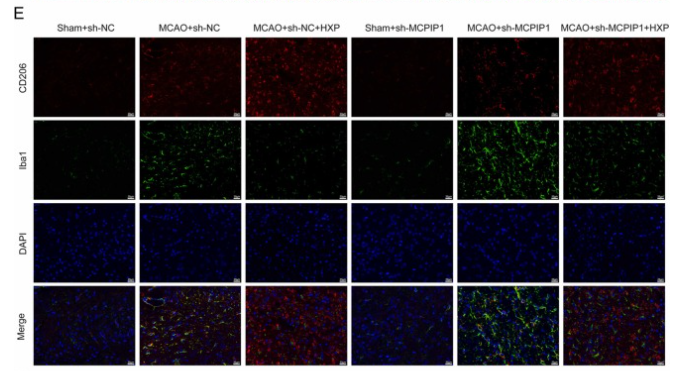
Application: IF/ICC    Species: Rat    Sample:

Fig. 3. HXP reduced neuroinflammation in MCAO rats by promoting MCPIP1-mediated microglia M2 polarization. A. After reperfusion for 7 d, brain water contents were examined. B. Brain infract volume was detected by 2,3,5-Triphentltetrazolium chloride staining. C. Changes in IL-1β, IL-6, iNOS, and TNF-α were measured via ELISA assy. D. Representative images of histopathological analysis. Scale bar, 20 µm. E. Representative immunofluorescence staining for CD206 (red) and Iba1 (green) in brain tissues. Scale bar, 20 µm. F. The expression of MCPIP1, CD16, iNOS, CD206, Arg1, and PPARγ was detected using Western blotting assay. Data are represented as mean ± standard deviation (N = 5). * * P 

3). Effects of hydrogen-rich saline in neuroinflammation and mitochondrial dysfunction in rat model of sepsis-associated encephalopathy. Journal of Translational Medicine, 2022 (PubMed: 36435787) [IF=7.4]

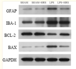
Application: WB    Species: Rat    Sample:

Fig. 8 Effects of HRS treatment on protein expressions in septic rats 48 h after LPS challenge. A Representative images of protein expression levels by western blot analysis in each group. B–E Statistical representation of relative protein expressions of GFAP, IBA-1, BCL-2 and BAX; respectively. All data are presented as mean ± SD ****p < 0.0001, ***p < 0.001, **p < 0.005, *p < 0.05. HRS Hydrogen-rich saline, LPS Lipopolysaccharide, GFAP Glial fibrillary acidic protein, IBA-1 Ionised calcium binding adaptor molecule 1, BCL-2 B-cell lymphoma 2

4). Cannabidivarin alleviates neuroinflammation by targeting TLR4 co-receptor MD2 and improves morphine-mediated analgesia. Frontiers in immunology, 2022 (PubMed: 36032146) [IF=7.3]

Application: IF/ICC    Species: Mouse    Sample:

Figure 8. CBDV inhibits chronic morphine treatment-induced glial activation and pro-inflammatory factors IL-6 and TNF-α mRNA expression in NAc. (A–C) Representative double immunofluorescent staining images of Iba1 and GFAP for the control group (A), morphine group (B), and morphine + CBDV group (C). NAc regions were collected following the final behavioral testing shown in Figure 7B . (D, F) The quantification of microglia (D) and astrocytes (F). (E, G) The size of the microglia (E) and astrocytes (G). (H, I) Total RNAs were extracted and qRT-PCR was performed to measure the expression of TNF-α (H) and IL-6 (I) in NAc. Scale bar: 200 μm. All the data represented mean ± S.E.M. ## P < 0.01, ### P < 0.001 versus the control group; *P < 0.05, **P < 0.01, ***P < 0.001 versus the morphine group.

5). Chitosan oligosaccharides exert neuroprotective effects via modulating the PI3K/Akt/Bcl-2 pathway in a Parkinsonian model. Food & Function, 2022 (PubMed: 35545086) [IF=6.1]

6). Inhibition of NLRP3 inflammasome activation and pyroptosis with the ethyl acetate fraction of Bungeanum ameliorated cognitive dysfunction in aged mice. Food & Function, 2021 (PubMed: 34231604) [IF=6.1]

7). Nattokinase’s Neuroprotective Mechanisms in Ischemic Stroke: Targeting Inflammation, Oxidative Stress, and Coagulation. ANTIOXIDANTS & REDOX SIGNALING, 2024 [IF=5.9]

8). High glucose impairs cognitive function through inducing mitochondrial calcium overload in Treg cells. iScience, 2024 (PubMed: 38226157) [IF=5.8]

9). Aucubin improves chronic unpredictable mild stress-induced depressive behavior in mice via the GR/NF-κB/NLRP3 axis. International Immunopharmacology, 2023 (PubMed: 37523973) [IF=5.6]

10). CTRP1 attenuates cerebral ischemia/reperfusion injury via the PERK signaling pathway. Frontiers in Cell and Developmental Biology, 2021 (PubMed: 34422821) [IF=5.5]

Application: IF/ICC    Species: Rat    Sample: Brain

FIGURE 2 The location and expression of CTRP1 in the brain analyzed by immunofluorescence (n = 3). (A) The expression of CTRP1 in neuron in the cortex, NeuN was used to label neuron. CTRP1 expression was observed by fluorescence microscope and is shown by green fluorescence. NeuN expression is shown by red fluorescence. The nuclei were stained with DAPI and is shown by blue fluorescence. (B) The expression of CTRP1 in astroglia in the cortex. GFAP was used to label astroglia. CTRP1 expression is shown by red fluorescence. GFAP expression is shown by green fluorescence. (C) The expression of CTRP1 in microglia in the cortex. IBA1 was used to label microglia. CTRP1 expression is shown by green fluorescence. IBA1 expression is shown by red fluorescence. (D) The intensity of CTRP1 and NeuN in the cortex. The representative images were acquired under × 400 magnification, scale bars = 50 μm. ****p < 0.0001 vs. sham group.

加载更多

限制条款

产品的规格、报价、验证数据请以官网为准,官网链接:www.affbiotech.com | www.affbiotech.cn(简体中文)| www.affbiotech.jp(日本語)

产品的数据信息为Affinity所有,未经授权不得收集Affinity官网数据或资料用于商业用途,对抄袭产品数据的行为我们将保留诉诸法律的权利。

产品相关数据会因产品批次、产品检测情况随时调整,如您已订购该产品,请以订购时随货说明书为准,否则请以官网内容为准,官网内容有改动时恕不另行通知。

Affinity保证所销售产品均经过严格质量检测。如您购买的商品在规定时间内出现问题需要售后时,请您在Affinity官方渠道提交售后申请。

产品仅供科学研究使用。不用于诊断和治疗。 

产品未经授权不得转售。

Affinity Biosciences将不会对在使用我们的产品时可能发生的专利侵权或其他侵权行为负责。Affinity Biosciences, Affinity Biosciences标志和所有其他商标所有权归Affinity Biosciences LTD.