
, using NFAT2 Antibody. The lane on the left was treated with blocking peptide.")


 by IF/ICC. The samples were fixed with PFA and permeabilized in 0.1% Triton X-100, then blocked in 10% serum for 45 minutes at 25°C. Samples were then incubated with primary Ab(#DF6446) and mouse anti-beta tubulin Ab(#T0023) for 1 hour at 37°C. An AlexaFluor594 conjugated goat anti-rabbit IgG Ab(Red) and an AlexaFluor488 conjugated goat anti-mouse IgG Ab(Green) were used as the secondary antibody.
The nuclear counter stain is DAPI (blue).")
 TRAP staining, Bar= 50 μm; (B) IF staining of TLR4, Bar=100 μm; (C) Protein expression of RANKL, RANK, OC-STAMP, AP-1, NFATc1, TRAF6, NF-κB, TLR4, MyD88, p-IκBα, TNF-α, and MMP-12 in NR8383 cells treated with SiO2 and Ac-SDKP or not. Data are presented as the mean ± SD. n = 3 per group.")
 were combined using Photoshop software. The blots were cropped using Photoshop and are compliant with the digital image and integrity polices of Oncotarget. c-Fos and NFATc1 protein levels as well as IkBα protein expression and activity (phosphorylation) were detected using Western blot in RAW264.7 cells treated with PE, low-dose curcumin, and high-dose curcumin (a, c) and air pouch tissue samples (b, d) treated with PE, solvent, or PE+ curcumin. Non-treated (blank) samples were used as controls. The upper panels show typical results for Western blot images")
 difference from other corresponding groups. Insets are representative bands")
 and RANKL (30 ng/ml) and then treated with or without rSj‑Cys (0.3 µM) for different time periods (4 or 24 h). The mRNA and protein expression levels of NF‑κB‑associated signaling molecules IκBα and p65, and of downstream targets NFATc1 and c‑Fos, were assessed by (A) reverse transcription‑quantitative PCR and (B) western blot analysis.")
 in parathyroid hormone (PTH)-induced NFAT nuclear translocation in human umbilical vein endothelial cells (HUVECs).(G,H) Representative Western blot images showing fractionation assay results indicating the presence of p-NFATC1 in the cytoplasmic")
 The expression of miR-939 and miR-376a in UC patient tissues and normal human tissues. B) The expressions of NF-κB and NFAT in UC patient tissues and normal human tissues. Compared with normal group; *p<0.05.")
, PI3K inhibitor LY294002 (B), and NFAT inhibitor 11R-VIVIT (C). D-E Concentrations of IL-5 and IL-13 in the culture supernatants of pulmonary ILC2s were determined by ELISA in the presence of MEK inhibitor U0126-EtOH (D), PI3K inhibitor LY294002 (E), and NFAT inhibitor 11R-VIVIT (F). G-H The expression of signal proteins in the isolated ILC2s were determined by Western blot under the conditions of the presence or absence of U0126-EtOH (G) or LY294002 (H). Data are representative of at least two individual experiments, error bars represent SEM;")
产品描述
*The optimal dilutions should be determined by the end user. For optimal experimental results, antibody reuse is not recommended.
*Tips:
WB: 适用于变性蛋白样本的免疫印迹检测. IHC: 适用于组织样本的石蜡(IHC-p)或冰冻(IHC-f)切片样本的免疫组化/荧光检测. IF/ICC: 适用于细胞样本的荧光检测. ELISA(peptide): 适用于抗原肽的ELISA检测.
引用格式: Affinity Biosciences Cat# DF6446, RRID:AB_2838409.
展开/折叠
cytoplasmic 1; MGC138448; NF ATc; NF ATc1; NF-ATc; NF-ATc1; NF-ATc1.2; NFAC1_HUMAN; NFAT 2; NFAT transcription complex cytosolic component; NFATC 1; NFATc; NFATc1; Nuclear factor of activated T cells cytoplasmic 1; Nuclear factor of activated T cells cytoplasmic calcineurin dependent 1; Nuclear factor of activated T cells cytosolic component 1; nuclear factor of activated T-cells 'c'; Nuclear factor of activated T-cells;
抗原和靶标
A synthesized peptide derived from human NFAT2, corresponding to a region within the internal amino acids.
Expressed in thymus, peripheral leukocytes as T-cells and spleen. Isoforms A are preferentially expressed in effector T-cells (thymus and peripheral leukocytes) whereas isoforms B and isoforms C are preferentially expressed in naive T-cells (spleen). Isoforms B are expressed in naive T-cells after first antigen exposure and isoforms A are expressed in effector T-cells after second antigen exposure. Isoforms IA are widely expressed but not detected in liver nor pancreas, neural expression is strongest in corpus callosum. Isoforms IB are expressed mostly in muscle, cerebellum, placenta and thymus, neural expression in fetal and adult brain, strongest in corpus callosum.
- O95644 NFAC1_HUMAN:
- Protein BLAST With
- NCBI/
- ExPASy/
- Uniprot
MPSTSFPVPSKFPLGPAAAVFGRGETLGPAPRAGGTMKSAEEEHYGYASSNVSPALPLPTAHSTLPAPCHNLQTSTPGIIPPADHPSGYGAALDGGPAGYFLSSGHTRPDGAPALESPRIEITSCLGLYHNNNQFFHDVEVEDVLPSSKRSPSTATLSLPSLEAYRDPSCLSPASSLSSRSCNSEASSYESNYSYPYASPQTSPWQSPCVSPKTTDPEEGFPRGLGACTLLGSPRHSPSTSPRASVTEESWLGARSSRPASPCNKRKYSLNGRQPPYSPHHSPTPSPHGSPRVSVTDDSWLGNTTQYTSSAIVAAINALTTDSSLDLGDGVPVKSRKTTLEQPPSVALKVEPVGEDLGSPPPPADFAPEDYSSFQHIRKGGFCDQYLAVPQHPYQWAKPKPLSPTSYMSPTLPALDWQLPSHSGPYELRIEVQPKSHHRAHYETEGSRGAVKASAGGHPIVQLHGYLENEPLMLQLFIGTADDRLLRPHAFYQVHRITGKTVSTTSHEAILSNTKVLEIPLLPENSMRAVIDCAGILKLRNSDIELRKGETDIGRKNTRVRLVFRVHVPQPSGRTLSLQVASNPIECSQRSAQELPLVEKQSTDSYPVVGGKKMVLSGHNFLQDSKVIFVEKAPDGHHVWEMEAKTDRDLCKPNSLVVEIPPFRNQRITSPVHVSFYVCNGKRKRSQYQRFTYLPANVPIIKTEPTDDYEPAPTCGPVSQGLSPLPRPYYSQQLAMPPDPSSCLVAGFPPCPQRSTLMPAAPGVSPKLHDLSPAAYTKGVASPGHCHLGLPQPAGEAPAVQDVPRPVATHPGSPGQPPPALLPQQVSAPPSSSCPPGLEHSLCPSSPSPPLPPATQEPTCLQPCSPACPPATGRPQHLPSTVRRDESPTAGPRLLPEVHEDGSPNLAPIPVTVKREPEELDQLYLDDVNEIIRNDLSSTSTHS
种属预测
score>80的预测可信度较高,可尝试用于WB检测。*预测模型主要基于免疫原序列比对,结果仅作参考,不作为质保凭据。
High(score>80) Medium(80>score>50) Low(score<50) No confidence
研究背景
Plays a role in the inducible expression of cytokine genes in T-cells, especially in the induction of the IL-2 or IL-4 gene transcription. Also controls gene expression in embryonic cardiac cells. Could regulate not only the activation and proliferation but also the differentiation and programmed death of T-lymphocytes as well as lymphoid and non-lymphoid cells. Required for osteoclastogenesis and regulates many genes important for osteoclast differentiation and function (By similarity).
Phosphorylated by NFATC-kinase and GSK3B; phosphorylation induces NFATC1 nuclear exit and dephosphorylation by calcineurin promotes nuclear import. Phosphorylation by PKA and DYRK2 negatively modulates nuclear accumulation, and promotes subsequent phosphorylation by GSK3B or casein kinase 1.
Cytoplasm. Nucleus.
Note: Cytoplasmic for the phosphorylated form and nuclear after activation that is controlled by calcineurin-mediated dephosphorylation. Rapid nuclear exit of NFATC is thought to be one mechanism by which cells distinguish between sustained and transient calcium signals. The subcellular localization of NFATC plays a key role in the regulation of gene transcription (PubMed:16511445). Nuclear translocation of NFATC1 is enhanced in the presence of TNFSF11. Nuclear translocation is decreased in the presence of FBN1 which can bind and sequester TNFSF11 (By similarity).
Expressed in thymus, peripheral leukocytes as T-cells and spleen. Isoforms A are preferentially expressed in effector T-cells (thymus and peripheral leukocytes) whereas isoforms B and isoforms C are preferentially expressed in naive T-cells (spleen). Isoforms B are expressed in naive T-cells after first antigen exposure and isoforms A are expressed in effector T-cells after second antigen exposure. Isoforms IA are widely expressed but not detected in liver nor pancreas, neural expression is strongest in corpus callosum. Isoforms IB are expressed mostly in muscle, cerebellum, placenta and thymus, neural expression in fetal and adult brain, strongest in corpus callosum.
Rel Similarity Domain (RSD) allows DNA-binding and cooperative interactions with AP1 factors.
The N-terminal transactivation domain (TAD-A) binds to and is activated by Cbp/p300. The dephosphorylated form contains two unmasked nuclear localization signals (NLS), which allow translocation of the protein to the nucleus.
Isoforms C have a C-terminal part with an additional trans-activation domain, TAD-B, which acts as a transcriptional activator. Isoforms B have a shorter C-terminal part without complete TAD-B which acts as a transcriptional repressor.
研究领域
· Cellular Processes > Cell growth and death > Cellular senescence. (View pathway)
· Environmental Information Processing > Signal transduction > MAPK signaling pathway. (View pathway)
· Environmental Information Processing > Signal transduction > cGMP-PKG signaling pathway. (View pathway)
· Environmental Information Processing > Signal transduction > cAMP signaling pathway. (View pathway)
· Environmental Information Processing > Signal transduction > Wnt signaling pathway. (View pathway)
· Human Diseases > Infectious diseases: Viral > Hepatitis B.
· Human Diseases > Infectious diseases: Viral > HTLV-I infection.
· Human Diseases > Immune diseases > Inflammatory bowel disease (IBD).
· Organismal Systems > Development > Osteoclast differentiation. (View pathway)
· Organismal Systems > Immune system > Natural killer cell mediated cytotoxicity. (View pathway)
· Organismal Systems > Immune system > Th1 and Th2 cell differentiation. (View pathway)
· Organismal Systems > Immune system > Th17 cell differentiation. (View pathway)
· Organismal Systems > Immune system > T cell receptor signaling pathway. (View pathway)
· Organismal Systems > Immune system > B cell receptor signaling pathway. (View pathway)
· Organismal Systems > Endocrine system > Oxytocin signaling pathway.
文献引用

Application: IF/ICC Species: Mouse Sample: BMMs

Application: WB Species: human Sample:
Application: WB Species: Mouse Sample:

Application: IHC Species: Rat Sample:
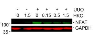
Application: WB Species: mouse Sample: Kidney
限制条款
产品的规格、报价、验证数据请以官网为准,官网链接:www.affbiotech.com | www.affbiotech.cn(简体中文)| www.affbiotech.jp(日本語)产品的数据信息为Affinity所有,未经授权不得收集Affinity官网数据或资料用于商业用途,对抄袭产品数据的行为我们将保留诉诸法律的权利。
产品相关数据会因产品批次、产品检测情况随时调整,如您已订购该产品,请以订购时随货说明书为准,否则请以官网内容为准,官网内容有改动时恕不另行通知。
Affinity保证所销售产品均经过严格质量检测。如您购买的商品在规定时间内出现问题需要售后时,请您在Affinity官方渠道提交售后申请。产品仅供科学研究使用。不用于诊断和治疗。
产品未经授权不得转售。
Affinity Biosciences将不会对在使用我们的产品时可能发生的专利侵权或其他侵权行为负责。Affinity Biosciences, Affinity Biosciences标志和所有其他商标所有权归Affinity Biosciences LTD.