产品: SP-C 抗体
货号: DF6647
描述: Rabbit polyclonal antibody to SP-C
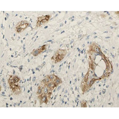
应用: WB IHC IF/ICC
反应: Human, Mouse, Rat
预测: Bovine, Horse, Rabbit
分子量: 21kDa; 21kD(Calculated).
蛋白号: P11686
RRID: AB_2838609

浏览相似产品>>

   规格 价格 库存
 50ul RMB¥ 1250 现货
 100ul RMB¥ 2300 现货
 200ul RMB¥ 3000 现货

货期: 当天发货

联系销售

产品描述

来源:
Rabbit
应用:
WB 1:500-1:2000, IHC 1:50-1:200, IF/ICC 1:100-1:500
*The optimal dilutions should be determined by the end user.
*Tips:

WB: 适用于变性蛋白样本的免疫印迹检测. IHC: 适用于组织样本的石蜡(IHC-p)或冰冻(IHC-f)切片样本的免疫组化/荧光检测. IF/ICC: 适用于细胞样本的荧光检测. ELISA(peptide): 适用于抗原肽的ELISA检测.

反应:
Human,Mouse,Rat
预测:
Bovine(100%), Horse(100%), Rabbit(92%)
克隆:
Polyclonal
特异性:
SP-C Antibody detects endogenous levels of total SP-C.
RRID:
AB_2838609
引用格式: Affinity Biosciences Cat# DF6647, RRID:AB_2838609.
偶联:
Unconjugated.
纯化:
The antiserum was purified by peptide affinity chromatography using SulfoLink™ Coupling Resin (Thermo Fisher Scientific).
保存:
Rabbit IgG in phosphate buffered saline , pH 7.4, 150mM NaCl, 0.02% sodium azide and 50% glycerol. Store at -20 °C. Stable for 12 months from date of receipt.
别名:

展开/折叠

BRICD6; BRICHOS domain containing 6; PSP C; PSPC; PSPC_HUMAN; Pulmonary surfactant apoprotein 2; Pulmonary surfactant apoprotein PSP C; pulmonary surfactant apoprotein-2 SP-C; Pulmonary surfactant associated protein C; Pulmonary surfactant associated proteolipid SPL pVal; Pulmonary surfactant associated proteolipid SPL(Val); Pulmonary surfactant protein SP5; Pulmonary surfactant-associated protein C; Pulmonary surfactant-associated proteolipid SPL(Val); SFTP 2; SFTP2; SFTPC; SFTPC surfactant pulmonary associated protein C; SMDP2; SP 5; SP C; SP-C; SP5; SPC; Surfactant associated protein pulmonary 2; Surfactant protein c; Surfactant proteolipid SPL-pVal; Surfactant pulmonary associated protein C;

抗原和靶标

免疫原:
Uniprot:
基因/基因ID:
描述:
This gene encodes the pulmonary-associated surfactant protein C (SPC), an extremely hydrophobic surfactant protein essential for lung function and homeostasis after birth. Pulmonary surfactant is a surface-active lipoprotein complex composed of 90% lipids and 10% proteins which include plasma proteins and apolipoproteins SPA, SPB, SPC and SPD. The surfactant is secreted by the alveolar cells of the lung and maintains the stability of pulmonary tissue by reducing the surface tension of fluids that coat the lung. Multiple mutations in this gene have been identified, which cause pulmonary surfactant metabolism dysfunction type 2, also called pulmonary alveolar proteinosis due to surfactant protein C deficiency, and are associated with interstitial lung disease in older infants, children, and adults. Alternatively spliced transcript variants encoding different protein isoforms have been identified.
序列:
MDVGSKEVLMESPPDYSAAPRGRFGIPCCPVHLKRLLIVVVVVVLIVVVIVGALLMGLHMSQKHTEMVLEMSIGAPEAQQRLALSEHLVTTATFSIGSTGLVVYDYQQLLIAYKPAPGTCCYIMKIAPESIPSLEALNRKVHNFQMECSLQAKPAVPTSKLGQAEGRDAGSAPSGGDPAFLGMAVNTLCGEVPLYYI

种属预测

种属预测:

score>80的预测可信度较高,可尝试用于WB检测。*预测模型主要基于免疫原序列比对,结果仅作参考,不作为质保凭据。

Species
Results
Score
Horse
100
Bovine
100
Rabbit
92
Xenopus
71
Pig
0
Sheep
0
Dog
0
Zebrafish
0
Chicken
0
Model Confidence:
High(score>80) Medium(80>score>50) Low(score<50) No confidence

翻译修饰 - P11686 作为底物

Site PTM Type Enzyme
K6 Ubiquitination
Y16 Phosphorylation
T187 Phosphorylation

研究背景

功能:

Pulmonary surfactant associated proteins promote alveolar stability by lowering the surface tension at the air-liquid interface in the peripheral air spaces.

细胞定位:

Secreted>Extracellular space>Surface film.

Extracellular region or secreted Cytosol Plasma membrane Cytoskeleton Lysosome Endosome Peroxisome ER Golgi apparatus Nucleus Mitochondrion Manual annotation Automatic computational assertionSubcellular location

文献引用

1). Extracellular Vesicle ITGAM and ITGB2 Mediate Severe Acute Pancreatitis-Related Acute Lung Injury. ACS Nano, 2023 (PubMed: 37022097) [IF=17.1]

2). Impaired autophagy-accelerated senescence of alveolar type II epithelial cells drives pulmonary fibrosis induced by single-walled carbon nanotubes. Journal of Nanobiotechnology, 2023 (PubMed: 36849924) [IF=10.2]

Application: WB    Species: Mouse    Sample: lung tissues

Fig. 1 Single-walled carbon nanotubes (SWCNTs) induced pulmonary fibrosis and senescence of AECIIs in mouse lung tissues. Mice were exposed to 40 μg SWCNT by intratracheal instillation, then lung tissues were collected on days 3, 7 and 28. A Scheme of workflow for evaluation of cellular senescence and pulmonary fibrosis induced by SWCNTs in vivo. B–D Western Blotting analysis of p21 and p16 protein expressions (n = 3) in lung tissues. Contents of TGF-β (E) and PAI-1 (F) in bronchoalveolar lavage fluid (BALF) quantified by ELISA (n = 5). HE staining (G) and Masson’s trichrome staining (H) of mouse lung tissues (200×). I The semiquantitative Ashcroft scores for the severity of pulmonary fibrosis (n = 4). J The hydroxyproline (HYP) level (n = 4) in lung tissues of mice. K Immunohistochemistry (IHC) of COL I expression (n = 5) in lung tissues of mice. L, M The correlation of hydroxyproline contents in mice lung tissues and senescence-associated secretory phenotype (SASP) factors (TGF-β and PAI-1) in BALF. N Immunostaining for p16 (a senescence-related marker) and SP-C (an AECIIs marker) from SWCNTs-exposed lung tissues of mice on day 28 was detected by immunofluorescence (IF) (400×). *P 

3). Insights into the mechanism of action of pterostilbene against influenza A virus-induced acute lung injury. Phytomedicine : international journal of phytotherapy and phytopharmacology, 2024 (PubMed: 38583346) [IF=7.9]

4). Novel genome-wide DNA methylation profiling reveals distinct epigenetic landscape, prognostic model and cellular composition of early-stage lung adenocarcinoma. Journal of translational medicine, 2024 (PubMed: 38711158) [IF=7.4]

Application: IHC    Species: Human    Sample:

Fig. 5 UXM analysis identified various cell populations in EM-seq and were verified by Immunohistochemistry (A) Cell proportion score between tumor (red) and normal (green) tissue analyzed by UXM. (B) The cellular composition of each tumor and normal tissue identified by UXM. (C) Representative immunohistochemistry image shows that CD3、CD19、SP-C were highly expressed in NSCLC tumor tissues (C-E); CD31were highly expressed in normal lung tissue (F); CD56 were weakly expressed in normal lung tissue (G).

5). Keratin 15 protects against cigarette smoke-induced epithelial mesenchymal transformation by MMP-9. Respiratory research, 2023 (PubMed: 38007424) [IF=5.8]

Application: IF/ICC    Species: Mouse    Sample:

Fig. 9 Krt15+ cells transform into AT2 cells to protect alveoli. A Immunofluorescence staining of lung tissue with Krt15 (Red) and Sftpc (Green). B The ratio of Krt15+Sftpc+ cells/Total cells. C Staining-intensity profiles showing signals from three fluorescent channels. D The ratio of TUNEL+ cells/Total cells. E TUNEL staining. N = 3 in each group.*p 

6). Establishment of Intestinal Organoid from Rousettus leschenaultii and the Susceptibility to Bat-Associated Viruses, SARS-CoV-2 and Pteropine Orthoreovirus. INTERNATIONAL JOURNAL OF MOLECULAR SCIENCES, 2021 (PubMed: 34639103) [IF=5.6]

7). Clevudine attenuates bleomycin-induced early pulmonary fibrosis via regulating M2 macrophage polarization. International Immunopharmacology, 2021 (PubMed: 34700113) [IF=5.6]

8). NLRP3 inflammasome mediates abnormal epithelial regeneration and distal lung remodeling in silica‑induced lung fibrosis. International journal of molecular medicine, 2024 (PubMed: 38240085) [IF=5.4]

9). Synergistic effect of constituent drugs of Baibutang on improving Yin-deficiency pulmonary fibrosis in rats. JOURNAL OF ETHNOPHARMACOLOGY, 2023 (PubMed: 36535334) [IF=5.4]

10). Rosmarinic acid treatment protects against lethal H1N1 virus-mediated inflammation and lung injury by promoting activation of the h-PGDS-PGD2-HO-1 signal axis. Chinese medicine, 2023 (PubMed: 37891648) [IF=4.9]

Application: IF/ICC    Species: Mouse    Sample:

Fig. 2 Effects of RosA on the H1N1 virus-triggered pro-inflammatory response. A Representative immunofluorescence images of IL-6, IL-8 (pink) and TNF-α, MCP-1 (red) in lung SpC+ (green) alveolar epithelial cells. B Quantitative analysis of fluorescence intensities for IL-6, TNF-α, IL-8 and MCP-1 in SpC+ alveolar epithelial cells. C Levels of pro-inflammatory mediators (IL-6, MCP-1, RANTES and TNF-α) in the lung homogenates were determined by Luminex assay. D Levels of pro-inflammatory mediators (IL-6, IL-8, IP-10, MCP-1, RANTES and TNF-α) were determined by Luminex assay.

加载更多

限制条款

产品的规格、报价、验证数据请以官网为准,官网链接:www.affbiotech.com | www.affbiotech.cn(简体中文)| www.affbiotech.jp(日本語)

产品的数据信息为Affinity所有,未经授权不得收集Affinity官网数据或资料用于商业用途,对抄袭产品数据的行为我们将保留诉诸法律的权利。

产品相关数据会因产品批次、产品检测情况随时调整,如您已订购该产品,请以订购时随货说明书为准,否则请以官网内容为准,官网内容有改动时恕不另行通知。

Affinity保证所销售产品均经过严格质量检测。如您购买的商品在规定时间内出现问题需要售后时,请您在Affinity官方渠道提交售后申请。

产品仅供科学研究使用。不用于诊断和治疗。 

产品未经授权不得转售。

Affinity Biosciences将不会对在使用我们的产品时可能发生的专利侵权或其他侵权行为负责。Affinity Biosciences, Affinity Biosciences标志和所有其他商标所有权归Affinity Biosciences LTD.