产品: CTLA4 抗体
货号: DF6793
描述: Rabbit polyclonal antibody to CTLA4
应用: WB IHC
反应: Human, Mouse
预测: Pig, Horse, Rabbit, Dog, Chicken
分子量: 25kDa; 25kD(Calculated).
蛋白号: P16410
RRID: AB_2838755

浏览相似产品>>

   规格 价格 库存
 50ul RMB¥ 1250 现货
 100ul RMB¥ 2300 现货
 200ul RMB¥ 3000 现货

货期: 当天发货

联系销售

产品描述

来源:
Rabbit
应用:
WB 1:500-1:2000, IHC 1:50-1:200
*The optimal dilutions should be determined by the end user.
*Tips:

WB: 适用于变性蛋白样本的免疫印迹检测. IHC: 适用于组织样本的石蜡(IHC-p)或冰冻(IHC-f)切片样本的免疫组化/荧光检测. IF/ICC: 适用于细胞样本的荧光检测. ELISA(peptide): 适用于抗原肽的ELISA检测.

反应:
Human,Mouse
预测:
Pig(100%), Horse(91%), Rabbit(91%), Dog(100%), Chicken(90%)
克隆:
Polyclonal
特异性:
CTLA4 Antibody detects endogenous levels of total CTLA4.
RRID:
AB_2838755
引用格式: Affinity Biosciences Cat# DF6793, RRID:AB_2838755.
偶联:
Unconjugated.
纯化:
The antiserum was purified by peptide affinity chromatography using SulfoLink™ Coupling Resin (Thermo Fisher Scientific).
保存:
Rabbit IgG in phosphate buffered saline , pH 7.4, 150mM NaCl, 0.02% sodium azide and 50% glycerol. Store at -20 °C. Stable for 12 months from date of receipt.
别名:

展开/折叠

ALPS5; CD 152; CD; CD152; CD152 antigen; CD152 isoform; Celiac disease 3; CELIAC3; CTLA 4; CTLA-4; CTLA4; CTLA4_HUMAN; Cytotoxic T cell associated 4; Cytotoxic T lymphocyte antigen 4; Cytotoxic T lymphocyte associated 4; Cytotoxic T lymphocyte associated 4, soluble isoform, included; Cytotoxic T lymphocyte associated antigen 4; Cytotoxic T lymphocyte associated antigen 4 short spliced form; Cytotoxic T lymphocyte associated protein 4; Cytotoxic T lymphocyte associated serine esterase 4; Cytotoxic T lymphocyte protein 4; Cytotoxic T-lymphocyte protein 4; Cytotoxic T-lymphocyte-associated antigen 4; GRD4; GSE; ICOS; IDDM12; insulin-dependent diabetes mellitus 12; Ligand and transmembrane spliced cytotoxic T lymphocyte associated antigen 4; OTTHUMP00000216623;

抗原和靶标

免疫原:
Uniprot:
基因/基因ID:
表达:
P16410 CTLA4_HUMAN:

Widely expressed with highest levels in lymphoid tissues. Detected in activated T-cells where expression levels are 30- to 50-fold less than CD28, the stimulatory coreceptor, on the cell surface following activation.

描述:
This gene is a member of the immunoglobulin superfamily and encodes a protein which transmits an inhibitory signal to T cells. The protein contains a V domain, a transmembrane domain, and a cytoplasmic tail. Alternate transcriptional splice variants, encoding different isoforms, have been characterized. The membrane-bound isoform functions as a homodimer interconnected by a disulfide bond, while the soluble isoform functions as a monomer. Mutations in this gene have been associated with insulin-dependent diabetes mellitus, Graves disease, Hashimoto thyroiditis, celiac disease, systemic lupus erythematosus, thyroid-associated orbitopathy, and other autoimmune diseases. [provided by RefSeq, Jul 2008]
序列:
MACLGFQRHKAQLNLATRTWPCTLLFFLLFIPVFCKAMHVAQPAVVLASSRGIASFVCEYASPGKATEVRVTVLRQADSQVTEVCAATYMMGNELTFLDDSICTGTSSGNQVNLTIQGLRAMDTGLYICKVELMYPPPYYLGIGNGTQIYVIDPEPCPDSDFLLWILAAVSSGLFFYSFLLTAVSLSKMLKKRSPLTTGVYVKMPPTEPECEKQFQPYFIPIN

种属预测

种属预测:

score>80的预测可信度较高,可尝试用于WB检测。*预测模型主要基于免疫原序列比对,结果仅作参考,不作为质保凭据。

Species
Results
Score
Pig
100
Dog
100
Horse
91
Rabbit
91
Chicken
90
Bovine
0
Sheep
0
Xenopus
0
Zebrafish
0
Model Confidence:
High(score>80) Medium(80>score>50) Low(score<50) No confidence

翻译修饰 - P16410 作为底物

Site PTM Type Enzyme
S55 Phosphorylation
Y60 Phosphorylation
N113 N-Glycosylation
N145 N-Glycosylation
Y165 Phosphorylation
Y182 Phosphorylation
Y201 Phosphorylation P06239 (LCK) , P42681 (TXK) , O60674 (JAK2) , P06241 (FYN)
Y218 Phosphorylation P06239 (LCK) , P06241 (FYN)

研究背景

功能:

Inhibitory receptor acting as a major negative regulator of T-cell responses. The affinity of CTLA4 for its natural B7 family ligands, CD80 and CD86, is considerably stronger than the affinity of their cognate stimulatory coreceptor CD28.

翻译修饰:

N-glycosylation is important for dimerization.

Phosphorylation at Tyr-201 prevents binding to the AP-2 adapter complex, blocks endocytosis, and leads to retention of CTLA4 on the cell surface.

细胞定位:

Cell membrane>Single-pass type I membrane protein.
Note: Exists primarily an intracellular antigen whose surface expression is tightly regulated by restricted trafficking to the cell surface and rapid internalisation;.

Extracellular region or secreted Cytosol Plasma membrane Cytoskeleton Lysosome Endosome Peroxisome ER Golgi apparatus Nucleus Mitochondrion Manual annotation Automatic computational assertionSubcellular location
组织特异性:

Widely expressed with highest levels in lymphoid tissues. Detected in activated T-cells where expression levels are 30- to 50-fold less than CD28, the stimulatory coreceptor, on the cell surface following activation.

亚基结构:

Homodimer; disulfide-linked Ref.23,. Binds to CD80/B7-1 and CD86/B7.2. Interacts with ICOSLG.

研究领域

· Environmental Information Processing > Signaling molecules and interaction > Cell adhesion molecules (CAMs).   (View pathway)

· Human Diseases > Immune diseases > Autoimmune thyroid disease.

· Human Diseases > Immune diseases > Rheumatoid arthritis.

· Organismal Systems > Immune system > T cell receptor signaling pathway.   (View pathway)

文献引用

1). Vascular endothelial growth factor A is a potential prognostic biomarker and correlates with immune cell infiltration in hepatocellular carcinoma. Journal of Cellular and Molecular Medicine (PubMed: 36729917) [IF=5.3]

2). GOLM1 is related to the inflammatory/immune nature of uveal melanoma and acts as a promising indicator for prognosis and immunotherapy response. Frontiers in Genetics (PubMed: 36468024) [IF=3.7]

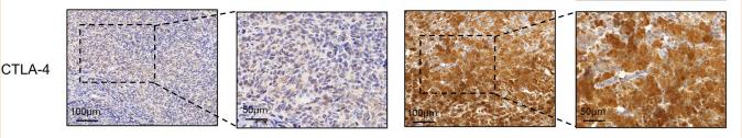
Application: IHC    Species: Human    Sample: tumor tissues

FIGURE 7 Correlation between GOLM1 expression and PD-1, PD-L1, CTLA4, and IFN-γ. (A) Immunohistochemistry assay revealed the protein expression of GOLM1, PD-1, PD-L1, CTLA4, and IFN-γ. (B,C) The expression difference of PD-1 among GOLM1-low (LEXP) and GOLM1-high (HEXP) subgroups (B) and the correlation between PD-1 and GOLM1 expression (C). (D,E) The expression difference of PD-L1 among the LEXP and HEXP groups (D) and the correlation between PD-L1 and GOLM1 expression (E). (F,G) The expression difference of CTLA4 among the LEXP and HEXP groups (F) and the correlation between CTLA4 and GOLM1 expression (G). (H,I) The expression difference of IFN-γ among the LEXP and HEXP groups (H) and the correlation between IFN-γ and GOLM1 expression (I).

3). High Expression of Ferritinophagy-Related RRM2 Gene in Relation to Unfavorable Outcome and Immune Cells Infiltration in Hepatocellular Carcinoma.

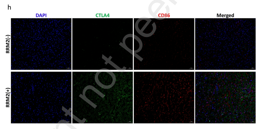
Application: IF/ICC    Species: Human    Sample: HCC tissues

Figure 7. Correlation analysis of RRM2 expression and immunotherapy markers in HCC. (a-d) Correlation between RRM2 expression level and PDCD1, CD274, CTLA4 and CD86 expression levels in HCC tissues based on TCGA database. (e) RRM2 mRNA expression in paracancerous and hepatocellular carcinoma tissues by PCR. (f) PD-L1 mRNA expression in paracancerous and hepatocellular carcinoma tissues by PCR. (g) Correlation between expression of RRM2 and PL This preprint research paper has not been peer reviewed. Electronic copy available at: https://ssrn.com/abstract=4072482 Correlations between overall survival and mRNA expression of RRM2 analyzed by and univariate Cox regression. (b) Correlations between overall survival and mRNA expression of RRM2 analyzed by multivariate Cox regression. (c-e) The expression level of RRM2 was negatively correlated with OS, DSS and PFI of HCC by TCGA database. (f-h) Kaplan-meier Plotter was used to analyze the expression levels of RRM2 in GEO, EGA and TCGA databases and there was a negative correlation with OS, DSS and PFI of HCC Figure 5. Enrichment analysis of RRM2 functional networks. (a-b) Enrichment plots by GSEA. (c-d) Enrichment of gene ontology (GO) terms and Kyoto Encyclopedia of Genes and Genomes (KEGG) for genes related to RRM2. (e) Protein–protein interaction network of RRM2. (f-i) Correlation between RRM2 expression levels and Top2A, BUB1B, KIF11 and CDC20 expression levels. (j) The heat map shows the top 50 genes positively related to RRM2 in the HCC cohort. (k) The heat map shows the top 50 genes negatively related to RRM2 in the HCC cohort. Figure 6. Correlation analysis of RRM2 expression and infiltration levels of immune cells in tumor tissues. (a) The relationship between RRM2 mRNA expression and CD8+ T cell and Regulatory T Cell in various tumors was evaluated using a variety of algorithms based on the TIMER database. (b) RRM2 expression was positively correlated with tumor purity and infiltration levels of B cells, CD8+ T cells, CD4+ T cells, macrophages, and DCs in HCC tissues based on the TIMER database. (c) Correlation between RRM2 expression level and infiltration of various immune cells (Th2 Cell、 T helper Cell、TFH、aDC、NK CD56bright Cell、Macrophages、Th1 cell、T cell、B cell、 Tcm、Treg、iDC、NK CD58dim Cell、Tem、Eosinophils、Tgd、Mast cell、Th17 Cell、 NK Cell、pDC、Cytotoxic Cell、CD8+ T cell、DC、Neutrophils) in HCC tissues. Figure 7. Correlation analysis of RRM2 expression and immunotherapy markers in HCC. (a-d) Correlation between RRM2 expression level and PDCD1, CD274, CTLA4 and CD86 expression levels in HCC tissues based on TCGA database. (e) RRM2 mRNA expression in paracancerous and hepatocellular carcinoma tissues by PCR. (f) PD-L1 mRNA expression in paracancerous and eprint not peer reviewe

限制条款

产品的规格、报价、验证数据请以官网为准,官网链接:www.affbiotech.com | www.affbiotech.cn(简体中文)| www.affbiotech.jp(日本語)

产品的数据信息为Affinity所有,未经授权不得收集Affinity官网数据或资料用于商业用途,对抄袭产品数据的行为我们将保留诉诸法律的权利。

产品相关数据会因产品批次、产品检测情况随时调整,如您已订购该产品,请以订购时随货说明书为准,否则请以官网内容为准,官网内容有改动时恕不另行通知。

Affinity保证所销售产品均经过严格质量检测。如您购买的商品在规定时间内出现问题需要售后时,请您在Affinity官方渠道提交售后申请。

产品仅供科学研究使用。不用于诊断和治疗。 

产品未经授权不得转售。

Affinity Biosciences将不会对在使用我们的产品时可能发生的专利侵权或其他侵权行为负责。Affinity Biosciences, Affinity Biosciences标志和所有其他商标所有权归Affinity Biosciences LTD.