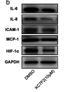
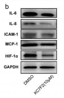
 qRT-PCR results showed that CD274 and IL8 were upregulated in the basal-like breast cancer cell lines BT549 and MDA-MB-231 (p < 0.0001). Similar to the qRT-PCR results, the western blot analysis (C–E) indicated that CD274 and IL8 protein were increased in the BT549 and MDA-MB-231 cell lines compared to the MCF7 and T47D cell lines. P < 0.001, p < 0.01, and p < 0.05")
 The levels of CXCL8 mRNA in the normal (N = 81), colorectal cancer (CRC)-early stage (N = 44)and CRC-advanced stage (N = 37) groups. (B) The levels of CXCL8 protein in normal (N = 87), CRC-early stage (N = 48), and CRC-advanced stage (N = 39)groups. (C) Immunohistochemical staining of normal, CRC-early stage and CRC-advanced stage groups.")
 Representative HE-staining images of prostate tissue in SD rats injected with normal saline, NC exosomes, prostatic fluid exosomes of CP/CPPS-A patients, and ultrasound-treated prostatic fluid exosomes of CP/CPPS-A patients. (B) Results of prostate tissue in different groups measured by MPO activity assay. (C–E) Expression of IL-8 (TRITC secondary antibodies), TNF-α (FITC secondary antibodies), and iNOS (TRITC secondary antibodies) in different groups of prostate tissue observed under a fluorescence microscope, scale bar =100 µm. *, P<0.05. NS, normal saline; NC, normal control; CP/CPPS-A, type IIIA chronic prostatitis/chronic pelvic pain syndrome; CP/CPPS-A + US, ultrasonic treatment in the CP/CPPS-A; MPO, myeloperoxidase.")

 or primary FLS from RA patients (B) were cultured with the indicated concentrations of alamandine in the presence of TNF-α (10 ng/ml) for 24 h, mRNA levels of inflammation related genes were analyzed with real time PCR and protein levels in MH7A cells were validated with western blotting (C). (D) MH7A cells were treated with alamandine 10 μg/ml for 6 h, and further stimulated with TNF-a (10 ng/ml) for 30 min. The phosphorylated expression of JNK, p38, ERK and NF-κB p65 were evaluated by wester blotting. (E) The expression of MrgD in MH7A cells (upper) and primary RA FLS (lower) were detected by immunofluorescent staining. The magnification is × 400. Results from three independent experiments are shown as mean ± SEM. * P < 0.05, * * P < 0.01.")
 show that CD274 and IL8 were upregulated in the basal-like breast cancer cell lines, BT549 and MDA-MB-231 (p")
产品描述
*The optimal dilutions should be determined by the end user.
*Tips:
WB: 适用于变性蛋白样本的免疫印迹检测. IHC: 适用于组织样本的石蜡(IHC-p)或冰冻(IHC-f)切片样本的免疫组化/荧光检测. IF/ICC: 适用于细胞样本的荧光检测. ELISA(peptide): 适用于抗原肽的ELISA检测.
引用格式: Affinity Biosciences Cat# DF6998, RRID:AB_2838954.
展开/折叠
(Ala-IL-8)77; (Ser-IL-8)72; 9E3; Beta thromboglobulin like protein; C-X-C motif chemokine 8; CEF-4; chemokine, CXC motif, ligand 8; CXCL8; Emoctakin; GCP-1; GCP/IL-8 protein I; GCP/IL-8 protein II; GCP/IL-8 protein III; GCP/IL-8 protein IV; GCP/IL-8 protein V; GCP/IL-8 protein VI; GCP1; Granulocyte chemotactic protein 1; IL-8; IL-8(1-77); IL-8(9-77); IL8; IL8/NAP1 form I; IL8/NAP1 form II; IL8/NAP1 form III; IL8/NAP1 form IV; IL8/NAP1 form V; IL8/NAP1 form VI; IL8_HUMAN; Inteleukin 8; LECT; LUCT; Lymphocyte-derived neutrophil-activating factor; LYNAP; MDNCF; MDNCF-b; MDNCF-c; MONAP; Monocyte-derived neutrophil chemotactic factor; Monocyte-derived neutrophil-activating peptide; NAF; NAP-1; NAP1; Neutrophil activating peptide 1; Neutrophil activating protein 1; Neutrophil-activating factor; Neutrophil-activating protein 1; Protein 3 10C; Protein 3-10C; SCYB8; Small inducible cytokine subfamily B member 8; T cell chemotactic factor; T-cell chemotactic factor; TSG1;
抗原和靶标
- P10145 IL8_HUMAN:
- Protein BLAST With
- NCBI/
- ExPASy/
- Uniprot
MTSKLAVALLAAFLISAALCEGAVLPRSAKELRCQCIKTYSKPFHPKFIKELRVIESGPHCANTEIIVKLSDGRELCLDPKENWVQRVVEKFLKRAENS
研究背景
IL-8 is a chemotactic factor that attracts neutrophils, basophils, and T-cells, but not monocytes. It is also involved in neutrophil activation. It is released from several cell types in response to an inflammatory stimulus. IL-8(6-77) has a 5-10-fold higher activity on neutrophil activation, IL-8(5-77) has increased activity on neutrophil activation and IL-8(7-77) has a higher affinity to receptors CXCR1 and CXCR2 as compared to IL-8(1-77), respectively.
Several N-terminal processed forms are produced by proteolytic cleavage after secretion from at least peripheral blood monocytes, leukcocytes and endothelial cells. In general, IL-8(1-77) is referred to as interleukin-8. IL-8(6-77) is the most promiment form.
Citrullination at Arg-27 prevents proteolysis, and dampens tissue inflammation, it also enhances leukocytosis, possibly through impaired chemokine clearance from the blood circulation.
Secreted.
Homodimer.
Belongs to the intercrine alpha (chemokine CxC) family.
研究领域
· Cellular Processes > Cell growth and death > Cellular senescence. (View pathway)
· Environmental Information Processing > Signaling molecules and interaction > Cytokine-cytokine receptor interaction. (View pathway)
· Environmental Information Processing > Signal transduction > NF-kappa B signaling pathway. (View pathway)
· Environmental Information Processing > Signal transduction > Phospholipase D signaling pathway. (View pathway)
· Human Diseases > Endocrine and metabolic diseases > Non-alcoholic fatty liver disease (NAFLD).
· Human Diseases > Infectious diseases: Bacterial > Epithelial cell signaling in Helicobacter pylori infection.
· Human Diseases > Infectious diseases: Bacterial > Shigellosis.
· Human Diseases > Infectious diseases: Bacterial > Salmonella infection.
· Human Diseases > Infectious diseases: Bacterial > Pertussis.
· Human Diseases > Infectious diseases: Bacterial > Legionellosis.
· Human Diseases > Infectious diseases: Parasitic > Chagas disease (American trypanosomiasis).
· Human Diseases > Infectious diseases: Parasitic > Malaria.
· Human Diseases > Infectious diseases: Parasitic > Amoebiasis.
· Human Diseases > Infectious diseases: Viral > Hepatitis C.
· Human Diseases > Infectious diseases: Viral > Hepatitis B.
· Human Diseases > Infectious diseases: Viral > Influenza A.
· Human Diseases > Cancers: Overview > Pathways in cancer. (View pathway)
· Human Diseases > Cancers: Overview > Transcriptional misregulation in cancer.
· Human Diseases > Cancers: Specific types > Bladder cancer. (View pathway)
· Human Diseases > Immune diseases > Rheumatoid arthritis.
· Organismal Systems > Immune system > Chemokine signaling pathway. (View pathway)
· Organismal Systems > Immune system > Toll-like receptor signaling pathway. (View pathway)
· Organismal Systems > Immune system > NOD-like receptor signaling pathway. (View pathway)
· Organismal Systems > Immune system > RIG-I-like receptor signaling pathway. (View pathway)
· Organismal Systems > Immune system > IL-17 signaling pathway. (View pathway)
文献引用
Application: WB Species: Human Sample: HUVECs
Application: WB Species: human Sample: HUVECs

Application: IHC Species: mouse Sample: brains
Application: WB Species: mouse Sample: brains
Application: IF/ICC Species: Mouse Sample:
Application: IHC Species: mouse Sample: tumor
Application: IHC Species: rat Sample: inflammatory cell

Application: IF/ICC Species: pig Sample: 3D4/2 cells
Application: WB Species: mouse Sample: TAMs
限制条款
产品的规格、报价、验证数据请以官网为准,官网链接:www.affbiotech.com | www.affbiotech.cn(简体中文)| www.affbiotech.jp(日本語)产品的数据信息为Affinity所有,未经授权不得收集Affinity官网数据或资料用于商业用途,对抄袭产品数据的行为我们将保留诉诸法律的权利。
产品相关数据会因产品批次、产品检测情况随时调整,如您已订购该产品,请以订购时随货说明书为准,否则请以官网内容为准,官网内容有改动时恕不另行通知。
Affinity保证所销售产品均经过严格质量检测。如您购买的商品在规定时间内出现问题需要售后时,请您在Affinity官方渠道提交售后申请。产品仅供科学研究使用。不用于诊断和治疗。
产品未经授权不得转售。
Affinity Biosciences将不会对在使用我们的产品时可能发生的专利侵权或其他侵权行为负责。Affinity Biosciences, Affinity Biosciences标志和所有其他商标所有权归Affinity Biosciences LTD.