


 and mouse anti-beta tubulin Ab(T0023 1:200) for 1 hour at 37°C. An AlexaFluor594 conjugated goat anti-rabbit IgG(H+L) Ab(Red) and an AlexaFluor488 conjugated goat anti-mouse IgG(H+L) Ab(Green) were used as the secondary antibody.
The nuclear counter stain is DAPI(blue).")

 Kaplan–Meier analysis of the correlation between PARP1 expression and overall survival (OS, n = 77) and (B) progression-free survival (PFS, n=33). (C–E) Scatter plots of PARP1 mRNA relative to expression of MYC paralogs in SCLC primary tumors (n=81) (C), CCLE cell lines (n=50) (D), murine SCLC tumors (n=14) (E). (F) Representative images of IHC analysis of PARP1 and c-MYC in two independent cases. Scale bar, 100 μm. (G) Heat map showing the correlation between PARP1 and c-MYC in 17 paraffin-embedded SCLC tumor tissues. The heat map was depicted according to the IOD value of each IHC slides (red indicates c-MYC and PARP1 positive staining, green indicates negative staining). The significance analysis was performed by Student’s t test. (H–I) ChIP-qRT-PCR experiment indicating the direct binding of c-MYC and MYCN to the PARP1 promoter in DMS273 (H) and H526 (I) cells. (J) Western blot analysis showing the downregulated proteins in DMS53 and DMS273 cells upon c-MYC knockdown. (K) Western blot analysis showing the upregulated proteins in SHP77 cells with ectopic c-MYC overexpression. GAPDH was used as a loading control. BS, binding site.")
 Kaplan–Meier analysis of the correlation between PARP1 expression and overall survival (OS, n = 77) and (B) progression-free survival (PFS, n=33). (C–E) Scatter plots of PARP1 mRNA relative to expression of MYC paralogs in SCLC primary tumors (n=81) (C), CCLE cell lines (n=50) (D), murine SCLC tumors (n=14) (E). (F) Representative images of IHC analysis of PARP1 and c-MYC in two independent cases. Scale bar, 100 μm. (G) Heat map showing the correlation between PARP1 and c-MYC in 17 paraffin-embedded SCLC tumor tissues. The heat map was depicted according to the IOD value of each IHC slides (red indicates c-MYC and PARP1 positive staining, green indicates negative staining). The significance analysis was performed by Student’s t test. (H–I) ChIP-qRT-PCR experiment indicating the direct binding of c-MYC and MYCN to the PARP1 promoter in DMS273 (H) and H526 (I) cells. (J) Western blot analysis showing the downregulated proteins in DMS53 and DMS273 cells upon c-MYC knockdown. (K) Western blot analysis showing the upregulated proteins in SHP77 cells with ectopic c-MYC overexpression. GAPDH was used as a loading control. BS, binding site.")
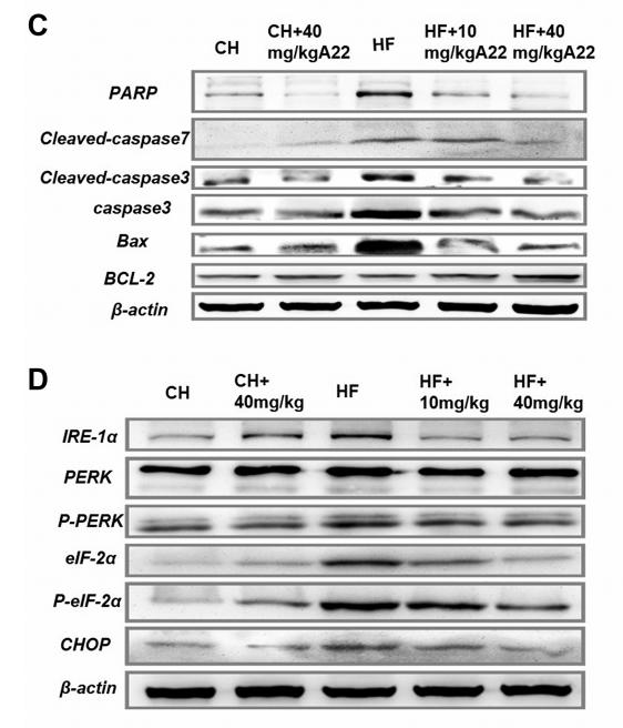
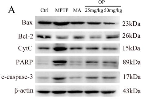
 The expression of apoptosis related proteins regulating the mitochondrial pathway (Bax, Bcl-2, PARP1,cleaved Caspase-9 and cleaved Caspase-3) in the tumor tissues was determined by western blot. * P<0.05, pcTERT-Noxa vs. pcTERT, pcTERT-Puma vs. pcTERT. Each experiment was performed with three mice. The IHC staining photos were collected from the tumor of each mouse and one representative picture of each group has been shown.")
 Western blot and (B) statistical results of western blots of PARP‐1 (n = 5, one‐way ANOVA followed by Bonferroni's post hoc test). (C) Immunofluorescence and (D) statistical analysis of immunofluorescence staining of PARP‐1 in nerve cells from each group (Scale bar = 50 μm, n = 3, one‐way ANOVA followed by Games–Howell post hoc test). (E) Mitochondrial membrane potential was measured with the JC‐1 probe (n = 5, one‐way ANOVA followed by Bonferroni's post hoc test). (F, H, J) Western blot and (G, I, K) statistical results of western blots of Mito‐AIF, Cyto‐AIF, and Nucleo‐AIF (n = 5, one‐way ANOVA followed by Bonferroni's post hoc test). The data represent means ± SD, * means, p")
 Western blot and (B) statistical results of western blots of PARP‐1 (n = 5, one‐way ANOVA followed by Bonferroni's post hoc test). (C) Immunofluorescence and (D) statistical analysis of immunofluorescence staining of PARP‐1 in nerve cells from each group (Scale bar = 50 μm, n = 3, one‐way ANOVA followed by Games–Howell post hoc test). (E) Mitochondrial membrane potential was measured with the JC‐1 probe (n = 5, one‐way ANOVA followed by Bonferroni's post hoc test). (F, H, J) Western blot and (G, I, K) statistical results of western blots of Mito‐AIF, Cyto‐AIF, and Nucleo‐AIF (n = 5, one‐way ANOVA followed by Bonferroni's post hoc test). The data represent means ± SD, * means, p")
 PI staining images of RAW264.7 cells. Scale bar = 200 μm. (B) PI-positive cell rate (n = 3). (C) DHE staining images of RAW264.7 cells. Scale bar = 200 μm. (D) Statistical analysis of relative fluorescence intensity in DHE staining (n = 3). (E) Levels of TNF-α in RAW264.7 cells (n = 8). (F) Levels of IL-6 in RAW264.7 cells (n = 8). (G) Levels of IL-1β in RAW264.7 cells (n = 8). (H–J) The protein levels of PARP1 and HMGB1 in RAW264.7 cells (n = 3). Quantitative data are shown as the mean ± SD, *P < 0.05, **P < 0.01, ***P < 0.001, ****P < 0.0001.")
 TUNEL staining of apoptosis in ovaries after intrabursal injection with miR-133a agomir. Representative ovarian tissue sections are shown. The right panel is a magnification of the left panel. Relative areas stained with TUNEL were quantified using Image-Pro Plus 6.0 and are shown as the mean±SEM in statistical charts ( n=3). ** P")
 Representative images and average fluorescence intensity analysis of PARP1 and GSK3β expression detected by immunofluorescence in rat ovarian tissue (scale = 50 μm). n = 6. (B) Representative images and semi-quantitative analysis of PARP1, GSK3β, Bax, Bcl-2, and Caspase-3 expression detected by WB in rat ovarian tissue. n = 3. *p")
 Representative images and average fluorescence intensity analysis of PARP1 and GSK3β expression detected by immunofluorescence in rat ovarian tissue (scale = 50 μm). n = 6. (B) Representative images and semi-quantitative analysis of PARP1, GSK3β, Bax, Bcl-2, and Caspase-3 expression detected by WB in rat ovarian tissue. n = 3. *p")
| 产品: | PARP1 抗体 |
| 货号: | DF7198 |
| 描述: | Rabbit polyclonal antibody to PARP1 |
| 应用: | WB IHC IF/ICC |
| 文献验证: | WB, IHC, IF/ICC |
| 反应: | Human, Mouse, Rat |
| 预测: | Pig, Zebrafish, Bovine, Horse, Sheep, Rabbit, Dog, Chicken, Xenopus |
| 分子量: | 89kDa(cleaved), 113kDa(precursor)(Observed); 113kD(Calculated). |
| 蛋白ID: | P09874 |
| RRID: | AB_2839150 |
产品描述
*The optimal dilutions should be determined by the end user. For optimal experimental results, antibody reuse is not recommended.
*Tips:
WB: 适用于变性蛋白样本的免疫印迹检测. IHC: 适用于组织样本的石蜡(IHC-p)或冰冻(IHC-f)切片样本的免疫组化/荧光检测. IF/ICC: 适用于细胞样本的荧光检测. ELISA(peptide): 适用于抗原肽的ELISA检测.
引用格式: Affinity Biosciences Cat# DF7198, RRID:AB_2839150.
展开/折叠
ADP-ribosyltransferase diphtheria toxin-like 1; ADPRT 1; ADPRT; ADPRT1; APOPAIN; ARTD1; NAD(+) ADP-ribosyltransferase 1; PARP; PARP-1; PARP1; PARP1_HUMAN; Poly [ADP-ribose] polymerase 1; Poly ADP ribose polymerase 1; Poly[ADP-ribose] synthase 1; PPOL; SCA1;
抗原和靶标
A synthesized peptide derived from human PARP1, corresponding to a region within the internal amino acids.
- P09874 PARP1_HUMAN:
- Protein BLAST With
- NCBI/
- ExPASy/
- Uniprot
MAESSDKLYRVEYAKSGRASCKKCSESIPKDSLRMAIMVQSPMFDGKVPHWYHFSCFWKVGHSIRHPDVEVDGFSELRWDDQQKVKKTAEAGGVTGKGQDGIGSKAEKTLGDFAAEYAKSNRSTCKGCMEKIEKGQVRLSKKMVDPEKPQLGMIDRWYHPGCFVKNREELGFRPEYSASQLKGFSLLATEDKEALKKQLPGVKSEGKRKGDEVDGVDEVAKKKSKKEKDKDSKLEKALKAQNDLIWNIKDELKKVCSTNDLKELLIFNKQQVPSGESAILDRVADGMVFGALLPCEECSGQLVFKSDAYYCTGDVTAWTKCMVKTQTPNRKEWVTPKEFREISYLKKLKVKKQDRIFPPETSASVAATPPPSTASAPAAVNSSASADKPLSNMKILTLGKLSRNKDEVKAMIEKLGGKLTGTANKASLCISTKKEVEKMNKKMEEVKEANIRVVSEDFLQDVSASTKSLQELFLAHILSPWGAEVKAEPVEVVAPRGKSGAALSKKSKGQVKEEGINKSEKRMKLTLKGGAAVDPDSGLEHSAHVLEKGGKVFSATLGLVDIVKGTNSYYKLQLLEDDKENRYWIFRSWGRVGTVIGSNKLEQMPSKEDAIEHFMKLYEEKTGNAWHSKNFTKYPKKFYPLEIDYGQDEEAVKKLTVNPGTKSKLPKPVQDLIKMIFDVESMKKAMVEYEIDLQKMPLGKLSKRQIQAAYSILSEVQQAVSQGSSDSQILDLSNRFYTLIPHDFGMKKPPLLNNADSVQAKVEMLDNLLDIEVAYSLLRGGSDDSSKDPIDVNYEKLKTDIKVVDRDSEEAEIIRKYVKNTHATTHNAYDLEVIDIFKIEREGECQRYKPFKQLHNRRLLWHGSRTTNFAGILSQGLRIAPPEAPVTGYMFGKGIYFADMVSKSANYCHTSQGDPIGLILLGEVALGNMYELKHASHISKLPKGKHSVKGLGKTTPDPSANISLDGVDVPLGTGISSGVNDTSLLYNEYIVYDIAQVNLKYLLKLKFNFKTSLW
种属预测
score>80的预测可信度较高,可尝试用于WB检测。*预测模型主要基于免疫原序列比对,结果仅作参考,不作为质保凭据。
High(score>80) Medium(80>score>50) Low(score<50) No confidence
研究背景
Poly-ADP-ribosyltransferase that mediates poly-ADP-ribosylation of proteins and plays a key role in DNA repair. Mainly mediates glutamate and aspartate ADP-ribosylation of target proteins: the ADP-D-ribosyl group of NAD(+) is transferred to the acceptor carboxyl group of glutamate and aspartate residues and further ADP-ribosyl groups are transferred to the 2'-position of the terminal adenosine moiety, building up a polymer with an average chain length of 20-30 units. Mediates the poly(ADP-ribosyl)ation of a number of proteins, including itself, APLF and CHFR. Also mediates serine ADP-ribosylation of target proteins following interaction with HPF1; HPF1 conferring serine specificity. Probably also catalyzes tyrosine ADP-ribosylation of target proteins following interaction with HPF1. Catalyzes the poly-ADP-ribosylation of histones in a HPF1-dependent manner. Involved in the base excision repair (BER) pathway by catalyzing the poly-ADP-ribosylation of a limited number of acceptor proteins involved in chromatin architecture and in DNA metabolism. ADP-ribosylation follows DNA damage and appears as an obligatory step in a detection/signaling pathway leading to the reparation of DNA strand breaks. In addition to base excision repair (BER) pathway, also involved in double-strand breaks (DSBs) repair: together with TIMELESS, accumulates at DNA damage sites and promotes homologous recombination repair by mediating poly-ADP-ribosylation. In addition to proteins, also able to ADP-ribosylate DNA: catalyzes ADP-ribosylation of DNA strand break termini containing terminal phosphates and a 2'-OH group in single- and double-stranded DNA, respectively. Required for PARP9 and DTX3L recruitment to DNA damage sites. PARP1-dependent PARP9-DTX3L-mediated ubiquitination promotes the rapid and specific recruitment of 53BP1/TP53BP1, UIMC1/RAP80, and BRCA1 to DNA damage sites. Acts as a regulator of transcription: positively regulates the transcription of MTUS1 and negatively regulates the transcription of MTUS2/TIP150. With EEF1A1 and TXK, forms a complex that acts as a T-helper 1 (Th1) cell-specific transcription factor and binds the promoter of IFN-gamma to directly regulate its transcription, and is thus involved importantly in Th1 cytokine production. Involved in the synthesis of ATP in the nucleus, together with NMNAT1, PARG and NUDT5. Nuclear ATP generation is required for extensive chromatin remodeling events that are energy-consuming.
Phosphorylated by PRKDC and TXK.
Poly-ADP-ribosylated on glutamate and aspartate residues by autocatalysis. Poly-ADP-ribosylated by PARP2; poly-ADP-ribosylation mediates the recruitment of CHD1L to DNA damage sites. ADP-ribosylated on serine by autocatalysis; serine ADP-ribosylation takes place following interaction with HPF1.
S-nitrosylated, leading to inhibit transcription regulation activity.
Nucleus. Nucleus>Nucleolus. Chromosome.
Note: Localizes to sites of DNA damage.
研究领域
· Cellular Processes > Cell growth and death > Apoptosis. (View pathway)
· Cellular Processes > Cell growth and death > Necroptosis. (View pathway)
· Environmental Information Processing > Signal transduction > NF-kappa B signaling pathway. (View pathway)
· Genetic Information Processing > Replication and repair > Base excision repair.
文献引用
Application: WB Species: mouse Sample: liver

Application: WB Species: rat Sample:
Application: IHC Species: rat Sample: brain
Application: WB Species: Rat Sample: NRK-52E cells
Application: WB Species: mouse Sample:
限制条款
产品的规格、报价、验证数据请以官网为准,官网链接:www.affbiotech.com | www.affbiotech.cn(简体中文)| www.affbiotech.jp(日本語)产品的数据信息为Affinity所有,未经授权不得收集Affinity官网数据或资料用于商业用途,对抄袭产品数据的行为我们将保留诉诸法律的权利。
产品相关数据会因产品批次、产品检测情况随时调整,如您已订购该产品,请以订购时随货说明书为准,否则请以官网内容为准,官网内容有改动时恕不另行通知。
Affinity保证所销售产品均经过严格质量检测。如您购买的商品在规定时间内出现问题需要售后时,请您在Affinity官方渠道提交售后申请。产品仅供科学研究使用。不用于诊断和治疗。
产品未经授权不得转售。
Affinity Biosciences将不会对在使用我们的产品时可能发生的专利侵权或其他侵权行为负责。Affinity Biosciences, Affinity Biosciences标志和所有其他商标所有权归Affinity Biosciences LTD.