


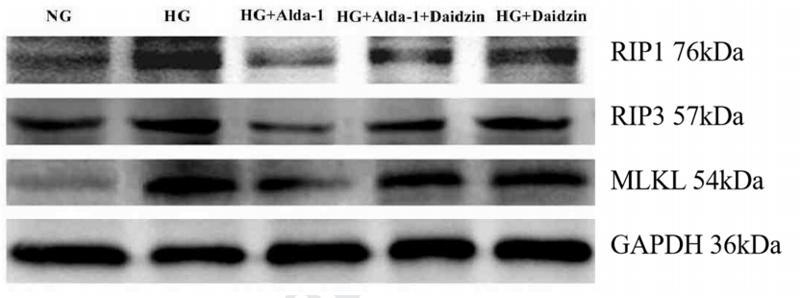
 Representative western blots of RIP1, RIP3, MLKL and cleaved caspase‑3 proteins.")
. Protein expression was assessed as positive immune response or negative immune response depending on the immunohistochemistry score of different groups (NS group: n=8; DEM group: n=7; RES group: n=8). Positive immune response slices had more brown-staining cells in particular areas than the negative ones (B–D).")
 and RIP3 (B)mRNA overexpression in the pericontusional cortex was inhibited by 2-BFI treatment. (C) Representative Western bands showing the protein expression of RIP1,RIP3, and their substrate MLKL in the pericontusional cortex at 72 h after TBI.")
 and Sham rats (S, open column) at the 8th week after the
operation. (A) Representative Western immunoblot images. (BeG) Semi-quantitative values of left ventricular phospho-RIP1 (B), RIP1 (C), phospho-RIP3 (D), RIP3 (E), phospho-
MLKL (F), MLKL (G), cleaved caspase-8 (H), and TNF- a (І) contents. Each value represents the mean ± SEM of 8 animals. *p < 0.05 vs. corresponding Sham group.
# p < 0.05 vs.
vehicle-treated CAL group.")
 Western blot analysis of protein expression of RIP1/RIP3/MLKL in rat myocardium during “early” and “late” stages. (B–D) Quantification of RIP1 (B), RIP3 (C), MLKL (D) in (A). (E) Western blot analysis of marker protein expression of autophagosome LC-3 I and LC-3 II in rat myocardium during “early” and “late” stages. (F) Quantification of LC-3 I and LC-3 II in e. All data are presented as mean ± SEM. A Student's t-test was performed. **p < 0.05 means compared with control group (3W); *p < 0.05 means vs. control Group (8 weeks) comparison.")
 Expression of RIPK1 protein in each group (RIPK1/β-actin). ##P < 0.01, vs. Sham; *P ≤ 0.05, vs. Model; &&P < 0.01, &P < 0.05, vs. EA. (B) Expression of RIPK3 protein in each group (RIPK3/β-actin). ##P < 0.01, vs. Sham; *P < 0.05, vs. Model; &P < 0.05, &&P< 0.01, vs. EA. (C) Expression of MLKL protein in each group (MLKL/β-actin). ##P < 0.01, vs. Sham; *P < 0.05, **P < 0.01, vs. Model; &&P < 0.01, vs. EA.")
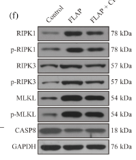
 Immunofluorescence staining for RIPK1 and NeuN colocalization in the spinal cords of the GDF-11, SCI, and sham groups (scale bar = 25 μm). (b) Quantification of the optical density of RIPK1 in neurons of spinal cord lesions. (c) Immunofluorescence staining for RIPK3 and NeuN colocalization in spinal cords belonging to the GDF-11, SCI, and sham groups (scale bar = 25 μm). (d) Quantification of the optical density of RIPK3 in neurons of spinal cord lesions. (e) Western blot assay for Caspase-8, MLKL, RIPK3, and RIPK1 expression levels in the GDF-11, SCI, and sham groups. Gels were subjected to identical experimental conditions, with cropped blots presented. (f) Optical densities of Caspase-8, MLKL, RIPK3, and RIPK1 expression levels were quantified and investigated in the respective groups. Data are expressed as the mean ± SEM, n = 6 per group. ∗∗p < 0.01 vs. the sham group. #p < 0.05 and ##p < 0.01 vs. the SCI group.")

. Values are presented as mean ± SD. *P")
 RIPK1 representative western blot image. Full-length blots/gels are presented in Supplementary Fig. 2. (B) RIPK1 expression levels in lung grafts (n = 3). (C) RIPK3 representative western blot image. Full-length blots/gels are presented in Supplementary Fig. 3. (D) RIPK3 expression levels in lung grafts (n = 3). (E) MLKL representative western blot image. Full-length blots/gels are presented in Supplementary Fig. 4. (F) MLKL expression levels in lung grafts (n = 3). Compared with CS group, *P")
 HTR8 cells were infected with ZIKV at a MOI of 1 post sic-FLIP transfection. On D1 and D2 post-infection, MLKL, pMLKL and LC3B levels were measured by western blot. (B-C) Caspase-8 (B) and caspase-3 (C) were measured by qPCR post sicaspase-8 or sicaspase-3 transfection on day 1. (D-E) Western blot assays of caspase-8 (D) or caspase-3 (E) expression in HTR8 cells post sicaspase-8 or sicaspase-3 transfection for 1 day and 2 days. (F-I) HTR8 cells were infected with ZIKV at a MOI of 1 post sicaspase-8 (F-G) or sicaspase-3 (H-I) transfection for 24 hours. The viral load was measured on D1, D2 and D3 post-infection by qPCR (F, H) and plaque assay (H, I). The data represent either a single experiment chosen as representative from three independent experiments (B-C, F-I) or the collective results of three independent experiments (D-E). All the data are analyzed by unpaired Student’s t test. Data are presented as means ± SD. *P")
产品描述
*The optimal dilutions should be determined by the end user.
*Tips:
WB: 适用于变性蛋白样本的免疫印迹检测. IHC: 适用于组织样本的石蜡(IHC-p)或冰冻(IHC-f)切片样本的免疫组化/荧光检测. IF/ICC: 适用于细胞样本的荧光检测. ELISA(peptide): 适用于抗原肽的ELISA检测.
引用格式: Affinity Biosciences Cat# DF7412, RRID:AB_2839350.
展开/折叠
9130019I15Rik; FLJ34389; hMLKL; Mixed lineage kinase domain like; Mixed lineage kinase domain like protein; Mixed lineage kinase domain like pseudokinase; Mixed lineage kinase domain-like protein; Mlkl; MLKL_HUMAN;
抗原和靶标
- Q8NB16 MLKL_HUMAN:
- Protein BLAST With
- NCBI/
- ExPASy/
- Uniprot
MENLKHIITLGQVIHKRCEEMKYCKKQCRRLGHRVLGLIKPLEMLQDQGKRSVPSEKLTTAMNRFKAALEEANGEIEKFSNRSNICRFLTASQDKILFKDVNRKLSDVWKELSLLLQVEQRMPVSPISQGASWAQEDQQDADEDRRAFQMLRRDNEKIEASLRRLEINMKEIKETLRQYLPPKCMQEIPQEQIKEIKKEQLSGSPWILLRENEVSTLYKGEYHRAPVAIKVFKKLQAGSIAIVRQTFNKEIKTMKKFESPNILRIFGICIDETVTPPQFSIVMEYCELGTLRELLDREKDLTLGKRMVLVLGAARGLYRLHHSEAPELHGKIRSSNFLVTQGYQVKLAGFELRKTQTSMSLGTTREKTDRVKSTAYLSPQELEDVFYQYDVKSEIYSFGIVLWEIATGDIPFQGCNSEKIRKLVAVKRQQEPLGEDCPSELREIIDECRAHDPSVRPSVDEILKKLSTFSK
研究背景
Pseudokinase that plays a key role in TNF-induced necroptosis, a programmed cell death process. Activated following phosphorylation by RIPK3, leading to homotrimerization, localization to the plasma membrane and execution of programmed necrosis characterized by calcium influx and plasma membrane damage. Does not have protein kinase activity. Binds to highly phosphorylated inositol phosphates such as inositolhexakisphosphate (InsP6) which is essential for its necroptotic function.
Phosphorylation by RIPK3 induces a conformational switch that is required for necroptosis. It also induces homotrimerization and localization to the plasma membrane.
Cytoplasm. Cell membrane.
Note: Localizes to the cytoplasm and translocates to the plasma membrane on necroptosis induction.
Homooligomer. Homotrimer; forms homotrimers on necroptosis induction. Interacts with RIPK3; the interaction is direct. Upon TNF-induced necrosis, forms in complex with PGAM5, RIPK1 and RIPK3. Within this complex, may play a role in the proper targeting of RIPK1/RIPK3 to its downstream effector PGAM5.
The protein kinase domain is catalytically inactive but contains an unusual pseudoactive site with an interaction between Lys-230 and Gln-356 residues. Upon phosphorylation by RIPK3, undergoes an active conformation (By similarity).
The coiled coil region 2 is responsible for homotrimerization.
Belongs to the protein kinase superfamily.
研究领域
· Cellular Processes > Cell growth and death > Necroptosis. (View pathway)
· Environmental Information Processing > Signal transduction > TNF signaling pathway. (View pathway)
文献引用

Application: WB Species: Mouse Sample: BLCA cells
Application: WB Species: rat Sample: cardiomyocytes
Application: WB Species: Mouse Sample: skin tissues
限制条款
产品的规格、报价、验证数据请以官网为准,官网链接:www.affbiotech.com | www.affbiotech.cn(简体中文)| www.affbiotech.jp(日本語)产品的数据信息为Affinity所有,未经授权不得收集Affinity官网数据或资料用于商业用途,对抄袭产品数据的行为我们将保留诉诸法律的权利。
产品相关数据会因产品批次、产品检测情况随时调整,如您已订购该产品,请以订购时随货说明书为准,否则请以官网内容为准,官网内容有改动时恕不另行通知。
Affinity保证所销售产品均经过严格质量检测。如您购买的商品在规定时间内出现问题需要售后时,请您在Affinity官方渠道提交售后申请。产品仅供科学研究使用。不用于诊断和治疗。
产品未经授权不得转售。
Affinity Biosciences将不会对在使用我们的产品时可能发生的专利侵权或其他侵权行为负责。Affinity Biosciences, Affinity Biosciences标志和所有其他商标所有权归Affinity Biosciences LTD.