
, using NLRP3 Antibody at 1/1000 dilution.
Observed bands:100kD.")


 by IF/ICC. The samples were fixed with PFA and permeabilized in 0.1% Triton X-100,then blocked in 10% serum for 45 minutes at 25°C. Samples were then incubated with primary Ab(DF7438) and mouse anti-beta tubulin Ab(T0023) for 1 hour at 37°C. An AlexaFluor594 conjugated goat anti-rabbit IgG(H+L) Ab(Red) and an AlexaFluor488 conjugated goat anti-mouse IgG(H+L) Ab(Green) were used as the secondary antibody.
The nuclear counter stain is DAPI(blue).")
 by IF/ICC. The samples were fixed with PFA and permeabilized in 0.1% Triton X-100,then blocked in 10% serum for 45 minutes at 25°C. Samples were then incubated with primary Ab(DF7438 1:200) and mouse anti-beta tubulin Ab(T0023 1:200) for 1 hour at 37°C. An AlexaFluor594 conjugated goat anti-rabbit IgG(H+L) Ab(Red) and an AlexaFluor488 conjugated goat anti-mouse IgG(H+L) Ab(Green) were used as the secondary antibody.")
, cleaved caspase-1 (c), and IL-18 e) were shown.")
 and IL-1β levels in the supernatant by ELISA (B). Summarized data showed cell viability of RAECs treated with acarbose at the concentration of 1, 3, 9µM (C), and IL-1β concentration secreted from RAECs treated with 3, 9µM acarbose under 30mM glucose stimulation (D). RAECs were incubated with 30mM glucose for 24 hours and pretreated with 3µM acarbose, the protein expression of NLRP3 (E) and p20/pro-caspase-1 (F), the activity of caspase-1 (G), and IL-1β concentration (H).")
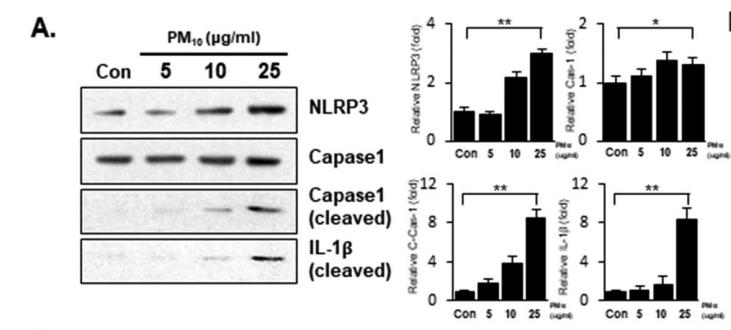
. Cell lysates were probed with antibodies
against NLRP3, cleaved-caspase-1 and cleaved IL-1β. β-actin was
used as a loading control for lysates.")

 The protein levels of cleaved caspase-1, GSDMD, ASC, NLRP3, IL-1β, and IL-18 were evaluated by Western blotting in brain tissues. (b–g) Analyses of cleaved caspase-1, GSDMD, ASC, NLRP3, IL-1β, and IL-18 (normalized to β-actin). ∗P < 0.05, ∗∗P < 0.01, and ∗∗∗P < 0.001. n = 4.")
 and AZA (9.0 mg/kg) on the protein levels of NLRP3, ASC, caspase-1 p10 and pro-caspase-1 in colons of TNBS-induced colitis in rats.")
 Lipopolysaccharide (LPS)-
induced acute lung injury in mice. Images show the protein levels of NLRP3, pro-interleukin 1β (pro-IL-1β), pro-caspase-1, cleaved-caspase-1. (B) Bleomycin (BLM)-
induced pulmonary inflammation in mice. Images show the protein levels of NLRP3, pro-IL-1β, pro-caspase-1, cleaved-caspase-1. Results are shown as mean ± SD (n
= 6). #P < 0.05; ##P < 0.01; ###P < 0.001; *P < 0.05; **P < 0.01; ***P < 0.001.")
 Representative images and histograms of AMPK, NF-κB p65 and NLRP3 in HUVECs determined by immunofluorescence. The data are representative of one experiment performed in triplicate and are expressed as the mean ± S.D. **P<0.01 vs. control group; ##P<0.01 vs. AGEs group.")
 Representative western blot results of proteins related to the AMPK and NF-κB p65/NLRP3 signaling pathway proteins in HUVECs with or without AGEs/salidroside treatment. GAPDH was used as a loading control. The data are representative of one experiment performed in triplicate and are expressed as the mean ± S.D. **P<0.01 vs. Control group; ##P<0.01 vs. AGEs group")
, saline group (B), and pranoprofen group (C) by performing western blot. The e proteinic expression of (a) NLRP3, (b) IL-1β, IL-1β17p, and (c) MMP-13 in the control group(0.72 ± 0.024, 0.11 ± 0.002, 1.10 ± 0.028, and 0.30 ± 0.008) appeared to be significantly lower than the other two groups.")
, ASC (B), and Caspase-1 (C). Data are shown as mean ± SD, n = 8. #p < 0.05 and ##p < 0.01 versus the Control group. ⁎p < 0.05 and ⁎⁎p < 0.01 versus the Model group.")
Representative Western blot analysis of NLRP3 inflammasome in Sham, SCI + vehicle and SCI + zinc group(n = 6).")
Representative Western blot analysis of NLRP3 inflammasome in Sham, SCI + vehicle and SCI + zinc group(n = 6). (B-E) The result of WB analysis of NLRP3, ASC, Caspase-1, IL-1β from each group. (F) Immunofluorescence staining was used to detect the level of NLRP3 from each group (n = 8, scale bar = 50 µm).")
. a. The mRNA expressions of key genes governing pyroptosis were detected after 24 h treatment, and β-ACTIN was used as an internal control. b. HepG2 cells were stained with anti-GSDMD (red) antibody and DAPI, and then visualized under a microscope at 200× magnification after 24 h treatment. c. Cell supernatants were analyzed for IL-1β secretion by ELISA. d and e. Representative western blots of NLRP3, GSDMD/−N, pro-CAS-1, P20 and IL-1β after 24 h treatment, and β-ACTIN was used as a protein-loading control. f. The morphology of pyroptotic cells was visualized by AO/EB staining. All groups were visualized under a microscope at 50× and 200× magnification after 24 h treatment. The data are presented as means ± SD for 3–5 biological replicates; **P < 0.01, ***, P < 0.001vs. BSA;#P < 0.05,##P < 0.01,vs. PA; ns no significant differences between two connected groups")
, anti-Caspase1 antibody (middle panel), and anti-IL-1β (lower panel) (magnification × 400). B. Western blot analysis of the protein expression of
NLRP3 (upper panel), Caspase1 (middle panel) and IL-1β (bottom panel). C-E. Densitometric analyses of the Western blotting results, NLRP3 to β-actin (C), Caspase1
to β-actin (D), IL-1β to β-actin (E) values are the mean ± SD, *P<0.05 vs. control groups, #P<0.05 vs. CP groups, n = 3.")
 The activation level of NLRP3
inflammasome and inflammatory factors in the brain of rats. After FMT from healthy rats, the expression of IL-1β, IL-18, NLRP3, and TLR4 in brain was significantly
downregulated, which were significantly different from that in Mn-treated group. *P < 0.05, **P < 0.01, ***P < 0.001.")
. *P < 0.05 and **P < 0.01 vs. control group. #P < 0.05 and ##P < 0.01 vs. LPS group.")
 Representative images of the normal, T2DM model and sericin groups are shown. Scale bar, 50 µm. (B) Relative staining intensities were calculated. *P<0.05 vs. the normal group; #P<0.05, vs. the T2DM model group. T2DM, type 2 diabetes mellitus; NLRP3, nucleotide-binding oligomerization domain-like receptor protein 3.")
 Representative western blotting results of NLRP3 and caspase-1 in the normal, T2DM model and sericin groups are shown. (B) Relative protein expression levels were calculated. (C) mRNA expression levels of NLRP3 and caspase-1 were detected by reverse transcription-quantitative PCR. *P<0.05 vs. the normal group; #P<0.05, vs. the T2DM model group. T2DM, type 2 diabetes mellitus; NLRP3, nucleotide-binding oligomerization domain-like receptor protein 3.")
.")
 Representative examples of Western blot analysis demonstrating the expression levels of NLRP3, ASC, procaspase-1, and caspase-1; quantification of the expression levels of (B) NLRP3, (C) ASC, and (D) procaspase-1 and (E) caspase-1; (F) representative examples of Western blot analysis demonstrating the expression levels of TLR4, MyD88, p-NF-κB, and NF-κB p65; quantification of the expression levels of (G) TLR4, (H) MyD88, (I) p-NF-κB, and NF-κB p65. Data presented are the means ± SD. ##p < 0.01 compared with Sham; *p < 0.05, **p < 0.01 compared with MI/R.")
 and ASC
(B). (C, D) The representative photographs and grayscale analysis of NLRP3, Pro-casp-1, casp-1, Pro-IL-1β, and IL-1β in the liver by Western blotting. Data was shown
as the mean ± SEM, n = 4, **p < 0.01, *p < 0.05, compared with control group")
. IL‐18 and IL‐1β in culture supernatant were measured by ELISA (C, D). The mRNA and proteins levels of pyroptosis markers in liver were detected in the three groups at the 24th week after HFD (E, F). Results are presented as means ± SD from three independent experiments, *P < .05 vs the control or BSA group; # P < .05 vs the PA or HFD group")
 Representative Western blotting images of NLRP3, ASC, Caspase-1and Caspase-1 p10 (B–E) The protein expression levels of NLRP3, ASC, Caspase-1 and Caspase-1 p10 in the NLRP3 inflammasome. Data are expressed as the means ± SD (n = 3). # p < 0.05, ## p < 0.01 vs. control group; *p < 0.05, **p < 0.01 vs. DSS group; & p < 0.05, && p < 0.01 vs. COP group.")
, miR‐302c‐3p inhibitor and miR‐302c‐3p inhibitor NC (in‐NC). F, Expression levels of NLRP3 are shown, as tested by qRT‐PCR after biotinylated miR‐302c‐3p or its mutant were transfected into HUVECs for 24 h. G, Luciferase activity in 293T cells cotransfected with the miR‐302c‐3p mimic or miR‐302c‐3p NC and NLRP3 3′ UTR wild‐type (WT) or mutant (Mut) recombinant plasmids containing the miR‐302c‐3p binding sites. *P < .05; **P < .01; ***P < .001. Error bars indicate the mean ± SEM of at least triplicate independent experiments")
.
(A) The mRNA expression of NLRP3. (B) The mRNA expression of ASC. (C) The mRNA expression of caspase-1. (D) Representative images of the western blotting analysis for the quantification of NLRP3, ASC, and caspase-1 p10. (E) The protein expression of NLRP3. (F) The protein expression of ASC. (G) The protein expression of caspase-1 p10. The data are expressed as the means ±SDs. ∗, P < 0.05 and ∗∗, P < 0.01 compared with the Sham group. #, P < 0.05 and ##, P < 0.01 compared with the AMI group. △, P < 0.05 and △△, P < 0.01 compared with the AMI group.")
 H&E stain of colon tissues; red arrow was muscular layer, black arrow was inflammation cells, green arrow was serosal layer, blue arrow was mucous layer, yellow arrow was gland hyperplasia; (B) the histology scoring of colon histology; (C) effects of OPA on claudin-1, ZO-1, occludin, and NLRP3 of colon tissue in colitis mice; The integrated optical density (IOD) of claudin-1 (D), ZO-1 (E), occluding (F), and NLRP3 (G) positive cells. The red arrows represent nucleus; the green arrows represent positive cells. Con was control group, M was model group, H was the high dose OPA group, L was low dose OPA group, P was positive group. Data were expressed as the mean ± SD (n = 3), the statistical analyses were done with t-test; ** p < 0.01, * p < 0.05 compared with the model group; ## p < 0.01, # p < 0.05 compared with the control group.")
 Representative Western blot bands; The protein expression of (B) NLRP3, (C) caspase-1, (D) ASC, (E) IL-1β; the gene expression of (F) NLRP3, (G) caspase-1, (H) ASC, (I) IL-1β; Protein levels were determined by Western blotting and the band optical densities were quantitated by Image J software. The renal protein levels were normalized to GAPDH. Relative gene levels were determined by RT-qPCR. The results are expressed as mean ± SD (n = 3–5). # p < 0.05, ## p < 0.01, compared with NC group; * p < 0.05, ** p < 0.01, compared with HUA group.")
 The effect of MEL on the protein levels of NLRP3, TLR4, NF-κB, and caspase-1 of the terminal ileum tissues in NEC mice was estimated by Western blot")
 = 5 mice per group, n(IRI) = 5 mice per group *indicates statistical difference at P < 0.05.")
 The immunofluorescence for NLRP3 in the uterus of the different groups. The
NLRP3 expression in the myometrium of the LPS-treated group significantly increased, when
compared to the PBS group. Scale bars = 60 µm. (B) The immunofluorescence for caspase-1 in
the uterus of the different groups. The caspase-1 expression both in the uterine decidua and
myometrium of the LPS-treated group was significantly higher, when compared to the PBS
group. Scale bar = 60 µm. All results were expressed relative to the data obtained from the
control samples (PBS group). The data are expressed as mean ± standard error of the mean
(SEM). *
P<0.05, **P<0.01. Five representative areas of each sample were analyzed (PBS,
n=10; LPS, n=10).")
 Representative images of the
western blot analyses from hepatic Bip, IRE1, phospho-IRE1, and NLRP3 expression. (B) The semi-quantitative analysis of Bip, and (C and D) representative images of the IHC analyses from hepatic phospho-IRE1 and NLRP3 expression (samples from 3 rats in each group were analyzed). Values are
expressed as means ± SEM (n = 3); ###P < 0.001, versus control; ***P < 0.001, versus HF-HCC.")
 Immunohistochemistry results of NLRP3 and caspase-1 in rats. Relative percentages of (B) NLRP3 and (C) caspase-1 positive area to total area. (D) Western blot and (E) ELISA results of IL-1β in kidney of rats. (F) Western blot and (G) ELISA results of IL-18 in kidney and serum of rats. Data are presented as mean ± SEM (n = 3–8 for each group). ††P < 0.01 vs. CON, ###P < 0.001 vs. CON, *P < 0.05, **P < 0.01, ***P < 0.001 vs. DKD.")
 The overexpression efficiency of circDIP2C was determined by qRT-PCR in HUVECs. (B-H)
HUVECs were singly treated with ox-LDL, ox-LDL + Vector and ox-LDL + circDIP2C, and untreated HUVECs served as a Control. (B) Cell viability was determined by
CCK-8 assay. (C) Tube formation was detected by tube formation assay. (D and E) The levels of ROS and MDA were measured by ROS and MDA determination assays.
(F and G) SOD and LDH activity were determined by SOD and LDH activity assay, respectively. (H) Western blot analysis was employed to detect the protein
expression of NLRP3, pro-Caspase 1, Caspase 1, pro-IL-1β, IL-1β and VEGF. *P < 0.05.")
 for 24 h and the expreimental mice was treated with L7 at different concentrations (25, 50 and 100 mg/kg). The quantification
of the proteins was exhibited following the immunoblotting analysis. (A) The expressions of NLRP3, caspase-1, GSDMD-N and IL-1β in vitro were determined by
Western blotting using specific antibodies. (B) The expressions of NLRP3, cleaved-caspase-1, GSDMD-N and IL-1β in vivo were determined by Western blotting using
specific antibodies. (C) The density ratio of NLRP3, caspase-1 p10, GSDMD-N and IL-1β to β-actin, after treatment with L7 in vitro. (D) The density ratio of NLRP3,
caspase-1 p10, GSDMD-N and IL-1β to β-actin, after treatment with L7 in vivo. Data is represented as mean ± SD of three independent experiments. #p < 0.05 or ###p
< 0.001 compared to the sham group. *p < 0.05, **p < 0.01 or ***p < 0.001 compared to the amyloid beta (Aβ) group.")
 co-localization of NLRP3 with F4/80 in lung tissues (400×). (B) Co-localization of cleaved-caspase-1 with F4/80 (400×).")
 NLRP3, (D) caspase-1, (E) IL-1β and (F) IL-18 mRNA levels on LPS-induced DPFs detected by qPCR and (G) their protein levels tested by western blotting (normalized to that of β-tubulin).")
 The protein expression levels of NLRP3 and Caspase-1.")
. DMSO, dimethyl sulfoxide; EAP,
experimental autoimmune prostatitis [Color figure can be viewed at wileyonlinelibrary.com]")
, Western blotting was detected the downregulated expression of NLRP3, ASC, caspase‐1, IL‐1β, and the
activation of Sirt1 by melatonin in the EAP group. (B–F), Relative density analyses of NLRP3, ASC, caspase‐1, IL‐1β, and Sirt1 bands compared
with the corresponding β‐actin band, respectively. ****p < .0001, ***p < .001, **p < .01, *p < .05. EAP, experimental autoimmune prostatitis; IL,
interleukin; Sirt1, silent information regulator 1 [Color figure can be viewed at wileyonlinelibrary.com]")
 through the activation of proliferator-activated receptor-γ (PPAR-γ) to inhibit the nuclear factor-kappa B (NF-κB)/nod-like receptor protein 3 (NLRP3) inflammatory axis in spinal cord neurons. (a–c) Tumor cell inoculation significantly induced the activation of NF-κB and NLRP3 in the spinal cord of BCP rats (∗∗∗p < 0.001 vs. the sham group; n = 4, one-way ANOVA (a–c)). (d, e) Immunofluorescence results showed that in the dorsal horn of the spinal cord of BCP rats, p-NF-κB (red) and NLRP3 (red) were primarily expressed in neurons (green) rather than astrocytes (green) or microglia (green). Lumbar enlargements were collected on day 18 after the operation or cancer cell inoculation. Sections were counterstained with DAPI (blue) to label cell nuclei. The white arrows indicate colocalization of p-NF-κB and NLRP3 with NeuN (neuronal nuclei, neuronal-specific marker), GFAP (glial fibrillary acidic protein, astrocyte specific marker), and IBA-1- (ionized calcium binding adapter molecule 1, microglial specific marker) immunoreactive cells in the spinal dorsal horn, respectively; n = 4. Scale bar = 50 μm. n represents the number of experimental animals in each group. (f–i) Repeated intrathecal injection with PDTC, an NF-κB inhibitor, significantly inhibited the established BCP (∗∗p < 0.01 and ∗∗∗p < 0.001 vs. the vehicle control group; n = 6, two-way repeated measures ANOVA (i)) and BCP-induced NF-κB/NLRP3 activation (∗∗∗p < 0.001 vs. the vehicle control group; n = 4, one-way ANOVA (f–h)). (j–l) Repeated intrathecal injection with MCC950, an NLRP3 inhibitor, significantly inhibited the established BCP (∗∗p < 0.01 and ∗∗∗p < 0.001 vs. the vehicle control group; n = 6, two-way repeated measures ANOVA (l)) and BCP-induced NLRP3 activation (∗∗∗p < 0.001 vs. the vehicle control group; n = 4, one-way ANOVA (j, k)). (m–o) Rosiglitazone inhibits the BCP-induced activation of the NF-κB/NLRP3 inflammatory axis, whereas GW9662 reversed this effect (∗p < 0.05 and ∗∗∗p < 0.001 vs. the vehicle control group; ∗p < 0.05 and ∗∗∗p < 0.001 vs. the BCP+RG group; n = 4, one-way ANOVA (m–o)). N.S: not statistically significant.")

The protein expression levels of Nrf2, Keap1, HO-1, NQO1, GCLM, GCLC, NLRP3, TXNIP and TXNRD1 were assessed. The levels were normalized to β-actin. (B) Nrf2
staining. The random grouping situation was WT group, APP/PS1 model group, TSG (60, 120 and 180 mg/kg) groups. P# < 0.05, P## < 0.01, P### < 0.001 vs WT
group. P* < 0.05, P** < 0.01, P*** < 0.001 vs APP/PS1 group. Scare bar: 500, 100 and 50 μm, n = 5/group.")
 The liver levels of IL-18 and IL-1β. (B) The hepatic
mRNA expression of NLRP3. (C) Representative bands of TXNIP, NLRP3, ASC and cleaved-caspase 1. (D) Quantitative results of Western blot bands densities of
TXNIP, NLRP3, ASC and Cleaved-caspase1. Data are presented as the mean ± SD (n = 3 ~ 8). ##P < 0.01 vs. NC group; **P < 0.01 vs. Model group.")
 Hippocampus slices were stained for NLRP3
(brown) and hematoxylin (blue) (40 × ), and
arrows showed high definition (100 × ) of
NLRP3 staining. (B) Co-localization of
NLRP3 with microglia, staining with Iba-1
(red) and NLRP3 (green), and arrows
showed the co-localization region (yellow)
in microglia. (C) Number of Iba-1 and
NLRP3 double-positive cells in DG region.
The data are expressed as means ± SEM. *p
< 0.05 vs. Con group, #p < 0.05 vs. Pb
group. (D) Protein expression of NLRP3 and
its molecules. (E) and (F) Gray analysis of
protein expression. (G) mRNA level of
NLRP3 in hippocampus tissues. Data are
expressed as means ± SEM. *p < 0.05 vs.
Con group, **p < 0.01 vs. Con group, #p <
0.05 vs. Pb group. (For interpretation of the
references to colour in this figure legend, the
reader is referred to the Web version of this
article.)")

 Gene transcription in the healthy (n = 10) and inflamed (n = 10) gingiva was measured by real time qPCR. (b and c) Samples from healthy (n = 6) and inflamed gingiva (n = 6) were stained by the immunolohistological chemistry. The scale bar represents 50 µm. (d) Protein levels in the gingival tissue from chronic periodontitis (CP, n = 3) or normal control (NC, n = 3) were evaluated by Western blot. *p < 0.05; **p < 0.01; ***p < 0.001.")
 Gene transcription in the healthy (n = 10) and inflamed (n = 10) gingiva was measured by real time qPCR. (b and c) Samples from healthy (n = 6) and inflamed gingiva (n = 6) were stained by the immunolohistological chemistry. The scale bar represents 50 µm. (d) Protein levels in the gingival tissue from chronic periodontitis (CP, n = 3) or normal control (NC, n = 3) were evaluated by Western blot. *p < 0.05; **p < 0.01; ***p < 0.001.")
 and NLRP3 was tagged by Cy3 (red). The white arrow pointed to the overlapped area of high fluorescence. Scale bar = 100 μm. C, D IL-1β level and IL-18 level in kidney tissue. Data were mean ± SD from five independent experiments. Each point represented an independent data. Group comparisons were performed by (A) two-way ANOVA followed by Tukey’s post hoc test and (C) (D) one-way ANOVA followed by Tukey’s post hoc test. (N = 5/group, #P < 0.05 vs Sham group, *P < 0.05 and **P < 0.01 vs CLP + DMSO group).")
 and NLRP3 was tagged by Cy3 (red). The white arrow pointed to the overlapped area of high fluorescence. Scale bar = 100 μm. C, D IL-1β level and IL-18 level in kidney tissue. Data were mean ± SD from five independent experiments. Each point represented an independent data. Group comparisons were performed by (A) two-way ANOVA followed by Tukey’s post hoc test and (C) (D) one-way ANOVA followed by Tukey’s post hoc test. (N = 5/group, #P < 0.05 vs Sham group, *P < 0.05 and **P < 0.01 vs CLP + DMSO group).")
")
 Western blotting of Calpain 1, Calpain 2, Klotho, AIM2, NLRP3, pro-Caspase 1, cleaved-Caspase 1, IL-1β and IL-18 in the kidney of all mice. (B) Quantitative determination of Calpain 1, Calpain 2, Klotho, AIM2, NLRP3, cleaved-Caspase 1 and IL-18. (C) The mRNA levels of Calpain 2, Klotho, AIM2, ASC and GSDMD in the kidney among different groups. (D) The calpain activity of renal issues was measured by the relative fluorescence units (400/505 nm). (E) The cathepsin B activity of kidney issues was tested by the relative fluorescence units (400/505 nm). (F) Representative immunohistochemical micrographs from the kidney issues of different groups stained with Calpain 1, Calpain 2 and AIM2. ×400, bar = 50 μm. All data were presented as mean ± SEM (n = 3). NS, no significance; *p < 0.05 vs. sham group; **p < 0.01 vs. sham group; ***p < 0.001 vs. sham group; #p < 0.05 vs. IR group; ##p < 0.01 vs. IR group; ###p < 0.001 vs. IR group. CP, calpeptin; IR, ischemia/reperfusion; RFU, relative fluorescence units.")
 RT-qPCR for examining the relative mRNA levels of IL-1β, NLRP3, ASC as well as Caspase-1 in the hippocampus in each group. (E) Representative blots showing IL-1β, NLRP3, ASC, pro-Caspase-1, and cleaved-Caspase-1 in the hippocampus. (F–J) Quantitative analysis of IL-1β, NLRP3, ASC, pro-Caspase-1, and cleaved-Caspase-1 expression according to the western blotting results. (K–M) IHC showing the expression IL-1β and NLRP3 in the hippocampus. Magnification, 200× (50 μm). N = 6 each group. P values were calculated with ANOVA followed by Turkey's post-hoc test. Ns, not significant; *p < 0.05; **p < 0.01; ***p < 0.001; ****p < 0.0001.")
 RT-qPCR for examining the relative mRNA levels of IL-1β, NLRP3, ASC as well as Caspase-1 in the hippocampus in each group. (E) Representative blots showing IL-1β, NLRP3, ASC, pro-Caspase-1, and cleaved-Caspase-1 in the hippocampus. (F–J) Quantitative analysis of IL-1β, NLRP3, ASC, pro-Caspase-1, and cleaved-Caspase-1 expression according to the western blotting results. (K–M) IHC showing the expression IL-1β and NLRP3 in the hippocampus. Magnification, 200× (50 μm). N = 6 each group. P values were calculated with ANOVA followed by Turkey's post-hoc test. Ns, not significant; *p < 0.05; **p < 0.01; ***p < 0.001; ****p < 0.0001.")
 pyroptosis by upregulating the expression of nucleotide-binding oligomerization segment-like receptor family 3 (NLRP3) inflammasome in FLS and the effect of monomeric derivatives of paeoniflorin (MDP). (a, b) shHIF-1α was transfected into FLS and exposed to hypoxia for 2 h. The expression of hypoxia-inducible factor (HIF)-1α, NLRP3, speck-like protein containing CARD (ASC), caspase-1, and cleaved-caspase-1 in FLS was detected by Western blot (n = 3). (c, d) NLRP3 short hairpin (shNLRP3) construct was transfected into FLS and exposed to hypoxia for 2 h. The expression of HIF-1α, NLRP3, and N-terminal domain of human gasdermin D (GSDMD-N) in FLS after exposure to hypoxia for 2 h was detected by Western blot (n = 3). (e, f) FLS were treated with or without MDP and exposed to hypoxia for 2 h. The expression of HIF-1α, NLRP3, ASC, caspase-1, cleaved-caspase-1, and GSDMD-N in FLS was detected by Western blot (n = 3). Data are expressed as mean ± SD. #p < 0.05, ##p < 0.01 vs. the control group; ∗p < 0.05, ∗∗p < 0.01 vs. the hypoxia group. (g) Analysis of expression levels of HIF-1α in the nucleus. (h) Quantitative PCR analysis and quantification of the mRNA levels of NLRP3, ASC, and caspase-1. Data are expressed as mean SD. #p < 0.05 vs. the control group; ∗p < 0.05 vs. the hypoxia group.")
 Immunofluorescence images showing the expression of GSDMD, Caspase-1, NLRP3, IL-1β in RAW-ASC cells. Quantitative analysis and comparison of fluorescence intensity of images between groups. N=3. *P < 0.05.")
 Expression of NLRP3, ASC, Pro-Caspase-1 (Pro-Casp1), P20, Pro-IL-1β, IL-1β in RAW-ASC cells. (B) Quantitative analysis and comparison of proteins related to pyroptosis in A. The expression of these proteins was quantified by normalizing to GAPDH. (C) Expression of NLRP1 and AIM2 in RAW-ASC cells. (D) Quantitative analysis and comparison of proteins related to pyroptosis in C. The expression of NLRP1 and AIM2 was quantified by normalizing to α-tubulin and GAPDH. N=3. *P < 0.05.")
 Western blot analysis of NLRP3, caspase1, cleaved-caspase1, GSDMD-N, ASC, IL-18, and IL-1β protein abundance. (f) The mRNA expression of IL-1β, IL-18, caspase1, and GSDMD genes. (g) Representative immunofluorescent images co-stained with PGC-1α (red), DAPI (blue) and both channels merged (400x magnification). The graphs (down panel) show the fluorescence intensity profiles in two fluorescence channels along the arrow and the white arrows represent nucleus PGC-1α; the yellow arrowheads show cytoplasm PGC-1α, whereas a clear nuclear translocation (white arrow) and shrinkage of PGC-1α (red) is seen in quercetin treatment and inhibited by ethanol. Data are expressed as mean ± SD (n = 6). Different subscript letters indicate significant differences among the groups (p < 0.05).")
 Cellular cytokine productions detected by ELISA. (e-f) Western blotting results of NLRP3, PYCARD, and caspase-1 proteins. ∗p < 0.05, ∗∗p < 0.01, ∗∗∗p < 0.001, compared with the specified group.")
 and p65 (B) in the liver tissues detected by immunohistochemical staining (×200 magnification ). The protein levels of NLRP3, TLR4, and pp65 were detected by Western blotting (C). Mean ± SEM (n = 3). *p < 0.05, **p < 0.01 and ***p < 0.001 vs. the ETH group. ##p < 0.01 vs. the CON group.")
 Pyroptosis-related proteins in the lung tissues of rats with CPB-induced acute lung injury, including NLRP3, ASC, and caspase 1 were examined by immunofluorescence analysis. (b) Quantitative analysis of NLRP3, ASC, and caspase 1 expression. Scare bar = 100 μm. Values are expressed as mean ± SD. ∗P < 0.05; ∗∗P < 0.01, vs. CPB group.")
 The mRNA expression levels of NLRP3, ASC, GSDMD, and caspase 1 in CPB-induced lung injury rats treated with XFZYD, Ac-YVAD-CMK, and Bay-11-7082 were measured via RT-qPCR. (b and c) western blots were performed to determine NLRP3, ASC, Caspase-1 p20, Pro-GSDMD, GSDMD p30, IL-18, IL-1β p-P65, P65, p-IKBα, and IKBα levels in lung tissues of rats with CPB-induced acute lung injury. β-actin was used as a loading control for the blots. All the data were presented as the means ± SD. from independent experiments performed in triplicate. ∗P < 0.05; ∗∗P < 0.01, vs. CPB group.")
 Pathological HandE staining of neurons in hippocampus. (B,D) TUNEL (green)/caspase-1 (red) double-labeled staining of neurons in hippocampus and quantitative analysis of double-labeled cells. (C,E) Double immunofluorescent labeling with caspase-1 (green)/NLRP3 (red) and quantitative analysis of colocalization percentage. Comparison within the same genotype: *p < 0.05 vs. Sham group; #p < 0.05 vs. I/R group. Comparison within the I/R group: ▴p < 0.05 vs. C57 mice. Comparison within the EA group: Δp < 0.05 vs. C57 mice. Data are representative of four independent experiments [(A–E) mean ± SD are representative of values from four independent experiments in (D,E)].")
 Western blotting analysis of NLRP3, pro-Casp-1, Casp-1 p20, IL-1β, cleaved IL-1β, and GSDMD in different treatment groups and quantitative analysis of bands. (C) Western blotting analysis of casp-11 in Cas-1 ko mice. Comparison within the same genotype: *p < 0.05 vs. Sham group; #p < 0.05 vs. I/R group. Comparison within the I/R group: ▴p < 0.05 vs. C57 mice. Comparison within the EA group: Δp < 0.05 vs. C57 mice. Data are representative of two (C) or four independent experiments [(A,B) mean ± SD are representative of values from four independent experiments in (B)].")
 The expression levels of iNOS, COX-2, ADAMTS-5, MMP-13, and NLRP3 in IVD tissues from the con group (n = 5), needle puncture + PBS injection group (n = 5) and needle puncture + scutellarin injection group (n = 5) were detected by immunohistochemistry (scale bar: 100 μm). (K,L) The expression levels of cleaved caspase-3 in IVD tissues from each indicated group were assayed by immunofluorescence staining (scale bar: 100 μm). Each experiment was performed three times independently. (*p < 0.05, **p < 0.01, ***p < 0.001).")
 HNPCs were treated with PBS, TNF-α (10 ng/ml), TNF-α (10 ng/ml) +scutellarin (1 μM) for 15 min, 30 min, and 1 h. Total protein was extracted, and the levels of p-p65, p65, p-p38, p38, p-JNK, JNK, p-ERK, and ERK were detected by Western blotting. (F) Nuclear translocation of p65 in HNPCs, which were treated as indicated for 6 h, was assayed through immunofluorescence staining (scale bar: 100 μm or 25 μm). (G,H) After culturing as indicated for 24 h, the mRNA expression of NLRP3 and IL-1β in each group was detected by real-time PCR. (I,J) Western blotting was performed to assay the protein levels of NLRP3 in each group treated for 48 h as indicated. (K) IL-1β secretion levels of each indicated group were detected by ELISA. (L–O) The expression levels of NLRP3 and IL-1β in HNPCs in the indicated groups were examined by immunofluorescence staining (scale bar: 100 μm). Each experiment was performed three times independently. (*p < 0.05, **p < 0.01, ***p < 0.001, ****p < 0.0001).")
 HNPCs were treated with PBS, TNF-α (10 ng/ml), TNF-α (10 ng/ml) +scutellarin (1 μM) for 15 min, 30 min, and 1 h. Total protein was extracted, and the levels of p-p65, p65, p-p38, p38, p-JNK, JNK, p-ERK, and ERK were detected by Western blotting. (F) Nuclear translocation of p65 in HNPCs, which were treated as indicated for 6 h, was assayed through immunofluorescence staining (scale bar: 100 μm or 25 μm). (G,H) After culturing as indicated for 24 h, the mRNA expression of NLRP3 and IL-1β in each group was detected by real-time PCR. (I,J) Western blotting was performed to assay the protein levels of NLRP3 in each group treated for 48 h as indicated. (K) IL-1β secretion levels of each indicated group were detected by ELISA. (L–O) The expression levels of NLRP3 and IL-1β in HNPCs in the indicated groups were examined by immunofluorescence staining (scale bar: 100 μm). Each experiment was performed three times independently. (*p < 0.05, **p < 0.01, ***p < 0.001, ****p < 0.0001).")
, NLRP3 (red), and DAPI (blue) post-NMDA injection. Scale bar = 50 μm. GCL: ganglion cell layer; INL: inner nuclear layer; ONL: outer nuclear layer; CTL: the control group. The results were recorded as mean ± SD from at least three independent experiments. *p < 0.05, **p < 0.01, ***p < 0.001, ****p < 0.0001 versus control or saline group. ns: not significant")
, NLRP3 (red), and DAPI (blue) post-NMDA injection. Scale bar = 50 μm. GCL: ganglion cell layer; INL: inner nuclear layer; ONL: outer nuclear layer; CTL: the control group. The results were recorded as mean ± SD from at least three independent experiments. *p < 0.05, **p < 0.01, ***p < 0.001, ****p < 0.0001 versus control or saline group. ns: not significant")
. Compared with control group, ∗P < 0.05; compared with model group, #P < 0.05. n = 6/group.")

 and GSDMD (red) in each group. F4/80 represented macrophages. C GSDMD in monocytes/macrophages as a proportion of total GSDMD was calculated. D The level of serum IL-1β in each group was detected by ELISA. n = 8 animals for each group. Each experiment was repeated three times. Significant differences were calculated using one-way ANOVA. Values were shown as mean ± SD. **p < 0.01, ***p < 0.001. PIL pristane-induced lupus. DAPI 4′, 6-diamidino-2-phenylindole.")
 for 4 h, and then treated with Aβ42 oligomers (1 μM) for 24 h, followed by treatment with stigmasterol (20 μM) for 4 h. (A,B) Representative western blot analysis of AMPK signaling. GAPDH immunoreactivity was used as a loading control. (C-E) Representative western blot analysis of NF-κB signaling. The cytosolic and nuclear fractions were prepared and analyzed with total NF-κB p65. α-Tubulin immunoreactivity was used as a loading control in the cytosolic fraction, Histone H3 was used as the loading control in the nuclear fraction. (F-H) Representative western blot analysis of NLRP3 signaling, including NLRP3 and Caspase-1, p20. GAPDH immunoreactivity was used as a loading control (I, J) Concentration of TNFα and IL-1β. Data were presented as the mean ± SEM from three independent experiments. One-way ANOVA with Tukey’s multiple comparison test revealed a difference between groups.")
. Brown cells are positive (as shown by the black arrow) and blue cells are negative.")
 Relative mRNA levels of NLRP3, ASC, and caspase-1 were measured via RT-qPCR. (d–f) Relative expression levels of NLRP3, ASC, and caspase-1 were measured via western blot assay. (NO, normoxia group; NA, normoxia + artesunate group; HO, hyperoxia group; HA, hyperoxia + artesunate group. P value was calculated by one-way ANOVA and SNK test.")
 Protein levels of NLRP3, caspase-1, GSDMD, and IL-1β in CD4 + T cells were quantified by western blotting. β-actin was used for standardization of protein concentration. **P < 0.01, ***P < 0.001, ****P < 0.0001. (F-J) Assessment of pyroptosis using flow cytometry. Propidium iodide (PI)-positive areas represent increased permeability of cell membrane, and caspase-1 serves as a marker of inflammatory reaction. CD4 +T cell subpopulations double-positive for caspase-1 and PI were regarded as pyroptotic cells. (K-N) Ultrastructure of CD4 +T cells observed using transmission electron microscopy (TEM). Red arrows indicate large bubbles emerging from the membranes of pyroptotic cells.")
 Results of Western blotting showing the expression of key factors of pyroptosis in brain tissue of rats in Sham group, CLP group, CLP + Pue group, and Pue group. D-I Relative expression levels of NLRP3, Caspase-1, cleaved-Caspase-1, GSDMD, cleaved-GSDMD, and ASC in each group, normalized to β-actin and GAPDH protein levels (mean ± SEM, n = 10), *P < 0.05, **P < 0.01, ***P < 0.0001. J-K Expression of NLRP3 and GSDMD mRNA in rat brain tissue (mean ± SEM, n = 6), *P < 0.05, **P < 0.01, ***P < 0.0001. The effect of puerarin on the expression of Caspase-1 in hippocampus and cortex was detected by immunohistochemistry. N1-4 Expression of Caspase-1 in the hippocampus of different groups of rats (magnification × 400; scale bar: 50 μm). P1-4 Expression of Caspase-1 in the cerebral cortex of different groups of rats (magnification × 400; scale bar: 50 μm). L, M Optical density of Caspase-1 in the hippocampus and cortex (mean ± SEM, n = 6) was significantly higher in the CLP group than in the other three groups (***P < 0.0001). The expression of Caspase-1 in the CLP + Pue group was similar to that in the Sham group and the Pue group.")
 Protein expression of NLRP3, CASP1, and ASC. (B,C,D) The analysis of protein expression of NLRP3, CASP1, and ASC. (## p < 0.01, vs. sham group;")
 After beta-TC-6 cells were treated with cholesterol, GRP78, GRP94, NLRP3, casp1, and IL-1β mRNA levels were detected using RT-qPCR. (b) After beta-TC-6 cells were treated with cholesterol, GRP78, GRP94, NLRP3, pro-casp1, pro-IL-1β, p20 (cleaved casp1), and p17 (cleaved IL-1β) protein levels were analyzed by western blot, using GAPDH as an internal reference. (c) Statistical analysis of the western blot experiments in (b). “MOCK” indicates the control group, *P < 0.05, **P < 0.01, ***P < 0.001, ****P < 0.0001.")
 Western blotting assay to measure the protein levels of NLRP3, cleaved-IL-1β, pro-caspase-1, and cleaved-caspase-1 in prostate tissues of EAP and RvD1-treated mice. Statistical analysis of expression levels of NLRP3 (B), cleaved-IL-1β (C), pro-caspase-1, and cleaved-caspase-1 (D) detected by Western blotting assay. The data are shown as the mean ± SD and were analyzed by one-way ANOVA analysis. **P < 0.01; ***P < 0.001; ****P < 0.0001.")
 Western blot results of TRPV1, UCP2, cleaved caspase-1, ASC and NLRP3 in control, LPS + ATP, LPS + ATP + capsaicin, and LPS + ATP + capsaicin + genipin groups. (b) Quantification of TRPV1 expression in each group. Data were presented as means ± SD, ****p < 0.0001 vs CON, #p < 0.05 vs LPS + ATP. (c) Quantification of UCP2 expression in each group. Data were presented as means ± SD, ****p < 0.0001 vs CON, ##p < 0.01 vs LPS + ATP, Δp < 0.05 vs LPS + ATP + capsaicin. (d) Quantification of cleaved caspase-1 expression in each group. Data were presented as means ± SD, ****p < 0.0001 vs CON, ###p < 0.001 vs LPS + ATP, Δp < 0.05 vs LPS + ATP + capsaicin. (e) Quantification of ASC expression in each group. Data were presented as means ± SD, ****p < 0.0001 vs CON, ###p < 0.001 vs LPS + ATP, Δp < 0.05 vs LPS + ATP + capsaicin + genipin. (f) Quantification of NLRP3 expression in each group. Data were presented as means ± SD, ****p < 0.0001 vs CON, ####p < 0.0001 vs LPS + ATP, ΔΔp < 0.01 vs LPS + ATP + capsaicin. (g) Immunofluorescence results of ASC in each group. (h) Quantification of the fluorescence density of ASC in each group")
 Blank control group; (B) model group; (C) L-T4 group; (D) low-dose BAR group; (E) medium-dose BAR group; (F) high-dose BAR group. NLRP3, nucleotide-binding oligomerization domain-like receptor protein 3; L-T4, levothyroxine; GSDMD, gasdermin D; BAR, Bushen Antai recipe.")
 with or without plasmon-activated water (PAW) against protein expression of (A) NLRP3 inflammasomes and (B) PYCARD in Caco-2 cells. Data are presented as mean ± SEM (n = 5 per group). The bars not sharing the same letter indicate statistical differences between the groups at p < 0.05.")
 Double immunofluorescence of NF-κB and NLRP3. Atherosclerotic tissue was stained with NF-κB (green), NLRP3 (red), and DAPI (blue) to mark atherosclerotic plaques and areas of inflammation, respective changes in color in the merged figures corresponded to Red + Blue = Magenta; and Red + Green = Yellow. (B) Immunohistochemistry of IL-1β. For the above staining, we made two slices, and three fields of view ( × 200) were randomly selected for each slice for gray value analysis. (C) Gray value analysis of double immunofluorescence (NF-κB & NLRP3). (D) Gray value analysis of immunohistochemistry (IL-1β). Bars represent the mean ± SD, n = 6. *p < 0.05 and **p < 0.01 compared to the model group, ##p < 0.01 compared to the model + ZL + atorvastatin group.")
 Western blot analysis of NLRP3 and cleaved-caspase-1 expression in mice’ prefrontal cortex. (B) Immunofluorescence staining of NLRP3 in mice’ prefrontal cortex (Thin arrows, cells expressing NLRP3). Cell nucleus was stained with DAPI. Scale bar, 50 µm; Original magnification, 400×. (C-E) ELISA analysis of the TNF-α, IL-6 and IL-1β expression levels in mice’ prefrontal cortex. (F-H) Detection of the ROS generation, MDA content and CAT activity in mice’ prefrontal cortex using the commercial reagent kits. ns, no significance")
, and incubation was continued for 24 h. Next, 3 mM ATP was added to the medium and incubated for 45 min to activate the expression of NLRP3, and then, indices reflecting oxidative stress (A: ROS, B: MDA, and C: SOD) were assessed. (D) GSDMD-N immunofluorescence staining was performed to detect cellular pyroptosis; (E) Western immunoblotting assay was performed to evaluate the level of expression of GSDMD-N, NLRP3, ASC, and C-caspase-1 proteins. (F) Protein quantitative analysis was performed. (G) Caspase-1 activity was determined using the Caspase 1 Activity Assay Kit ; ELISA was performed to determine the level of expression of IL-1β (H) and IL-18 (I) to evaluate the inhibitory effect of irisin on HG-induced oxidative stress and pyroptosis in Min6 cells. Scale bar = 130 μm")
 Representative immunofluorescence staining images and quantitative analysis of spinal NLRP3 and caspase-1 in the control, CFA and CFA + emodin groups. Scale bar, 20 µm. (E) Representative western blots and (F) quantitation examination of NLRP3 and cleaved caspase-1 expression levels the in spinal cords of the control, CFA and CFA + emodin groups. Data are presented as the mean ± SD (n=5). *P")
 Immunofluorescence for NLRP3 and IL-1β expression in vitro. (B) Relative protein expression of NLRP3, Cleaved-CASP1, and Cleaved-IL-1β in vitro. *P")
 Atherosclerosis and Artemisia annua L. related target genes and intersecting genes. (B) Pathway enrichment analysis of intersecting genes. (C) Relationship diagram of enrichment pathways and related genes. (D) Serum concentrations of TNF-α and IL-1β in the mice. (E) Immunohistochemistry for NF-κB and IL-1β expression in the aorta. (F) Immunofluorescence for NLRP3 expression in the aorta. **P")
. Values are presented as mean ± SD. *P")
 NLRP3, ASC, caspase-1, IL-18, and IL-1β protein expression levels were evaluated by Western blotting in THP-1 cells. (B) The concentrations of LDH released by THP-1-derived MΦs were determined using an LDH assay kit to evaluate cell integrity. n = 3 in each group. *p < 0.05 between the indicated groups. (C) Mice were treated with LAA for 1 h before S. aureus infection. The protein expression of ASC, caspase-1, IL-18, and IL-1β in the lung tissue was measured by Western blotting. THP-1-derived MΦs were primed with LPS and then stimulated with LAA for 6 h with or without the NLRP3 inflammasome activator nigericin or ATP for 0.5 h before the end of the experiment. (D) NLRP3, caspase-1 and IL-1β protein expression levels were evaluated by Western blotting. (E) The levels of IL-1β were tested by ELISA. n = 4 in each group. **p < 0.01 between the indicated groups.")
 NLRP3 (A), ASC (B), GSDMD (C), and Caspase-1 (D) mRNA expression levels were measured by quantitative reverse transcription-polymerase chain reaction. (E) Western blot analysis of inflammation- and pyroptosis-associated protein expression levels. β-Actin was used as an internal reference for NLRP3, ASC, GADMD, IL-18, and IL-1β. GAPDH was used as an internal reference for TLR4, P65, p-P65, IκBα, and p-IκBα. (F–M) NLRP3 (F), ASC (G), GSDMD (H), IL-18 (I), IL-1β (J), TLR4 (K), p-P65/P65 (L), and p-IκBα/IκBα (M) protein expression levels were measured by Western blot assay. All western blot data were normalized to the sham group. Data are expressed as mean ± SD (n = 3). *P < 0.05, **P < 0.01, vs. sham group; #P < 0.05, ##P < 0.01, vs. model group (one-way analysis of variance followed by Tukey's post hoc test). ASC: Apoptosis-associated speck-like protein containing CARD; BA: Biochanin A; GAPDH: glyceraldehyde-3-phosphate dehydrogenase; GSDMD: gasdermin D; IL-18: interleukin-18; IL-1β: interleukin-1β; NLRP3: NOD-like receptor thermal protein domain associated protein 3; PC: positive control; p-P65: phospho-NF-κB P65; p-IκBα: phospho-NF-kappa-B inhibitor alpha; SCI: spinal cord injury; TLR4: Toll-like receptor 4.")
 Representative images of H&E staining (A) and quantitative analysis (B) of inflammation in the spinal cord. (C and D) Representative images of immunofluorescence staining and quantitative analysis of GFAP in the spinal cord. (E and F) Representative images of immunofluorescence staining and quantitative analysis of IL-1β in spinal cord sections. (G and H) Representative images of immunofluorescence staining and quantitative analysis of NLRP3 in the spinal cord. Scale bar = 20 μm. (I and J) Representative bands of western blotting and quantitative analysis of GFAP, IL-1β, NLRP3 and Caspase-1 expression levels in the spinal cord. Data are presented as the mean ± SD (n = 3). *P < 0.05 vs. control group, #P < 0.05 vs. OXA group. H&E, hematoxylin and eosin; GFAP, glial fibrillary acidic protein; NLRP3, NOD-like receptor protein 3 inflammasome; IL-1β, interleukin-1β; OXA, oxaliplatin; Pue, puerarin.")
. According to the integrity of the lung tissues and the degree of inflammatory cell aggregation, the degree of lung damage was scored, being proportional to the score. C-H. The NLRP3 inflammasome and autophagy-related protein levels of the lung were measured by Western blot (n=3). The levels of ASC, pro-caspase 1, and NLRP3 protein in the lungs of CLP rats were reduced by Rap treatment, and the Beclin-1 and ratio of LC3Ⅱ to Ⅰ levels were increased by Rap treatment, while 3-MA treatment had the contrary effect. Data are presented as the mean ± standard deviation. ##P")
; semiquantitative analysis of fluorescence intensity (B). Scale bar = 20 μm (magnification: ×400). The number of samples was 6. One‐way ANOVA and Tukey tests were applied for multigroup comparisons. # p")
 Expression of intestinal barrier markers (claudin-1, occludin, and ZO-l) and TLR4/NF-κB/NLRP3/IL1-β (IHC staining ×200); (B) mRNA expression of IL-6, TNF-α, IL-17A, ICAM-1, MCP-1, and MIP-2 in the colon tissue of rat in each treatment group (nsp > .05 vs. sham group; **p < .01 vs. sham group; ##p < .01 vs. AKI group).")
. Note: Panel A depicts a representative western blot, while Panels B, C, D, and E present the protein levels of NLRP3, caspase-1-2/caspase-1-1, IL-1β-2/IL-1β-1, and IL-18. Notably, NLRP3 and IL-18 are depicted as ratios relative to GAPDH protein levels; caspase-1 and IL-1β exhibit both total and activated forms, with the comparison method being the ratio of activated form to total protein. *P")
产品描述
*The optimal dilutions should be determined by the end user.
*Tips:
WB: 适用于变性蛋白样本的免疫印迹检测. IHC: 适用于组织样本的石蜡(IHC-p)或冰冻(IHC-f)切片样本的免疫组化/荧光检测. IF/ICC: 适用于细胞样本的荧光检测. ELISA(peptide): 适用于抗原肽的ELISA检测.
引用格式: Affinity Biosciences Cat# DF7438, RRID:AB_2839376.
展开/折叠
AGTAVPRL; AII/AVP; Angiotensin/vasopressin receptor AII/AVP like; Angiotensin/vasopressin receptor AII/AVP-like; C1orf7; Caterpiller protein 1.1; CIAS 1; CIAS1; CLR1.1; Cold autoinflammatory syndrome 1; Cold autoinflammatory syndrome 1 protein; Cryopyrin; Familial cold autoinflammatory syndrome; FCAS; FCU; LRR and PYD domains-containing protein 3; Muckle-Wells syndrome; MWS; NACHT; NACHT LRR and PYD containing protein 3; NALP 3; NALP3; NALP3_HUMAN; NLR family pyrin domain containing 3; NLRP3; PYPAF 1; PYPAF1; PYRIN containing APAF1 like protein 1; PYRIN-containing APAF1-like protein 1;
抗原和靶标
Predominantly expressed in macrophages. Also expressed in dendritic cells, B- and T-cells (at protein level) (PubMed:11786556) (PubMed:17164409). Expressed in LPS-treated granulocytes, but not in resting cells (at protein level) (PubMed:17164409). Expression in monocytes is very weak (at protein level) (PubMed:17164409). Expressed in stratified non-keratinizing squamous epithelium, including oral, esophageal and ectocervical mucosa and in the Hassall's corpuscles in the thymus. Also, detected in the stratified epithelium covering the bladder and ureter (transitional mucosa) (at protein level) (PubMed:17164409). Expressed in lung epithelial cells (at protein level) (PubMed:23229815). Expressed in chondrocytes (PubMed:12032915). Expressed at low levels in resting osteoblasts (PubMed:17907925).
- Q96P20 NLRP3_HUMAN:
- Protein BLAST With
- NCBI/
- ExPASy/
- Uniprot
MKMASTRCKLARYLEDLEDVDLKKFKMHLEDYPPQKGCIPLPRGQTEKADHVDLATLMIDFNGEEKAWAMAVWIFAAINRRDLYEKAKRDEPKWGSDNARVSNPTVICQEDSIEEEWMGLLEYLSRISICKMKKDYRKKYRKYVRSRFQCIEDRNARLGESVSLNKRYTRLRLIKEHRSQQEREQELLAIGKTKTCESPVSPIKMELLFDPDDEHSEPVHTVVFQGAAGIGKTILARKMMLDWASGTLYQDRFDYLFYIHCREVSLVTQRSLGDLIMSCCPDPNPPIHKIVRKPSRILFLMDGFDELQGAFDEHIGPLCTDWQKAERGDILLSSLIRKKLLPEASLLITTRPVALEKLQHLLDHPRHVEILGFSEAKRKEYFFKYFSDEAQARAAFSLIQENEVLFTMCFIPLVCWIVCTGLKQQMESGKSLAQTSKTTTAVYVFFLSSLLQPRGGSQEHGLCAHLWGLCSLAADGIWNQKILFEESDLRNHGLQKADVSAFLRMNLFQKEVDCEKFYSFIHMTFQEFFAAMYYLLEEEKEGRTNVPGSRLKLPSRDVTVLLENYGKFEKGYLIFVVRFLFGLVNQERTSYLEKKLSCKISQQIRLELLKWIEVKAKAKKLQIQPSQLELFYCLYEMQEEDFVQRAMDYFPKIEINLSTRMDHMVSSFCIENCHRVESLSLGFLHNMPKEEEEEEKEGRHLDMVQCVLPSSSHAACSHGLVNSHLTSSFCRGLFSVLSTSQSLTELDLSDNSLGDPGMRVLCETLQHPGCNIRRLWLGRCGLSHECCFDISLVLSSNQKLVELDLSDNALGDFGIRLLCVGLKHLLCNLKKLWLVSCCLTSACCQDLASVLSTSHSLTRLYVGENALGDSGVAILCEKAKNPQCNLQKLGLVNSGLTSVCCSALSSVLSTNQNLTHLYLRGNTLGDKGIKLLCEGLLHPDCKLQVLELDNCNLTSHCCWDLSTLLTSSQSLRKLSLGNNDLGDLGVMMFCEVLKQQSCLLQNLGLSEMYFNYETKSALETLQEEKPELTVVFEPSW
种属预测
score>80的预测可信度较高,可尝试用于WB检测。*预测模型主要基于免疫原序列比对,结果仅作参考,不作为质保凭据。
High(score>80) Medium(80>score>50) Low(score<50) No confidence
研究背景
As the sensor component of the NLRP3 inflammasome, plays a crucial role in innate immunity and inflammation. In response to pathogens and other damage-associated signals, initiates the formation of the inflammasome polymeric complex, made of NLRP3, PYCARD and CASP1 (and possibly CASP4 and CASP5). Recruitment of proCASP1 to the inflammasome promotes its activation and CASP1-catalyzed IL1B and IL18 maturation and secretion in the extracellular milieu. Activation of NLRP3 inflammasome is also required for HMGB1 secretion. The active cytokines and HMGB1 stimulate inflammatory responses. Inflammasomes can also induce pyroptosis, an inflammatory form of programmed cell death. Under resting conditions, NLRP3 is autoinhibited. NLRP3 activation stimuli include extracellular ATP, reactive oxygen species, K(+) efflux, crystals of monosodium urate or cholesterol, amyloid-beta fibers, environmental or industrial particles and nanoparticles, cytosolic dsRNA, etc. However, it is unclear what constitutes the direct NLRP3 activator. Activation in presence of cytosolic dsRNA is mediated by DHX33. Independently of inflammasome activation, regulates the differentiation of T helper 2 (Th2) cells and has a role in Th2 cell-dependent asthma and tumor growth (By similarity). During Th2 differentiation, required for optimal IRF4 binding to IL4 promoter and for IRF4-dependent IL4 transcription. Binds to the consensus DNA sequence 5'-GRRGGNRGAG-3'. May also participate in the transcription of IL5, IL13, GATA3, CCR3, CCR4 and MAF (By similarity).
The disulfide bond in the pyrin domain might play a role in reactive oxygen species-mediated activation.
Ubiquitinated; undergoes both 'Lys-48'- and 'Lys-63'-linked polyubiquitination. Ubiquitination does not lead to degradation, but inhibits inflammasome activation (By similarity). Deubiquitination is catalyzed by BRCC3 and associated with NLRP3 activation and inflammasome assembly. This process can be induced by the activation of Toll-like receptors (by LPS), through a non-transcriptional pathway dependent on the mitochondrial production of reactive oxygen species, and by ATP.
Cytoplasm>Cytosol. Inflammasome. Endoplasmic reticulum. Secreted. Nucleus.
Note: In macrophages, under resting conditions, mainly located in the cytosol, on the endoplasmic reticulum. After stimulation with inducers of the NLRP3 inflammasome, mitochondria redistribute in the vicinity of the endoplasmic reticulum in the perinuclear region, which results in colocalization of NLRP3 on the endoplasmic reticulum and PYCARD on mitochondria, allowing the activation of inflammasome assembly. After the induction of pyroptosis, inflammasome specks are released into the extracellular space where they can further promote IL1B processing and where they can be engulfed by macrophages. Phagocytosis induces lysosomal damage and inflammasome activation in the recipient cells (PubMed:24952504). In the Th2 subset of CD4(+) helper T-cells, mainly located in the nucleus. Nuclear localization depends upon KPNA2. In the Th1 subset of CD4(+) helper T-cells, mainly cytoplasmic (By similarity).
Golgi apparatus membrane.
Note: (Microbial infection) Upon HRSV infection, the protein is mainly located in lipid rafts in the Golgi membrane.
Predominantly expressed in macrophages. Also expressed in dendritic cells, B- and T-cells (at protein level). Expressed in LPS-treated granulocytes, but not in resting cells (at protein level). Expression in monocytes is very weak (at protein level). Expressed in stratified non-keratinizing squamous epithelium, including oral, esophageal and ectocervical mucosa and in the Hassall's corpuscles in the thymus. Also, detected in the stratified epithelium covering the bladder and ureter (transitional mucosa) (at protein level). Expressed in lung epithelial cells (at protein level). Expressed in chondrocytes. Expressed at low levels in resting osteoblasts.
Sensor component of NLRP3 inflammasomes. Inflammasomes are supramolecular complexes that assemble in the cytosol in response to pathogens and other damage-associated signals and play critical roles in innate immunity and inflammation. The core of NLRP3 inflammasomes consists of a signal sensor component (NLRP3), an adapter (ASC/PYCARD), which recruits an effector proinflammatory caspase (CASP1 and, possibly, CASP4 and CASP5). Within the complex, NLRP3 and PYCARD interact via their respective DAPIN/pyrin domains. This interaction initiates speck formation (nucleation) which greatly enhances further addition of soluble PYCARD molecules to the speck in a prion-like polymerization process. NLRP3 localizes at the end of each PYCARD filament. Clustered PYCARD nucleates the formation of CASP1 filaments through the interaction of their respective CARD domains, acting as a platform for CASP1 polymerization. CASP1 filament formation increases local enzyme concentration, resulting in trans-autocleavage and activation. Active CASP1 then processes IL1B and IL18 precursors, leading to the release of mature cytokines in the extracellular milieu and inflammatory response. Reconstituted ternary inflammasomes show star-shaped structures, in which multiple filaments, containing CASP1, protrude radially from a single central hub, containing the sensor protein and PYCARD. In this complex, the sensor protein is sub-stoichiometric to PYCARD, and PYCARD is further substoichiometric to CASP1, suggesting amplifications of signal transduction from the sensor, via the adapter, to the effector. Interacts with MEFV; this interaction targets NLRP3 to degradation by autophagy, hence preventing excessive IL1B- and IL18-mediated inflammation. Interacts with GBP5 (via DAPIN domain); this interaction promotes inflammasome assembly in response to microbial and soluble, but not crystalline, agents. Interacts with EIF2AK2/PKR; this interaction requires EIF2AK2 activity, is accompanied by EIF2AK2 autophosphorylation and promotes inflammasome assembly in response to specific stimuli. Interacts with PML (isoform PML-1) (via the leucine-rich repeat (LRR) domain); PML-mediated increase in NLRP3 inflammasome activation does not depend upon this interaction. Directly interacts with IRF4 (via the LRR domain); this interaction is required for optimal IRF4 binding to IL4 promoter and efficient IL4 transactivation during differentiation of Th2 helper T-cells (By similarity). Interacts (via NACHT domain) with DHX33 (via DEAH box). Interacts with PYDC5.
The pyrin domain (also called DAPIN domain or PYD) is involved in PYCARD-binding.
The LRR domain mediates the interaction with IRF4 and PML.
Intramolecular interactions between NACHT and leucine-rich repeat (LRR) domains may be important for autoinhibition in the absence of activating signal.
Belongs to the NLRP family.
研究领域
· Cellular Processes > Cell growth and death > Necroptosis. (View pathway)
· Human Diseases > Infectious diseases: Bacterial > Pertussis.
· Human Diseases > Infectious diseases: Viral > Influenza A.
· Organismal Systems > Immune system > NOD-like receptor signaling pathway. (View pathway)
文献引用
Application: WB Species: mouse Sample: BV2 cells
限制条款
产品的规格、报价、验证数据请以官网为准,官网链接:www.affbiotech.com | www.affbiotech.cn(简体中文)| www.affbiotech.jp(日本語)产品的数据信息为Affinity所有,未经授权不得收集Affinity官网数据或资料用于商业用途,对抄袭产品数据的行为我们将保留诉诸法律的权利。
产品相关数据会因产品批次、产品检测情况随时调整,如您已订购该产品,请以订购时随货说明书为准,否则请以官网内容为准,官网内容有改动时恕不另行通知。
Affinity保证所销售产品均经过严格质量检测。如您购买的商品在规定时间内出现问题需要售后时,请您在Affinity官方渠道提交售后申请。产品仅供科学研究使用。不用于诊断和治疗。
产品未经授权不得转售。
Affinity Biosciences将不会对在使用我们的产品时可能发生的专利侵权或其他侵权行为负责。Affinity Biosciences, Affinity Biosciences标志和所有其他商标所有权归Affinity Biosciences LTD.