产品: CD81 抗体
货号: DF2306
描述: Rabbit polyclonal antibody to CD81
应用: WB IHC IF/ICC
反应: Human, Mouse, Rat
预测: Pig, Bovine, Horse, Sheep, Dog
分子量: 26 kDa; 26kD(Calculated).
蛋白号: P60033
RRID: AB_2839530

浏览相似产品>>

   规格 价格 库存
 50ul RMB¥ 1250 现货
 100ul RMB¥ 2300 现货
 200ul RMB¥ 3000 现货

货期: 当天发货

联系销售

产品描述

来源:
Rabbit
应用:
WB 1:500-1:2000, IF/ICC 1:100-1:500, IHC 1:50-1:200
*The optimal dilutions should be determined by the end user.
*Tips:

WB: 适用于变性蛋白样本的免疫印迹检测. IHC: 适用于组织样本的石蜡(IHC-p)或冰冻(IHC-f)切片样本的免疫组化/荧光检测. IF/ICC: 适用于细胞样本的荧光检测. ELISA(peptide): 适用于抗原肽的ELISA检测.

反应:
Human,Mouse,Rat
预测:
Pig(92%), Bovine(92%), Horse(100%), Sheep(92%), Dog(92%)
克隆:
Polyclonal
特异性:
CD81 Antibody detects endogenous levels of total CD81.
RRID:
AB_2839530
引用格式: Affinity Biosciences Cat# DF2306, RRID:AB_2839530.
偶联:
Unconjugated.
纯化:
The antiserum was purified by peptide affinity chromatography using SulfoLink™ Coupling Resin (Thermo Fisher Scientific).
保存:
Rabbit IgG in phosphate buffered saline, pH 7.4, 150mM NaCl, 0.02% sodium azide and 50% glycerol. Store at -20 °C. Stable for 12 months from date of receipt.
别名:

展开/折叠

26 kDa cell surface protein TAPA 1; 26 kDa cell surface protein TAPA-1; 26 kDa cell surface protein TAPA1; CD 81; CD81; CD81 antigen (target of antiproliferative antibody 1); CD81 antigen; CD81 molecule; CD81_HUMAN; CVID6; S5.7; TAPA 1; TAPA1; Target of the antiproliferative antibody 1; Tetraspanin 28; Tetraspanin-28; Tetraspanin28; Tspan 28; Tspan-28; Tspan28;

抗原和靶标

免疫原:
Uniprot:
基因/基因ID:
表达:
P60033 CD81_HUMAN:

Expressed on B cells (at protein level) (PubMed:20237408). Expressed in hepatocytes (at protein level) (PubMed:12483205). Expressed in monocytes/macrophages (at protein level) (PubMed:12796480). Expressed on both naive and memory CD4-positive T cells (at protein level) (PubMed:22307619).

描述:
May play an important role in the regulation of lymphoma cell growth. Interacts with a 16-kDa Leu-13 protein to form a complex possibly involved in signal transduction. May acts a the viral receptor for HCV
序列:
MGVEGCTKCIKYLLFVFNFVFWLAGGVILGVALWLRHDPQTTNLLYLELGDKPAPNTFYVGIYILIAVGAVMMFVGFLGCYGAIQESQCLLGTFFTCLVILFACEVAAGIWGFVNKDQIAKDVKQFYDQALQQAVVDDDANNAKAVVKTFHETLDCCGSSTLTALTTSVLKNNLCPSGSNIISNLFKEDCHQKIDDLFSGKLYLIGIAAIVVAVIMIFEMILSMVLCCGIRNSSVY

种属预测

种属预测:

score>80的预测可信度较高,可尝试用于WB检测。*预测模型主要基于免疫原序列比对,结果仅作参考,不作为质保凭据。

Species
Results
Score
Horse
100
Pig
92
Bovine
92
Sheep
92
Dog
92
Chicken
40
Xenopus
0
Zebrafish
0
Rabbit
0
Model Confidence:
High(score>80) Medium(80>score>50) Low(score<50) No confidence

翻译修饰 - P60033 作为底物

Site PTM Type Enzyme
K8 Ubiquitination
K11 Ubiquitination
K124 Ubiquitination
K144 Ubiquitination
K148 Ubiquitination
S183 Phosphorylation
K187 Ubiquitination

研究背景

功能:

Structural component of specialized membrane microdomains known as tetraspanin-enriched microdomains (TERMs), which act as platforms for receptor clustering and signaling. Essential for trafficking and compartmentalization of CD19 receptor on the surface of activated B cells. Upon initial encounter with microbial pathogens, enables the assembly of CD19-CR2/CD21 and B cell receptor (BCR) complexes at signaling TERMs, lowering the threshold dose of antigen required to trigger B cell clonal expansion and antibody production. In T cells, facilitates the localization of CD247/CD3 zeta at antigen-induced synapses with B cells, providing for costimulation and polarization toward T helper type 2 phenotype. Present in MHC class II compartments, may also play a role in antigen presentation. Can act both as positive and negative regulator of homotypic or heterotypic cell-cell fusion processes. Positively regulates sperm-egg fusion and may be involved in acrosome reaction (By similarity). In myoblasts, associates with CD9 and PTGFRN and inhibits myotube fusion during muscle regeneration (By similarity). In macrophages, associates with CD9 and beta-1 and beta-2 integrins, and prevents macrophage fusion into multinucleated giant cells specialized in ingesting complement-opsonized large particles. Also prevents the fusion of mononuclear cell progenitors into osteoclasts in charge of bone resorption (By similarity). May regulate the compartmentalization of enzymatic activities. In T cells, defines the subcellular localization of dNTPase SAMHD1 and permits its degradation by the proteasome, thereby controlling intracellular dNTP levels. Also involved in cell adhesion and motility. Positively regulates integrin-mediated adhesion of macrophages, particularly relevant for the inflammatory response in the lung (By similarity).

(Microbial infection) Acts as a receptor for hepatitis C virus (HCV) in hepatocytes. Association with CLDN1 and the CLDN1-CD81 receptor complex is essential for HCV entry into host cell.

(Microbial infection) Involved in SAMHD1-dependent restriction of HIV-1 replication. May support early replication of both R5- and X4-tropic HIV-1 viruses in T cells, likely via proteasome-dependent degradation of SAMHD1.

(Microbial infection) Specifically required for Plasmodium falciparum infectivity of hepatocytes, controlling sporozoite entry into hepatocytes via the parasitophorous vacuole and subsequent parasite differentiation to exoerythrocytic forms.

翻译修饰:

Not glycosylated.

Likely constitutively palmitoylated at low levels. Protein palmitoylation is up-regulated upon coligation of BCR and CD9-C2R-CD81 complexes in lipid rafts.

细胞定位:

Cell membrane>Multi-pass membrane protein. Basolateral cell membrane>Multi-pass membrane protein.
Note: Associates with CLDN1 and the CLDN1-CD81 complex localizes to the basolateral cell membrane.

Extracellular region or secreted Cytosol Plasma membrane Cytoskeleton Lysosome Endosome Peroxisome ER Golgi apparatus Nucleus Mitochondrion Manual annotation Automatic computational assertionSubcellular location
组织特异性:

Expressed on B cells (at protein level). Expressed in hepatocytes (at protein level). Expressed in monocytes/macrophages (at protein level). Expressed on both naive and memory CD4-positive T cells (at protein level).

亚基结构:

Homodimer. Part of a complex composed of CD19, CR2/CD21, CD81 and IFITM1/CD225 in the membrane of mature B cells. Interacts (via the second extracellular domain) with CD19; this interaction is initiated early during biosynthesis in the ER and enables trafficking of only properly folded CD19. Part of a complex that includes MHC class II/HLA-DR molecules and IFITM1. Interacts with IFITM1. Interacts with IFITM2 and IFITM3 (By similarity). Part of integrin-tetraspanin complex composed of CD9, CD81, beta-1 and beta-2 integrins in the membrane of monocyte/macrophages. Interacts (via the second extracellular domain) with integrin ITGAV:ITGB3. Interacts with CD247/CD3 zeta, ICAM1 and CD9 at the immune synapse on T cell membrane. Part of a GPCR-tetraspanin complex consisting at least of ADGRG1, CD81, possibly CD9, and GNA11 in which CD81 enhances the association of ADGRG1 with GNA11. Part of a complex composed of CD9, CD81, PTGFRN and IGSF8 (By similarity). Interacts directly with IGSF8. Interacts with CD53 and SCIMP. Interacts with SAMHD1 (via its C-terminus). Interacts with glypican GPC3 and with the transcriptional repressor HHEX; binding to GPC3 decreases the availability of free CD81 for binding to HHEX, resulting in nuclear translocation of HHEX and transcriptional repression (By similarity). Interacts with CLDN1. Interacts with CLDN6 and CLDN9.

(Microbial infection) Plays a critical role in HCV attachment and/or cell entry by interacting with HCV E1/E2 glycoproteins heterodimer.

蛋白家族:

Binds cholesterol in a cavity lined by the transmembrane spans.

Belongs to the tetraspanin (TM4SF) family.

研究领域

· Human Diseases > Infectious diseases: Parasitic > Malaria.

· Human Diseases > Infectious diseases: Viral > Hepatitis C.

· Organismal Systems > Immune system > B cell receptor signaling pathway.   (View pathway)

文献引用

1). Iron Oxide Nanoparticles Engineered Macrophage-Derived Exosomes for Targeted Pathological Angiogenesis Therapy. ACS nano, 2024 (PubMed: 38412252) [IF=17.1]

Application: WB    Species: Mouse    Sample:

Figure 2 Characterization of ESIONPs@EXO derived from ESIONPs engineered macrophages. (A) The morphology of EXO and ESIONPs@EXO determined by TEM. Scale bar: 200 nm (left) and 100 nm (right). (B) The size distribution of EXO and ESIONPs@EXO evaluated by NTA. (C) Western blot analysis of CD9, CD63, CD81, TSG101, and calnexin. (D) Relaxation properties of ESIONPs@EXO. (E) T1 and T2 weighted MR images of ESIONPs@EXO at different concentrations (measured on a 3 T MR scanner). 1/T1 (F) and 1/T2 (G) relaxation rates of ESIONPs@EXO at different concentrations.

2). Neddylation of Coro1a determines the fate of multivesicular bodies and biogenesis of extracellular vesicles. Journal of Extracellular Vesicles, 2021 (PubMed: 34623756) [IF=16.0]

Application: WB    Species: Mice    Sample: HEK293 cells

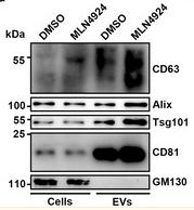
FIGURE 1 Neddylation inhibits EV secretion. (a) Flow cytometric analysis of EVs in the supernatants of HEK293 cells treated with DMSO or 100 nM MLN4924 for 12 h. Left, representative dot plots showing CD9 and CD81 staining of EVs captured with anti‑CD63‑coated beads after incubation with cell culture supernatants. Right, the ratio of CD9+ and CD81+ EVs. (b–d) EVs were purified from equal numbers of HEK293 cells treated with DMSO or 100 nM MLN4924 for 12 h. The BCA assay was used to determine the amount of EV protein (b). NTA to determine the EV concentration (c). WB analysis to detect the indicated EV markers (d). (e) WB analysis to detect the indicated proteins in HEK293 cells overexpressing NEDD8. (f) Flow cytometric ratio of CD9+ EVs in the supernatants of HEK293 cells overexpressing NEDD8. (g) ELISA analysis of CD9+ and CD81+ EVs in sera of mice intraperitoneally injected with MLN4924 at the indicated dose for 72 h. Representative results from three independent experiments are shown. n, sample number; *P < 0.05; **P < 0.01; ***P < 0.001 (unpaired two‐tailed Student's t‐test except for one‐way ANOVA followed by Tukey test in G; mean ± s.d.)

3). Mesenchymal Stem Cell Derived Exosomes as Nanodrug Carrier of Doxorubicin for Targeted Osteosarcoma Therapy via SDF1-CXCR4 Axis. International Journal of Nanomedicine, 2022 (PubMed: 35959282) [IF=8.0]

Application: WB    Species: Mouse    Sample: BM-MSCs cells

Figure 1 Characterization of exosomes: the size distributions of blank exosome (A) and exosome-doxorubicin (B) measured by NTA. The mean particle diameters were 141.6 nm for free exosome and 178.1 nm for exosome-doxorubicin. The morphology of blank exosome (C) and exosome-doxorubicin (D) as observed by TEM. (E) Western blotting analysis of the exosomal proteins CD81 and TSG101.

4). Anti-Tim4 Grafting Strongly Hydrophilic Metal–Organic Frameworks Immunoaffinity Flake for High-Efficiency Capture and Separation of Exosomes. ANALYTICAL CHEMISTRY, 2021 (PubMed: 33851819) [IF=7.4]

Application: WB    Species: Human    Sample: H1299 cells

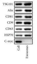
Figure 2. Characterization the captured exosomes: (A) TEM image, (B) size distribution via NTA analysis, and (C) Western blot analysis of exosomes marker proteins. Scale bar: 500 nm.

5). Focal ischemic stroke modifies microglia-derived exosomal miRNAs: potential role of mir-212-5p in neuronal protection and functional recovery. Biological Research, 2023 (PubMed: 37789455) [IF=6.7]

Application: WB    Species: Rat    Sample:

Fig. 2 MiRNA sequencing of microglial exosomes 3 days after MCAO/R and qRT–PCR validation. A, B Characterisation of microglial exosomes using nanoparticle tracking analysis and transmission electron microscopy scanning. Scale bar = 200 nm. C The exosome markers CD9, CD63, and CD81 were detected using western blot analysis. D Microglia-derived exosomes were detected by western blot analysis using the microglia marker CD11b. E Heatmap showing the levels of miRNAs in microglial exosomes at 3 days after MCAO/R. F Expression of miR-30c-5p, miR-126a-5p, miR-128-3p, miR-212-5p and miR-1949 in the ischemic penumbra of the cortex at 3 days after MCAO/R was determined using qRT–PCR. G Expression levels of the miRNA-targeting genes PLXNA2, PTEN and FOXO3 in the ischemic penumbra of the cortex at 3 days after MCAO/R. H The target sites of miR-212-5p in PLXNA2 mRNA 3’ untranslated region (3’UTR). I Map of the pmirGLO luciferase reporter vector. J Dual luciferase assays revealed the binding of miR-212-5p to the 3’UTR of PLXNA2. The data are presented as the means ± SEM (n = 5 per group). *P 

6). Fluoride Interferes with the Sperm Fertilizing Ability via Downregulated SPAM1, ACR, and PRSS21 Expression in Rat Epididymis. JOURNAL OF AGRICULTURAL AND FOOD CHEMISTRY, 2019 (PubMed: 31008594) [IF=6.1]

Application: WB    Species: rat    Sample: caudal epididymides

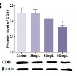
Figure 7.| Effects of fluoride on CD9 and CD81 expression in caudal epididymides. (A and B) Changes in CD9 and CD81 expression in caudal epididymides of rats treated with 0, 25, 50, and 100 mg/L NaF by western blot. β-actin was used as the control. The values are presented as the mean ± SEM. (∗) p < 0.05, (∗∗) p < 0.001, and(∗∗∗) p < 0.001 indicate significant differences compared to the control.

7). Mammary Epithelial Cell-Derived Exosomal miR-221-3p Regulates Macrophage Polarization by Targeting Igf2bp2 during Mastitis. Journal of Agricultural and Food Chemistry, 2023 (PubMed: 37757458) [IF=6.1]

8). Chemerin-Induced Down-Regulation of Placenta-Derived Exosomal miR-140-3p and miR-574-3p Promotes Umbilical Vein Endothelial Cells Proliferation, Migration, and Tube Formation in Gestational Diabetes Mellitus. Cells, 2022 (PubMed: 36359855) [IF=6.0]

9). CD73-Positive Small Extracellular Vesicles Derived From Umbilical Cord Mesenchymal Stem Cells Promote the Proliferation and Migration of Pediatric Urethral Smooth Muscle Cells Through Adenosine Pathway. Frontiers in Bioengineering and Biotechnology, 2022 (PubMed: 35573239) [IF=5.7]

Application: WB    Species: Human    Sample: PUSMCs

FIGURE 1 Identification of PUMSCs, UCMSCs and UCMSC-sEV. (A) Morphology of PUMSCs. (B) Immunofluorescence staining results show that the isolated PUMSCs express α-SMA, a smooth muscle cell surface marker. (C) Growth curve of PUMSCs. (D) Flow cytometric analysis of MSC surface markers shows that UCMSCs express high levels = of CD29, CD44, CD73, and CD90 but do not express CD45 and HLA-DR. (E) Transmission electron microscopy shows that the UCMSC-sEV are cup-shaped vesicles. Scale bar = 100 nm. (F) NanoFCM analysis of the particle size of UCMSC-sEV. (G) Western blotting was used to detect the expression of the sEV marker proteins CD9, CD63, CD81, and TSG101.

10). Exosomes secreted from osteocalcin-overexpressed endothelial progenitor cells promoted endothelial cell angiogenesis. AMERICAN JOURNAL OF PHYSIOLOGY-CELL PHYSIOLOGY, 2019 (PubMed: 31411920) [IF=5.5]

Application: WB    Species: rat    Sample: RAOECs

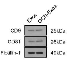
Figure 1| OCN overexpression and exosome identification. Rat EPCs were transfected with plasmid carrying the gene of OCN. The mRNA levels of OCN were analyzed by PCR (A) and the protein levels were detected by western blot method (B and D). The exosomes were isolated from cell culture medium and analyzed by western blot analysis to detect exosome marker proteins, CD9 and CD81 (C, E and F).

加载更多

限制条款

产品的规格、报价、验证数据请以官网为准,官网链接:www.affbiotech.com | www.affbiotech.cn(简体中文)| www.affbiotech.jp(日本語)

产品的数据信息为Affinity所有,未经授权不得收集Affinity官网数据或资料用于商业用途,对抄袭产品数据的行为我们将保留诉诸法律的权利。

产品相关数据会因产品批次、产品检测情况随时调整,如您已订购该产品,请以订购时随货说明书为准,否则请以官网内容为准,官网内容有改动时恕不另行通知。

Affinity保证所销售产品均经过严格质量检测。如您购买的商品在规定时间内出现问题需要售后时,请您在Affinity官方渠道提交售后申请。

产品仅供科学研究使用。不用于诊断和治疗。 

产品未经授权不得转售。

Affinity Biosciences将不会对在使用我们的产品时可能发生的专利侵权或其他侵权行为负责。Affinity Biosciences, Affinity Biosciences标志和所有其他商标所有权归Affinity Biosciences LTD.