.
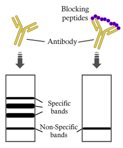
Bands result from membrane strip incubation.")
.
Bands result from membrane strip incubation.")
 production and ROS pathway related protein expressions. The 2′,7′-dich lorodihydrofluorescein diacetate assay was used for the detection of ROS in SW480 (A) and SW620 cells (B). ROS levels were significantly increased after DMA and 5-Fu treatment in both cell lines. The absolute intensity values of CHOP and Noxa were significantly different between cell lines (C). Results were obtained from three independent experiments and expressed as the means SD. Compared with SW480 (or SW620)")
 RT‑qPCR and (D) western blotting. A549 cells were transfected with GRP78‑siRNA or negative control, hyperoxia was established subsequently for 24, 48 and 72 h after transfection. Sham siRNA: A549 cells treated with negative control siRNA; siRNA+N: A549 cells were treated with GRP78‑siRNA and nomaxia for 24 h; siRNA+H 24 h, siRNA+H 48 h, siRNA+H 72 h: A549 cells were treated with GRP78‑siRNA and hyperoxia for 24, 48 and 72 h. CHOP protein expression was slightly increased after 24 h under hypoxia after (E and F) shamRNA treatment.")
.")

 Representative blots and histograms showing (B) p‑PERK/PERK ratio; (C) p‑eIF2α/eIF2α ratio; (D) ATF4 expression; and (E) CHOP expression.")
after TGF-β1 treatment.c–e Quantitative analysis of the relative protein level in (b).")
 Representative western blots.Results are expressed as the mean ± standard deviation. *P<0.05 vs. Con; #P<0.05 vs. the H/R group; &P<0.05 vs. The DEX+H/R group.")
 Protein expression levels of CHOP, ATF6, p-PERK, PERK, p-IRE1, IRE1 and β-actin in H9C2 cells treated with LPS, TA and TA + LPS, as determined by western blot analysis. Semi-quantification of the protein expression levels of (B) CHOP and ATF6, and (C) p-PERK, PERK and p-IRE1/IRE1 in the LPS, TA and TA + LPS groups. Data are presented as the mean ± SEM (n=3). *P<0.05 and **P<0.01 vs. control group; #P<0.05 vs. LPS group. TA, tannic acid; LPS, lipopolysaccharide; CHOP, C/EBP-homologous protein; ATF6, activating transcription factor 6; p, phosphorylated; PERK, protein kinase-like endoplasmic reticulum kinase; IRE1, inositol-requiring enzyme 1.")
 GRP78, (B) CHOP and (C) caspase-12, and protein expression levels of (D) GRP78, (E) CHOP and (F) caspase-12. (G) Representative western blots. Results are expressed as the mean ± standard deviation. *P<0.05 vs. Con; #P<0.05 vs. the H/R group; &P<0.05 vs. the DEX+H/R group. Con, control group; 1, normoxic incubation with DEX; 2, H/R incubation; 3, H/R incubation with dexmedetomidine; 4, normoxic incubation with 4-PBA; 5, H/R incubation with 4-PBA; 6, H/R incubation with DEX and 4-PBA; DEX, dexmedetomidine; GRP78, glucose-regulated protein 78; CHOP, C/EBP homologous protein; H/R, hypoxia/reoxygenation; 4-PBA, 4-phenylbutyric acid.")
 Representative western blots showed that GSK2606414 reduced the expression of CHOP, Bim, and cleaved caspase-3 in hippocampal tissue.")
 Representative blots of SIRT1, GRP78, p‑PERK, p‑eIF2α, CHOP and caspase‑12. Semiquantitative analysis of (B) SIRT1, (C) GRP78, (D) p‑PERK, (E) p‑eIF2α, (F) CHOP and (G) caspase‑12.")
 Levels of GRP78 and CHOP protein expression in Lv-Sig-1R cells and Lv-Ctrl cells.")
 Protein expression levels of CHOP, ATF6, p-PERK, PERK, p-IRE1, IRE1 and β-actin in H9C2 cells treated with LPS, TA and TA + LPS, as determined by western blot analysis. Semi-quantification of the protein expression levels of (B) CHOP and ATF6, and (C) p-PERK, PERK and p-IRE1/IRE1 in the LPS, TA and TA + LPS groups. Data are presented as the mean ± SEM (n=3). *P<0.05 and **P<0.01 vs. control group; #P<0.05 vs. LPS group. TA, tannic acid; LPS, lipopolysaccharide; CHOP, C/EBP-homologous protein; ATF6, activating transcription factor 6; p, phosphorylated; PERK, protein kinase-like endoplasmic reticulum kinase; IRE1, inositol-requiring enzyme 1.")
 Protein levels of ATF4, CHOP and p‑eIF2A/eIF2A were determined using western blotting. *P<0.05, **P<0.01 and ***P<0.001 vs. 0 mg/dl UA control group. FGF21, fibroblast growth factor 21; UA, uric acid; Sirt1,sirtuin 1; CHOP, eIF2A, eukaryotic initiation factor 2; ATF4, activating transcription factor 4; p‑, phosphorylated.")
 Representative western blots showed that GSK2606414 reduced the expression of CHOP, Bim, and cleaved caspase-3 in hippocampal tissue. The pooled data from three mice for each group are summarized in (b). (c) The western blot results showed that the expression of Bax and Bak in the mitochondria was obviously increased and that this effect was accompanied by a reduction in cytochrome c. These effects were prevented by the administration of GSK2606414. (d) Statistical data from three animals for each group are summarized. (e) Representative western blots showing that the cytoplasmic expression of Bax and Bak was obviously reduced and that of cytochrome C was significantly increased. GSK2606414 increased the expression of Bax and Bak and reduced cytochrome C. (f) Statistical data from three animals for each group are summarized. (g) The morphology of mitochondria in the hippocampal CA1 region in the (i) control group, (ii) GSK2606414 group, (iii) IH group, and (iv) IH + GSK2606414 group. In the control and GSK2606414 groups, intact mitochondria (indicated by arrowheads) with clear cristae were found. Much fewer cristae were found in the mitochondria from the IH group. GSK2606414 treatment preserved more intact mitochondria with discernable cristae. Scale bar: 0.25 μm. (h) The mitochondrial membrane potential (JC-1 fluorescence intensity ratio) was impaired by IH treatment and rescued by GSK2606414 treatment. ∗P < 0.05; ∗∗P < 0.01; ns: not significant.")
 on key proteins involved in endoplasmic reticulum stress in oleic acid-treated HepG2 cells.
Data are presented as mean ± SD. Different lowercase alphabet letters were significantly different at level of p < .05")
 Representative western blots showed that GSK2606414 reduced the expression of CHOP, Bim, and cleaved caspase-3 in hippocampal tissue. The pooled data from three mice for each group are summarized in (b). (c) The western blot results showed that the expression of Bax and Bak in the mitochondria was obviously increased and that this effect was accompanied by a reduction in cytochrome c. These effects were prevented by the administration of GSK2606414. (d) Statistical data from three animals for each group are summarized. (e) Representative western blots showing that the cytoplasmic expression of Bax and Bak was obviously reduced and that of cytochrome C was significantly increased. GSK2606414 increased the expression of Bax and Bak and reduced cytochrome C. (f) Statistical data from three animals for each group are summarized. (g) The morphology of mitochondria in the hippocampal CA1 region in the (i) control group, (ii) GSK2606414 group, (iii) IH group, and (iv) IH + GSK2606414 group. In the control and GSK2606414 groups, intact mitochondria (indicated by arrowheads) with clear cristae were found. Much fewer cristae were found in the mitochondria from the IH group. GSK2606414 treatment preserved more intact mitochondria with discernable cristae. Scale bar: 0.25 μm. (h) The mitochondrial membrane potential (JC-1 fluorescence intensity ratio) was impaired by IH treatment and rescued by GSK2606414 treatment. ∗P < 0.05; ∗∗P < 0.01; ns: not significant.")
 Representative blots of SIRT1, GRP78, p-PERK, p-eIF2α, CHOP and caspase-12. Semiquantitative analysis of (B) SIRT1, (C) GRP78, (D) p-PERK, (E) p-eIF2α, (F) CHOP and (G) caspase-12. (H) The mRNA levels of caspase-12 in the myocardium. Data are presented as the mean ± standard deviation. n=3. ##P<0.01 vs. Sham. *P<0.05 and **P<0.01 vs. MI/R. IOE, Inonotus obliquus extract; SIRT1, NAD-dependent protein deacetylase sirtuin-1; GRP78, glucose-regulated protein 78; PERK, protein kinase R-like endoplasmic reticulum kinase; eIF2α, eukaryotic translation initiation factor 2 subunit α; CHOP, C/EBP homologous protein; p, phosphorylated; MI/R, myocardial ischemia/reperfusion.")
 and C/EBP homologous protein (CHOP) in HK-2 cells were detected by western blot and quantitative real-time polymerase chain reaction (qRT-PCR). β-actin was used as a housekeeping gene. The results were expressed as the mean±standard deviation (SD) of three independent experiments.")
 Levels of insulin secretionby Lv-Sig-1R cells and Lv-Ctrl cells. (B) Levels of ATP production by Lv-Sig-1R cells and Lv-Ctrl cells. (C) Levels of GRP78 and CHOP protein expression in Lv-Sig-1R cells and Lv-Ctrl cells. (D) Expression of mitochondria-associated apoptotic proteins Bax, Bcl-2 and Cytochrome c in Lv-Sig-1R cells and Lv-Ctrl cells. The results are means ± SD for three observations; *P<0.05 and #P<0.05. ER, endoplasmic reticulum; GRP78, glucose-regulated protein 78; CHOP, C/EBP homologous protein; Sig-1R, Sigma-1 receptor; Lv, lentiviral; Ctrl, control; PA, palmitic acid.")
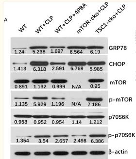
 patterns of mTOR pathway proteins, including mTOR, P-mTOR, downstream effectors p70s6k, p-p70s6k, 4EBP, and P-4EBP; (b) ERS-associated proteins, including GRP78 and CHOP; (c) apoptosis-associated proteins, including caspase-3, Bax, and Bcl-2. The protein expression was detected by immunoblotting. Data are mean ± SD. n = 4 biologically independent experiments (one-way ANOVA Tukey's post hoc test). ∗P < 0.05, ∗∗P < 0.01, and ∗∗∗P < 0.001.")
 and NeuN (red) immunofluorescence. B Statistical analysis of the fluorescence intensity of GRP78. C The expression of ER stress signaling-related proteins, including GRP78, ATF-6, p-PERK/PERK, and CHOP. D–G Expression histograms. ∗p")
 and CHOP (B) in mice kidney tissue Data are expressed as mean±SD, n=6, 200 or 400×). ##P")
 and CHOP (B) in mouse kidney tissue Data are expressed as mean±SD, n=6, 400×. ##P")
 staining for neuronal apoptosis. b Statistical analysis of the proportion of TUNEL-positive cells. c Expression levels of the ER stress-related apoptosis markers Caspase-12 and CHOP. d, e Protein expression levels. *P")
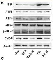
. C: the mRNA levels of ER stress-related genes were measured via quantitative PCR (n = 3, 1-way ANOVA with Duncan’s post hoc test). D: images of immunofluorescence staining of BiP in HUVECs in different groups. E: cellular ultrastructure in different groups. The arrow indicates endoplasmic reticulum. Data are shown as the means ± SD (*P < 0.05 vs. Con; **P < 0.01 vs. Con; #P < 0.05 vs. PA; ##P < 0.01 vs. PA). ATF-4, activating transcription factor 4; ATF-6, activating transcription factor 6; BiP, binding immunoglobulin protein; CHOP, C/EBP-homologous protein; ER, endoplasmic reticulum; HUVECs, human umbilical vein endothelial cells; MSCs, mesenchymal stem cells; PA, palmitic acid.")
 Representative western blot bands of GRP78, ATF4, CHOP, Cleaved caspase-12, PHLDA1, and PPARγ. n = 3. (B) The expression of CHOP in OGD/R-treated cells was detected by immunofluorescence staining. Red fluorescence: CHOP, blue fluorescence: nucleus.")
 Representative western blot bands of GRP78, ATF4, CHOP, Cleaved caspase-12, PHLDA1, and PPARγ. n = 3. (B) The expression of CHOP in OGD/R-treated cells was detected by immunofluorescence staining. Red fluorescence: CHOP, blue fluorescence: nucleus.")
产品描述
*The optimal dilutions should be determined by the end user. For optimal experimental results, antibody reuse is not recommended.
*Tips:
WB: 适用于变性蛋白样本的免疫印迹检测. IHC: 适用于组织样本的石蜡(IHC-p)或冰冻(IHC-f)切片样本的免疫组化/荧光检测. IF/ICC: 适用于细胞样本的荧光检测. ELISA(peptide): 适用于抗原肽的ELISA检测.
引用格式: Affinity Biosciences Cat# DF6025, RRID:AB_2838000.
展开/折叠
C/EBP homologous protein; C/EBP Homology Protein; C/EBP zeta; C/EBP-homologous protein 10; C/EBP-homologous protein; CCAAT/enhancer binding protein homologous protein; CEBPZ; CHOP 10; CHOP; CHOP-10; CHOP10; DDIT 3; DDIT-3; Ddit3; DDIT3_HUMAN; DNA Damage Inducible Transcript 3; DNA damage-inducible transcript 3 protein; GADD 153; GADD153; Growth Arrest and DNA Damage Inducible Protein 153; Growth arrest and DNA damage inducible protein GADD153; Growth arrest and DNA damage-inducible protein GADD153; MGC4154;
抗原和靶标
A synthesized peptide derived from human DDIT3, corresponding to a region within N-terminal amino acids.
- P35638 DDIT3_HUMAN:
- Protein BLAST With
- NCBI/
- ExPASy/
- Uniprot
MAAESLPFSFGTLSSWELEAWYEDLQEVLSSDENGGTYVSPPGNEEEESKIFTTLDPASLAWLTEEEPEPAEVTSTSQSPHSPDSSQSSLAQEEEEEDQGRTRKRKQSGHSPARAGKQRMKEKEQENERKVAQLAEENERLKQEIERLTREVEATRRALIDRMVNLHQA
种属预测
score>80的预测可信度较高,可尝试用于WB检测。*预测模型主要基于免疫原序列比对,结果仅作参考,不作为质保凭据。
High(score>80) Medium(80>score>50) Low(score<50) No confidence
研究背景
Multifunctional transcription factor in ER stress response. Plays an essential role in the response to a wide variety of cell stresses and induces cell cycle arrest and apoptosis in response to ER stress. Plays a dual role both as an inhibitor of CCAAT/enhancer-binding protein (C/EBP) function and as an activator of other genes. Acts as a dominant-negative regulator of C/EBP-induced transcription: dimerizes with members of the C/EBP family, impairs their association with C/EBP binding sites in the promoter regions, and inhibits the expression of C/EBP regulated genes. Positively regulates the transcription of TRIB3, IL6, IL8, IL23, TNFRSF10B/DR5, PPP1R15A/GADD34, BBC3/PUMA, BCL2L11/BIM and ERO1L. Negatively regulates; expression of BCL2 and MYOD1, ATF4-dependent transcriptional activation of asparagine synthetase (ASNS), CEBPA-dependent transcriptional activation of hepcidin (HAMP) and CEBPB-mediated expression of peroxisome proliferator-activated receptor gamma (PPARG). Inhibits the canonical Wnt signaling pathway by binding to TCF7L2/TCF4, impairing its DNA-binding properties and repressing its transcriptional activity. Plays a regulatory role in the inflammatory response through the induction of caspase-11 (CASP4/CASP11) which induces the activation of caspase-1 (CASP1) and both these caspases increase the activation of pro-IL1B to mature IL1B which is involved in the inflammatory response.
Ubiquitinated, leading to its degradation by the proteasome.
Phosphorylation at serine residues by MAPK14 enhances its transcriptional activation activity while phosphorylation at serine residues by CK2 inhibits its transcriptional activation activity.
Cytoplasm. Nucleus.
Note: Present in the cytoplasm under non-stressed conditions and ER stress leads to its nuclear accumulation.
The N-terminal region is necessary for its proteasomal degradation, transcriptional activity and interaction with EP300/P300.
Belongs to the bZIP family.
研究领域
· Cellular Processes > Cell growth and death > Apoptosis. (View pathway)
· Environmental Information Processing > Signal transduction > MAPK signaling pathway. (View pathway)
· Genetic Information Processing > Folding, sorting and degradation > Protein processing in endoplasmic reticulum. (View pathway)
· Human Diseases > Endocrine and metabolic diseases > Non-alcoholic fatty liver disease (NAFLD).
· Human Diseases > Cancers: Overview > Transcriptional misregulation in cancer.
文献引用
Application: WB Species: mouse Sample: liver
Application: IHC Species: Mice Sample: ileum
Application: WB Species: Human Sample: HaCaT cells
Application: WB Species: Human Sample: HaCaT cells

Application: WB Species: human Sample: MDA-MB-231 cells

Application: WB Species: Human Sample: HepG-2 cells
Application: WB Species: Mice Sample: CD4+ T cells

Application: WB Species: Rat Sample: nigral tissue

Application: WB Species: human Sample: HepG2 cells
Application: WB Species: Human Sample: HepG2 cells
限制条款
产品的规格、报价、验证数据请以官网为准,官网链接:www.affbiotech.com | www.affbiotech.cn(简体中文)| www.affbiotech.jp(日本語)产品的数据信息为Affinity所有,未经授权不得收集Affinity官网数据或资料用于商业用途,对抄袭产品数据的行为我们将保留诉诸法律的权利。
产品相关数据会因产品批次、产品检测情况随时调整,如您已订购该产品,请以订购时随货说明书为准,否则请以官网内容为准,官网内容有改动时恕不另行通知。
Affinity保证所销售产品均经过严格质量检测。如您购买的商品在规定时间内出现问题需要售后时,请您在Affinity官方渠道提交售后申请。产品仅供科学研究使用。不用于诊断和治疗。
产品未经授权不得转售。
Affinity Biosciences将不会对在使用我们的产品时可能发生的专利侵权或其他侵权行为负责。Affinity Biosciences, Affinity Biosciences标志和所有其他商标所有权归Affinity Biosciences LTD.