 antibody.")
 The mRNA levels of FUNDC1, LC3A, LC3B and PGAM5. (B) The western blot results of FUNDC1, p-FUNDC1 and LC3. (C) The quantification of FUNDC1, p-FUNDC1 (Ser17) and LC3II/LC3I protein levels. (D) Immunofluorescence co-location of COX IV and LC3 at day 50. In the images, the nucleus staining is shown in blue, COX IV staining is shown in green, LC3 staining is shown in red, and the signals of colocalization are shown in merged images. (E) Pearson coefficient of the colocalization of COX IV and LC3. Data are expressed as means ± SD (n = 6). “*” indicates significant difference compared to the corresponding control (*P < 0.05, **P < 0.01). “#” indicates statistically significant difference between corresponding groups (#P < 0.05, ##P < 0.01).")
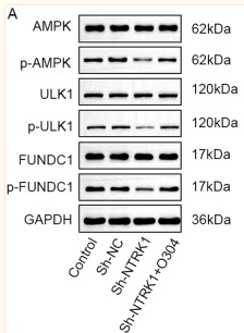
 The expression levels of ULK1, PGAM5, FUNDC1, p-FUNDC1 and GAPDH. (B) ULK1 relative to GAPDH, (C) PGAM5 relative to GAPDH, (D) FUNDC1 relative to GAPDH. (E) p-FUNDC1 relative to GAPDH. (F) ULK mRNA levels. (G) PGAM5 mRNA levels. (H) FUNDC1 mRNA levels. **P<0.05 vs. C group and #P<0.05 vs. DOX group. Protein levels were measured by western blotting. ULK1, PGAM5 and FUNDC1 mRNA levels were measured by RT-qPCR. Data are mean ± standard deviation from five independent experiments (n=13-20). MOX moxibustion; FUNDC1, FUN14 domain-containing protein 1; ULK1, Unc-51 Like Autophagy Activating Kinase 1; PGAM5, phosphoglycerate mutase family member 5; p-, phosphorylated; DOX, doxorubicin; BEN, benazepril C, control.")
 mRNA expression of IL-1β, IL-6, IL-8 and TNF-α in human healthy and pulpitis tissues. mRNA expression of (B) HIF-1α and (C) FUNDC1 in human healthy and pulpitis tissues. (D) Representative immunostaining images of HIF-1α and FUNDC1 in human healthy or inflamed dental pulp tissues. Scale bars are 100 and 25 µm, respectively. Results are presented as the means ± SD from ≥ three independent experiments. *P<0.05, **P<0.01 and ***P<0.001 vs. healthy. HIF-1α, hypoxia-inducible factor-1α; FUNDC1, FUN14 domain-containing 1.")
. The serum AMH (B), E2 (C), FSH (D), and LH (E) levels. F Ki67 expression in ovarian GCs; scale bar: 100 μm. G The Western blotting images and quantitation of functional protein expression in GCs. H The fertility and litter size.")
| 产品: | 磷酸化 FUNDC1 (Ser17) 抗体 |
| 货号: | AF0001 |
| 描述: | Rabbit polyclonal antibody to Phospho-FUNDC1 (Ser17) |
| 应用: | WB |
| 文献验证: | WB |
| 反应: | Human |
| 预测: | Pig, Bovine, Horse, Sheep, Rabbit, Dog, Chicken |
| 分子量: | 17KD(Observed); 17kD(Calculated). |
| 蛋白ID: | Q8IVP5 |
| RRID: | AB_2846773 |
产品描述
*The optimal dilutions should be determined by the end user. For optimal experimental results, antibody reuse is not recommended.
*Tips:
WB: 适用于变性蛋白样本的免疫印迹检测. IHC: 适用于组织样本的石蜡(IHC-p)或冰冻(IHC-f)切片样本的免疫组化/荧光检测. IF/ICC: 适用于细胞样本的荧光检测. ELISA(peptide): 适用于抗原肽的ELISA检测.
引用格式: Affinity Biosciences Cat# AF0001, RRID:AB_2846773.
展开/折叠
FUN14 domain containing 1; FUN14 domain containing protein 1; FUN14 domain-containing protein 1; FUND1_HUMAN; fundc1;
抗原和靶标
A synthesized peptide derived from human FUNDC1 around the phosphorylation site of Serine 17.
- Q8IVP5 FUND1_HUMAN:
- Protein BLAST With
- NCBI/
- ExPASy/
- Uniprot
MATRNPPPQDYESDDDSYEVLDLTEYARRHQWWNRVFGHSSGPMVEKYSVATQIVMGGVTGWCAGFLFQKVGKLAATAVGGGFLLLQIASHSGYVQIDWKRVEKDVNKAKRQIKKRANKAAPEINNLIEEATEFIKQNIVISSGFVGGFLLGLAS
种属预测
score>80的预测可信度较高,可尝试用于WB检测。*预测模型主要基于免疫原序列比对,结果仅作参考,不作为质保凭据。
High(score>80) Medium(80>score>50) Low(score<50) No confidence
研究背景
Acts as an activator of hypoxia-induced mitophagy, an important mechanism for mitochondrial quality control.
Phosphorylation at Tyr-18 by SRC inhibits activation of mitophagy. Following hypoxia, dephosphorylated at Tyr-18, leading to interaction with MAP1 LC3 family proteins and triggering mitophagy.
Mitochondrion outer membrane>Multi-pass membrane protein.
Widely expressed.
The YXXL motif mediates the interaction with MAP1 LC3 family proteins MAP1LC3A, MAP1LC3B and GABARAP.
Belongs to the FUN14 family.
文献引用

Application: WB Species: Mouse Sample:

Application: WB Species: rat Sample:
Application: WB Species: Mouse Sample:
限制条款
产品的规格、报价、验证数据请以官网为准,官网链接:www.affbiotech.com | www.affbiotech.cn(简体中文)| www.affbiotech.jp(日本語)产品的数据信息为Affinity所有,未经授权不得收集Affinity官网数据或资料用于商业用途,对抄袭产品数据的行为我们将保留诉诸法律的权利。
产品相关数据会因产品批次、产品检测情况随时调整,如您已订购该产品,请以订购时随货说明书为准,否则请以官网内容为准,官网内容有改动时恕不另行通知。
Affinity保证所销售产品均经过严格质量检测。如您购买的商品在规定时间内出现问题需要售后时,请您在Affinity官方渠道提交售后申请。产品仅供科学研究使用。不用于诊断和治疗。
产品未经授权不得转售。
Affinity Biosciences将不会对在使用我们的产品时可能发生的专利侵权或其他侵权行为负责。Affinity Biosciences, Affinity Biosciences标志和所有其他商标所有权归Affinity Biosciences LTD.