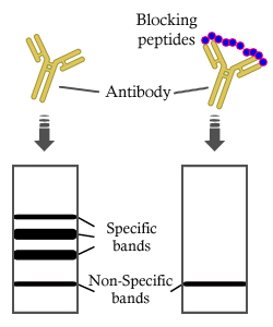
, using GPX4 Antibody. The lane on the left was treated with blocking peptide.")
, using GPX4 Antibody. The lane on the left was treated with blocking peptide.")
, using GPX4 Antibody at 1/1000 dilution.
5ug/NC membrane strip.
Exposure for 5s with Affinity™ ECL Kit(#KF8003).
Bands result from membrane strip incubation.")


.")
, using GPX4 Antibody. The lane on the left was treated with blocking peptide.")


 and mouse anti-beta tubulin Ab(T0023 1:200) for 1 hour at 37°C. An AlexaFluor594 conjugated goat anti-rabbit IgG(H+L) Ab(Red) and an AlexaFluor488 conjugated goat anti-mouse IgG(H+L) Ab(Green) were used as the secondary antibody.
The nuclear counter stain is DAPI(blue).")
 The expressions of NRF2,
HO-1, GPX4, and 4HNE were evaluated by Western blotting in VSC4.1 in each group (n =6). (B-E) Quantification of NRF2, HO-1, GPX4,
and 4HNE expressions (data shown as mean ± SEM, two-way ANOVA with Tukey's post hoc test, n = 6). (F) Immunofluorescence staining
was used to detect the level of GPX4 from each group (n = 6, scale bar = 50 µm). (G) Statistical analysis of immunofluorescence staining
for positive expression of GPX4 in VSC4.1 from each group (n = 6, all the data are expressed as means ± SD, two-way ANOVA followed by
Tukey's post hoc test was applied). (H) Confocal microscopy showed the non-oxidized lipid (red) and oxidized lipid (green) in VSC4.1 that
were pretreated with ZnG (90 μmol/L) or not (n = 6, scale bar = 250 µm). (I) Statistical analysis of the fluorescence intensity of oxidized lipid
expression in VSC4.1 from each group (n = 6, all the data are expressed as means ± SD, two-way ANOVA followed by Tukey's post hoc test
was applied). * means p < 0.05; ** means p < 0.01; and *** means p < 0.001")
 Representative
western blot bands of GPX4, TfR1, FTL, COX2 and GAPDH at
the indicated time point in the injured cortex, with bar graphs
(B - E). The relative densities of each protein were normalized
against GAPDH. ImageJ software was used for western blot
band quantification. The statical differences between the two
relevant groups have been shown in the bar graphs. One-way
ANOVA followed by post-hoc analysis. n = 7 animals per
group at each time point. **p < 0.01 ***p < 0.001 ****p <
0.0001 vs the Sham group.")
. The levels of oxidative indicators and antioxidant enzymes, such as (A) SOD, (B) GSH, (C) MDA,
(D) NO, (E) ROS and (FeG) GPX4, were detected by ELISAs or immunofluorescence. Data are expressed as the mean ± SD.
& P < 0.05 and && P < 0.01 vs SAMR1 group; *P < 0.05 and
**P < 0.01 vs. SAMP8 group.")
 GPX4 and (B) xCT. The random grouping situation was WT group, APP/PS1 model group,
TSG (60, 120 and 180 mg/kg) groups. Scare bar: 500, 100 and 50 μ m, n = 5/group.")
, FAC (b), and erastin (c) at concentrations of 0, 5, 10, 20, 40, 60, 80, and 100 μg/mL for 24 h. The data were analyzed with GraphPad Prism 5.02 software. d–f The protein levels of GPX4, Bcl-2, and Bax in HNPCs treated with heme (d), FAC (e), and erastin (f) at concentrations of 0, 5, 10, and 20 μg/mL for 24 h. g Viability of HNPCs was measured by CCK-8 after 20 μg/mL heme treatment for 24 h with or without 30 μg/mL DFO pretreatment for 1 h (upper), and the protein levels of GPX4 in HNPCs were measured after 20 μg/mL heme treatment for 24 h with or without 30 μg/mL DFO pretreatment for 1 h (lower). h Identification of ROS and ferroptosis-related metabolites in HNPCs after treatment with 20 μg/mL heme, 20 μg/mL FAC, and 10 μg/mL erastin for 24 h using high-resolution MALDI-TOF MS. i ROS levels in HNPCs after 20 μg/mL heme treatment for 24 h with or without 30 μg/mL DFO pretreatment for 1 h. The data were analyzed with GraphPad Prism 5.02 software. *P < 0.05, **P < 0.01")
 TGL shows inhibition of telomere shortening in the liver. Data are presented as the means ± SD of more than six independent experiments and more than three independent experiments in Western blot.")
 dose-dependent and (B) time-dependent manner. Herceptin (C) decreased GSH content whilst (D) increasing GSSG content, (E) consequently reducing the GSH/GSSG ratio, in H9c2 cells in a dose-dependent manner. Herceptin (F) decreased GSH content whilst (G) increasing GSSG content, (H) consequently reducing the GSH/GSSG ratio, in H9c2 cells. *P<0.05, **P<0.01 and ***P<0.001 vs. 0 µM or 12 h. GPX, glutathione peroxidase; GSH, reduced glutathione; GSSG, oxidized glutathione.")
 dose‑dependent and (B) time‑dependent manner.Herceptin (C) decreased GSH content whilst (D) increasing GSSG content, (E) consequently reducing the GSH/GSSG ratio, in H9c2 cells in a dose‑dependent manner.")
 Immunoblots of apoptosis- and ferroptosis-related proteins in hSOD1WT and hSOD1G93A mice, with or without treatment of ABAH in male (left) and female (right) mice. (b) The plasma MDA levels of male (left) and female (right) mice. (c) The motor performance of mice (left: male; right: female). Quantified graphs were shown as means and SEM. ∗P < 0.05, ∗∗P < 0.01, and∗∗∗P < 0.001. The significant difference in two datasets was analyzed by Student's t-test.")
 The mRNA and protein levels of NRF2 and GPX4 in human myocardial tissue samples. (e–g) The protein expression of HO-1, ACSL4, and NOX4 in human myocardial tissue samples. (h) The MDA levels in human myocardial tissue samples. In all panels, the data were obtained from fetuses with CHD (n = 7) and normal heart structure (n = 6). The data are presented as the mean ± SD. Statistical significance is shown as ∗P < 0.05 vs. controls, ∗∗P < 0.01 vs. controls, and ∗∗∗P < 0.001 vs. controls.")
 and pathological evidence (g–j) of ferroptosis in CKD rats. Green arrow, normal mitochondria; red arrow, MOM rupture; blue arrow, reduction or loss of MC; yellow arrow, MC swelling. **P < 0.01, ***P < 0.001 vs. Sham group; #P < 0.05, ##P < 0.01, ###P < 0.001 vs. CKD group. Scale bar in (a) = 50 μm. Scale bar in (f) = 500 μm.")
 and pathological evidence (g–j) of ferroptosis in CKD rats. Green arrow, normal mitochondria; red arrow, MOM rupture; blue arrow, reduction or loss of MC; yellow arrow, MC swelling. **P < 0.01, ***P < 0.001 vs. Sham group; #P < 0.05, ##P < 0.01, ###P < 0.001 vs. CKD group. Scale bar in (a) = 50 μm. Scale bar in (f) = 500 μm.")
 (n = 3, scale bar: 25 μm). The expression quantity of TFR was assessed by immunofluorescence staining of HOS cells after the indicated treatment (b, e) (n = 3, scale bar: 50 μm). The expression of iron metabolism-related proteins in UA- and/or Cis-treated HOS cells was determined by western blotting (c) (n = 3). The data are presented as the mean ± SD. ∗p < 0.05 vs. the control group.")
. Macroscopic view of xenograft tumours at the endpoint of the experiment (b) (n = 3). The average tumour volume and weight and body weight of the different groups (c–e) (n = 3). H&E staining and immunofluorescence staining of autophagy-, ferroptosis-, and iron metabolism-related proteins in xenograft tissue sections (f) (n = 3, scale bar: 50 μm). The data are presented as the mean ± SD. ∗p < 0.05 vs. the control group.")
 Western blotting assay for ACSL4 expression in the temporal cortex after SAH. (b) Quantification of ACSL4 expression. (c) Representative images of IF staining for ACSL4. (d) Western blotting assay for TFR, DMT1, ferritin, and FPN expression in the temporal cortex after SAH. (e–h) Quantification of the expression of TFR, DMT1, ferritin, and FPN. (i) Representative images of IF staining for FTH. (j) Quantification of ferrous iron content. (k) Western blotting assay for XCT and GPX4 expression. (l, m) Quantification of the expression of XCT and GPX4. (n) Representative images of IF staining for GPX4. (o) Western blotting assay for FSP1 and CoQ10B expression. (p, q) Quantification of the expression of FSP1 and CoQ10B. n = 6/group. Bars represent the mean ± SEM. ∗P < 0.05, ∗∗P < 0.01, and ∗∗∗P < 0.001. Scale bars = 50 μm. ASCL4: acyl-CoA synthetase long-chain family member 4; TFR: transferrin receptor; DMT1: divalent metal transporter 1; FPN: ferroportin; FTH: ferritin heavy chain; GPX4: glutathione peroxidase 4; FSP1: ferroptosis suppressor protein 1.")
 Renal cortex expression of SLC7A11, GPX4, HO-1 and SOD2 determined by immunofluorescence staining in mice after glyoxylic acid treatment (magnification, ×400). (B-E) Quantification of (B) SLC7A11, (C) GPX4, (D) HO-1 and (E) SOD2 in mouse kidneys assessed using immunofluorescence. (F) Western blot analysis of SLC7A11, GPX4, HO-1 and SOD2 in renal tissue lysates from these groups. (G-J) The ratio of the optical density of (G) SLC7A11, (H) GPX4, (I) HO-1, and (J) SOD2 to GAPDH was statistically analyzed. (K) Representative images of mitochondrial injury under glyoxylic acid treatment were observed by TEM. White triangles represent the injured mitochondria. The data are presented as the mean ± SD; n=6. *P<0.05 vs. the control group. CaOx, calcium oxalate; SLC7A11, solute carrier family 7 member 11; GPX4, glutathione peroxidase 4; HO-1, heme oxygenase 1; SOD2, superoxide dismutase 2; TEM, transmission electron microscopy.")
 Renal cortex expression of SLC7A11, GPX4, HO-1 and SOD2 determined by immunofluorescence staining in mice after glyoxylic acid treatment (magnification, ×400). (B-E) Quantification of (B) SLC7A11, (C) GPX4, (D) HO-1 and (E) SOD2 in mouse kidneys assessed using immunofluorescence. (F) Western blot analysis of SLC7A11, GPX4, HO-1 and SOD2 in renal tissue lysates from these groups. (G-J) The ratio of the optical density of (G) SLC7A11, (H) GPX4, (I) HO-1, and (J) SOD2 to GAPDH was statistically analyzed. (K) Representative images of mitochondrial injury under glyoxylic acid treatment were observed by TEM. White triangles represent the injured mitochondria. The data are presented as the mean ± SD; n=6. *P<0.05 vs. the control group. CaOx, calcium oxalate; SLC7A11, solute carrier family 7 member 11; GPX4, glutathione peroxidase 4; HO-1, heme oxygenase 1; SOD2, superoxide dismutase 2; TEM, transmission electron microscopy.")
. The western blot results of MAD2L2 and mTOR signaling relative proteins. (B. C. D). The predictive correlation of mTOR and ferroptosis relative genes. (E). The western blot results of mTOR and ferroptosis relative proteins.")
 The MDA level in brain tissues. (B, C) Western blot of 4-HNE and GPX4 in brain tissues. β-Actin was used as an internal reference. (D, E) Quantitative analyses of 4-HNE and GPX4 (normalized to β-actin). Data are presented as mean ± SEM (n = 4). **P < 0.01, vs. Sham + NS group; #P < 0.05, ##P < 0.01, vs. HI + NS group (one-way analysis of variance followed by Tukey’s post hoc test). 4-HNE: 4-Hydroxynonenal; CGA: chlorogenic acid; GPX4: glutathione peroxidase 4; HI: hypoxic-ischemic brain injury; MDA: malondialdehyde; NS: normal saline.")
) DHE staining showed that myo-inositol alleviates cisplatin-induced ROS generation in renal tubules. (Panels (D–F)) Immunofluorescence staining of 4-HNE revealed that the increased lipid peroxidation induced by cisplatin was attenuated by myo-inositol. (Panels (G–L)) Immunohistochemistry analyses demonstrated that CHIP was down-regulated while NOX4 was up-regulated in cisplatin-treated kidneys, and their expression was partially restored by myo-inositol treatment. (Panel (M)) Immunoblot analyses revealed that cisplatin treatment led to decreased expression of NCOA4, GPX4, and CHIP, while the expression of FTH1 and NOX4 increased. These changes were largely normalized with the supplementation of myo-inositol. (Panels (N,O)) qRT-PCR analysis showed that the mRNA levels of ACSL4 and PTGS2 have increased in mice kidneys following cisplatin treatment (n = 4), which was attenuated by myo-inositol treatment.")
 of ferroptosis between the KLF11-NC, KLF11-OE and KLF11-KO of A549 and PC9 cell lines. f, g Western blot results showed differences in protein levels of GPX4, SLC7A11, and ASCL4 of ferroptosis between the KLF11-NC, KLF11-OE, and KLF11-KO of A549 and PC9 cell lines.")
 Relative endoscopic presentation of normal esophagus and that of patients with ESCC. (B) Immunohistochemistry was used to detect the expression of TAGLN, GPX4 and ACSL4 in normal esophageal tissues and in ESCC stages I-II and III-IV tumoral tissues. (C) Statistical analysis of TAGLN, GPX4 and ACSL4 expression in normal esophagus and ESCC tissues. Data are presented as the mean ± SD of three independent experiments. (D) Statistical analysis of the expression of TAGLN, GPX4 and ACSL4 in normal esophagus, stage I-II and III-IV ESCC tissues. Data are presented as the mean ± SD of three independent experiments. *P")
 representative results of immunohistochemistry (IHC) and relative expression of GPX4; (B) representative results of IHC and relative expression of ACSL4; and (C) representative results of IHC and relative expression of SLC7A11. *p < 0.05 compared with the control group")
, TU212 cells (Fig. 8B) and tumour-bearing nude mice (Fig. 8C). Immunohistochemical results of p53 and GPX4 proteins in xenograft tumours (Fig. 8D). After treatment with 10 μM chidamide, the immunofluorescence signal of p53 was enhanced, and that of GPX4 protein was decreased in HN8 cells (Fig. 8E). Immunofluorescence images of TU212 cells (Fig. 8F).")
, TU212 cells (Fig. 8B) and tumour-bearing nude mice (Fig. 8C). Immunohistochemical results of p53 and GPX4 proteins in xenograft tumours (Fig. 8D). After treatment with 10 μM chidamide, the immunofluorescence signal of p53 was enhanced, and that of GPX4 protein was decreased in HN8 cells (Fig. 8E). Immunofluorescence images of TU212 cells (Fig. 8F).")
, TU212 cells (Fig. 8B) and tumour-bearing nude mice (Fig. 8C). Immunohistochemical results of p53 and GPX4 proteins in xenograft tumours (Fig. 8D). After treatment with 10 μM chidamide, the immunofluorescence signal of p53 was enhanced, and that of GPX4 protein was decreased in HN8 cells (Fig. 8E). Immunofluorescence images of TU212 cells (Fig. 8F).")
 Western blot assay used to detect the protein expression levels of CX43, aggrecan, Col2, Sox9, MMP13, BCL-2, and cleaved caspase-3 in OA and normal cartilage samples. (b) The expression levels of CX43 and antiferroptosis proteins (GPX4, SLC7A11) in chondrocytes transfected with or without Agomir-1 and CX43. (c) C11 BODIPY fluorescent probe used to detect the level of intracellular lipid-ROS in chondrocytes. Red indicated unoxidized dye, green indicated oxidized dye. (d) Intracellular ROS in chondrocytes detected by DCFHDA. (e–f) Fe2+ and MDA concentrations in chondrocytes were measured using the iron assay kit and MDA assay kit. Scale bar = 200 µm. Data were presented as the mean ± SD of at least three replicates.")

 Fe2+ concentrations, (B and C) lipid ROS levels, and (D) mRNA levels of Tfrc, Slc11a2, Slc7a11, Gpx4 and Ptgs2, and (E) protein expression of GPX4 and PTGS2 were measured. ***P")
 IEC-6 cells and Caco-2 cells were incubated with or without 10 ng/mL LPS for 12 h, followed by 200 μM DCA for 2 h. They were divided into four group (n = 6 in each group): control group, DCA group, LPS group and LPS + DCA group. (A) The relative mRNA expression level of GPX4 in each group of IEC-6 cells and Caco-2 cells (n = 6 in each group). (B) The relative mRNA expression level of ACSL4 in each group of IEC-6 cells and Caco-2 cells (n = 6 in each group). (C) The protein level of GPX4, ACSL4 and β-actin in each group of IEC-6 cells and Caco-2 cells. (D) Representative transmission electron microscopy images (scale bars, 1 μm) of cell and mitochondria, and Fe2+/Hoechst immunofluorescence in each group of IEC-6 cells. Mitochondria was showed by white arrows in the pictures. The red fluorescence is indicative of the presence of ferrous ions. (E) The content of Fe2+ in each group of IEC-6 cells and Caco-2 cells (n = 6 in each group). (F) The content of GSH in each group of IEC-6 cells and Caco-2 cells (n = 6 in each group). (G) The content of ROS in each group of IEC-6 cells and Caco-2 cells (n = 6 in each group). (H) The content of MDA in each group of IEC-6 cells and Caco-2 cells (n = 6 in each group). (I-K) IEC-6 cells were incubated 2 μM ferrostatin-1 (Fer-1) for 16 h, and then incubated with or without 10 ng/mL LPS for 12 h, followed by 200 μM DCA for 2 h. They were divided into six group (n = 5 or 6 in each group): control group, LPS group, LPS + DCA group, Fer-1 group, LPS + Fer-1 group and LPS + DCA + Fer-1 group. (I) The relative mRNA expression level of GPX4, ACSL4 and DMT1 in each group of IEC-6 cells (n = 6 in each group). (J) The content of GSH in each group of IEC-6 cells (n = 5 in each group). (K) The content of MDA in each group of IEC-6 cells (n = 5 in each group). Data are represented as mean ± SEM. ns P > 0.05, ∗P < 0.05, ∗∗P < 0.01, ∗∗∗P < 0.001, ∗∗∗∗P < 0.0001. ##P < 0.01, ####P < 0.0001. ∗ LPS group vs control group, LPS + DCA group vs LPS group. # LPS + Fer-1 group vs LPS group, LPS + DCA + Fer-1 group vs LPS + DCA group.")
 IEC-6 cells and Caco-2 cells were incubated with or without 10 ng/mL LPS for 12 h, followed by 200 μM DCA for 2 h. They were divided into four group (n = 6 in each group): control group, DCA group, LPS group and LPS + DCA group. (A) The relative mRNA expression level of GPX4 in each group of IEC-6 cells and Caco-2 cells (n = 6 in each group). (B) The relative mRNA expression level of ACSL4 in each group of IEC-6 cells and Caco-2 cells (n = 6 in each group). (C) The protein level of GPX4, ACSL4 and β-actin in each group of IEC-6 cells and Caco-2 cells. (D) Representative transmission electron microscopy images (scale bars, 1 μm) of cell and mitochondria, and Fe2+/Hoechst immunofluorescence in each group of IEC-6 cells. Mitochondria was showed by white arrows in the pictures. The red fluorescence is indicative of the presence of ferrous ions. (E) The content of Fe2+ in each group of IEC-6 cells and Caco-2 cells (n = 6 in each group). (F) The content of GSH in each group of IEC-6 cells and Caco-2 cells (n = 6 in each group). (G) The content of ROS in each group of IEC-6 cells and Caco-2 cells (n = 6 in each group). (H) The content of MDA in each group of IEC-6 cells and Caco-2 cells (n = 6 in each group). (I-K) IEC-6 cells were incubated 2 μM ferrostatin-1 (Fer-1) for 16 h, and then incubated with or without 10 ng/mL LPS for 12 h, followed by 200 μM DCA for 2 h. They were divided into six group (n = 5 or 6 in each group): control group, LPS group, LPS + DCA group, Fer-1 group, LPS + Fer-1 group and LPS + DCA + Fer-1 group. (I) The relative mRNA expression level of GPX4, ACSL4 and DMT1 in each group of IEC-6 cells (n = 6 in each group). (J) The content of GSH in each group of IEC-6 cells (n = 5 in each group). (K) The content of MDA in each group of IEC-6 cells (n = 5 in each group). Data are represented as mean ± SEM. ns P > 0.05, ∗P < 0.05, ∗∗P < 0.01, ∗∗∗P < 0.001, ∗∗∗∗P < 0.0001. ##P < 0.01, ####P < 0.0001. ∗ LPS group vs control group, LPS + DCA group vs LPS group. # LPS + Fer-1 group vs LPS group, LPS + DCA + Fer-1 group vs LPS + DCA group.")
 WB results for GPX4, NFκB, pMAPK/MAPK, Aggrecan, CollagenⅡ, SOX9, MMP3, MMP13 and Tubulin; (J) Quantitive analysis of relative Glutathione Peroxidase Activity; (K-L) GSH contents and ratio of GSH/GSSG; (M) Quantitative results of MDA content assay.")
 Changes in the concentration of MDA and Fe2+ in MDA-MB-23 and SK-BR-3 cells were detected after NGR1 treatment. (B, C) Changes in the expression of ACSL4, COX2, FIH1, GPX4, and NOX1 proteins were detected by Western blot in MDA-MB-231 and SK-BR-3 cells. (* P < 0.05, ** P < 0.01, *** P < 0.001, **** P < 0.0001).")
 affects ferroptosis of triple‐negative breast cancer (TNBC) cells by targeting GPX4. (A) Western blotting showing increased GPX4 expression in PTGER3‐knockdown MDA‐MB‐231 cells and decreased GPX4 expression in PTGER3‐overexpression MDA‐MB‐231 cells. RIPK1 expression showed no significant changes. (B) Subcellular localization of PTGER3 expression. (C) PTGER3 binding site sequence as predicted from JASPAR. (D) The luciferase activity of cells cotransfected with the ex‐PTEGR3 plasmid and GPX4 promoter was significantly lower compared with other groups. (E) With increasing ex‐PTEGR3 plasmid dose, the luciferase activity gradually declined. This dose–response pattern suggests that PTGER3 binds to the GPX4 promoter. (F–M) GSH (F, G), ROS (H, I), MDA (J, K), and ferrous ion (L, M) levels in cells treated with DMSO, erastin, and erastin+Fer‐1 (scale bars: 100 μm). Data are presented as the mean ± SD. One‐way ANOVA.")
 Representative Prussian blue staining images of DOX-induced rats treated with or without dioscin. The arrows indicate iron deposition in cardiac tissue. (B) Bar charts indicate cardiac tissue iron in DOX-induced rats treated with or without dioscin. Data are mean ± SD (n = 6). (C–G) Bar charts indicate serum 4-HNE levels and cardiac tissue MDA, glutathione (GSH), superoxide dismutase (SOD), and catalase (CAT) levels of DOX-induced rats treated with or without dioscin. Data are mean ± SD (n = 6). (H) The mitochondrial structure of cardiac tissues of DOX-induced rats treated with or without dioscin. (I) Representative images of immunofluorescence analysis of GPX4 expression were captured by fluorescence microscopy. (J) The bar chart indicates the GPX4 intensity in cardiac tissue. Data are mean ± SD (n = 3). (K) Representative Western blotting images of GPX4 and ACSL4 in DOX-induced rats treated with or without dioscin. (L,M) Bar charts indicate the protein expression levels of ACSL4 and GPX4 in cardiac tissues. Data are mean ± SD (n = 3). (N,O) Bar charts indicate the mRNA expression levels of ACSL4 and GPX4 in cardiac tissues. Data are mean ± SD (n = 6). * p < 0.05, ** p < 0.01, *** p < 0.001 vs. DOX treatment group; # p < 0.05, ## p < 0.01, ### p < 0.001 vs. control group. (K) Original western blotting figures can be found in Figure S3.")
 cells. (a, b) Bavachin induces GSH depletion in a dose-related manner over 24 h. (c, d) Bavachin greatly induces ROS generation in OS cells over 24 h. (e, f) Bavachin increases MDA accumulation in a dose-related manner. (g, h) Western blotting analysis shows the expression of p-STAT3, STAT3, P53, SLC7A11, and GPX4 in OS cells treated with bavachin (10 μM, 20 μM, and 40 μM) for 24 h. &p < 0.01 vs. the control group, #p < 0.001 vs. the control group, ∗p < 0.05, ∗∗p < 0.01, and ∗∗∗p < 0.001.")
 SK-Hep1 and HepG2 cells were treated with AO (0, 50, 100 or 200 μM) for 24 h, and the protein expression levels of GPX4 were measured by western blotting. (C) SK-Hep1 and (D) HepG2 cells were treated with AO for 24 h and lipid peroxidation levels were detected. (E-G) SK-Hep1 and HepG2 cells were treated with AO (0, 50, 100 or 200 μM) for 24 h, and the protein expression levels of Nrf2 and HO-1 were measured by western blotting. *P")
 combined with doxorubicin (DOX) regulated the intracellular iron level and of ferroptosis-related proteins. A and B, Cellular iron levels were measured using the iron ion detection kit after treatment for 48 h. C-F, Western blot detection and quantification analysis of the SLC7A11 and GPX4 protein in 8505C (C and D) and CAL62 cells (E and F) after treatment for 48 h. Data are reported as mean and SD. **P")
 The MDA level in spinal cord of mice. Scale bar = 20 μm. (B and C) Representative bands of western blotting and quantitative analysis for GPX4 and Nrf2 expression levels in the spinal cord. (D) The molecular docking images of Nrf2 with Pue. (E and F) Representative images of immunofluorescence staining and quantitative analysis for GPX4 in the spinal cord. (G and H) Representative bands of immunoprecipitation and quantitative analysis for the interaction between Nrf2 and GPX4. (I and J) Representative bands of western blotting and quantitative analysis for GPX4 and NLRP3 expression levels in the spinal cord after Fer-1 treatment. Data are presented as the mean ± SD (n = 3). *P < 0.05 vs. control group, #P < 0.05 vs. OXA group. GPX4, glutathione peroxidase 4; Nrf2, nuclear factor erythroid 2-related factor 2; NLRP3, NOD-like receptor protein 3 inflammasome; MDA, malondialdehyde; Fer-1, ferrostatin-1; OXA, oxaliplatin; Pue, puerarin.")
 Western blot assay was applied to determine the expressions of SLC7A11, COX-2, ACSL4, FTH1, GPX4 in FHC cells. (D) Western blot assay was conducted to detect the expressions of p-IκBα, IκBα, p-p65 and p65 in FHC cells. n = 3, **P < 0.01.")
 Intracellular chelatable iron in GC-2 cells treated with or without ZnO NPs stained with PGSK (green). Statistical analysis of MFI of PGSK was shown. (B) Representative FACS data for lipid peroxidation level in GC-2 cells following ZnO NPs treatment using C11 BODIPY. Statistical analysis of MFI of the ratio of green/red was shown. (C) qRT-PCR analysis of ferroptosis-related gene expression in GC-2 cells after ZnO NPs treatment. (D) Western blot of ferroptosis-related protein levels in GC-2 cells treated with ZnO NPs. Statistical analysis of mean grey values ratios of the corresponding proteins/β-actin was shown, the same as below. (E) The cell viability of GC-2 cells following ZnO NPs treatment with or without Fer-1 (3.5 µM). (F) Intracellular chelatable iron in GC-2 cells following ZnO NPs treatment with or without Fer-1 stained with PGSK. Statistical analysis of MFI of PGSK was shown. (G) Representative FACS data of lipid peroxidation level in GC-2 cells following ZnO NPs treatment with or without Fer-1 stained with C11 BODIPY. Statistical analysis of MFI of the ratio of green/red was shown. (H and I) The levels of GSH and MDA in GC-2 cells following ZnO NPs treatment with or without Fer-1. (J) qRT-PCR analysis of ferroptosis-related gene expression in GC-2 cells following ZnO NPs treatment with or without Fer-1. (K) Western blot of ferroptosis-related protein levels in GC-2 cells following ZnO NPs treatment with or without Fer-1")
 Representative fluorescence images (left) and fluorescence intensity quantification (right) of FerroOrange indicating the intracellular Fe2+ content of CD4+ T cells in EUA mice with or without PM2.5 exposure (n=6/ group; unpaired student’s t-test; scale bar: 25μm). (B) Representative flow cytometry images (left) and quantification (right) of ROS level in CD4+ T cells of EUA mice with or without PM2.5 exposure (n=5/ group; unpaired student’s t-test). (C) Representative flow cytometry images (left) and quantification (right) of oxidized and non-oxidized lipid production of CD4+ T cells in EUA mice with or without PM2.5 exposure (n=5/ group; unpaired student’s t-test). (D) The concentration of MDA in serum of EAU mice with or without PM2.5 exposure (n=3/ group; unpaired student’s t-test). (E) The concentration of MDA in CD4+ T cells of EAU mice with or without PM2.5 exposure (n=5/ group; unpaired student’s t-test). (F) GSH/GSSH ratio in CD4+ T cells of EUA mice with or without PM2.5 exposure (n=5/ group; unpaired student’s t-test). (G) Representative Western blotting images of TFRC, FTH1, FTL1, ACSL1, HMOX1, GPX4 and β-actin in vivo (left) and in vitro (right). (H) Quantification of relative expression of TFRC, FTH1, FTL1, ACSL1, HMOX1, GPX4 in vivo and in vitro (n=3/ group; one-way ANOVA). β-actin served as an internal control. Data represent mean ± SD; *, P < 0.05; **, P < 0.01; ***, P < 0.001.")
 Tumor volume curves were monitored during different treatments by intratumoral injection. CP, 5 mg/kg; CQ, 8.56 mg/kg; wortmannin (Wort), 0.35 mg/kg. n=5. (B and C) Photograph images and average tumor weights of the dissected tumors after the indicated treatments. n=5. (Dand E) Western blot analysis (D) of CT-26 cells exposed to the indicated treatments in the presence or absence of ATG5 expression attenuation. Image J software was used (E). CP, 15 μg/mL; CQ, 50 μM. Triple individual experiments were performed. (F) Cell viability analysis of CT-26 cells after the indicated treatments for 24 h in the presence or absence of ATG5 expression attenuation. CP, 15 μg/mL; CQ, 50 μM. Triple individual experiments were performed. n=3. (G and H) ROS analysis by flow cytometry after CT-26 after exposed to different treatments for 24 h and then stained with DCFH-DA (10 μM; 20 min). Triple individual experiments were performed. (I and J) Cell viability analysis (24 h treatment) and ROS analysis (6 h treatment) of CT-26 cells after the indicated treatments. CP, 15 μg/mL; CQ, 50 μM; NAC, 5 mM. (K) Total copper contents in CT-26 cells after the indicated treatments for 12 h was quantified by ICP-OES. CQ, 50 μM. (L) Fluorescence images stained by R6G (10 μΜ; 20 min) for CT-26 after the indicated treatments for 6 h. CP, 15 μg/mL; CQ, 50 μΜ. λex= 495 nm. (M) Western blot analysisof CT-26 cells after the indicated treatments for 24 h. CP, 15 μg/mL; CQ, 50 μM.")
, with a total of 15 fields of view for each indicator in each set. The images indicate a significant reduction in Plp1 in the PD group. B. The results include the levels of Ca2+ ions, iron ions, and malondialdehyde (MDA) in the midbrain tissue. The testing results indicate a significant increase in the levels of Ca2+ ions, iron ions, and MDA in the PD group compared to the Ctrl group. C. The protein expression levels of tyrosine hydroxylase (TH), a marker for dopaminergic neurons, and the core regulatory protein GPX4 in ferroptosis were examined in the midbrain tissue. From the graph, it can be visually observed that there is a significant reduction in dopaminergic neurons in the PD group compared to the Ctrl group, indicating that the model mice meet the modeling standards. The decrease in GPX4 and the increase in ferroptosis-related factors in Fig. A suggest that a large number of cells in the midbrain tissue have undergone ferroptosis.")
 After the drug treatment, LN229 and U251 cells showed decreased expression of xCT, FHL and GPX 4, Accumulation of TFRC, DMT 1 and p53 (Flubendazole, 0, 0.125, 0.25, 0.5 μM, 24 h); (C, D) GPX4 depletion in U251 and LN229 cells treated with drugs (Flubendazole, 0.5 μM, 24 h; 400X, 1: 50 μm). (E–G) Flubendazole binds to two sites 229 Gly and 314 Arg in xCT protein. (2D diagram); (F, H) Flubendazole binds to two sites 457 Thr and 516 Gly in TFRC protein. (Student's t‐test was used.).")
 The concentration of total Fe, MDA and GSH in mice serum after PS-NPs exposure. (D-F) Effect of PS-NPs on total Fe, MDA and GSH in mice testes. (G) Gene expression of ferroptosis-associated genes in isolated testes using qRT-PCR after treatment. (H and I) Western blotting and quantitative analysis of ferroptosis-related protein expression. (J and K) Immunofluorescence images of GPX4 and SLC7A11 in testes following PS-NPs treatment for 35 days")
 rats. (A) Rotameter test. (B) Rotarod test. (C)–(E) Open field test. (F), (G) Immunohistochemistry results showed tyrosine hydroxylase (TH) positive dopaminergic neurons. (H), (I) Western blotting and quantification of TH, hypoxia-inducible factor 1α (HIF1α), solute carrier family 7 member 11 (SLC7A11), nuclear factor-erythroid 2-related factor 2 (Nrf2), and glutathione peroxidase 4 (GPX4) levels. (J) Fe2+ detection. (K), (L) Ferroptosis detection by transmission electron microscope (TEM). Red arrow indicated abnormal mitochondria with higher membrane density and smaller volume. n = 12 for behavior tests, n = 6 for western blotting experiment and TEM, n = 5 for Fe2+ detection. Scale bar = 2 μm in (K) and (L), Scale bar = 200 μm in (F) and (G). ∗p < 0.05, ∗∗p < 0.01, ∗∗∗p < 0.001.")

 Electron microscopy was used to observe the morphology of chondrocytes following LPS and SAL treatment. Scale bar, 500 nm. (B) Representative western blot analysis of (C) Gpx4 and (D) SLC7A11. (E) GSH and (F) MDA content determination in chondrocytes. (G) BODIPY C11 probe incubation of chondrocytes to detect intracellular LPO fluorescence intensity. (H) DCFH-DA probe incubated chondrocytes to detect intracellular ROS fluorescence intensity. Scale bar, 100 µm. ****P")

 Brain sections were subjected to Prussian blue staining to detect the deposition of iron in the perihematoma of brain tissues, Bar = 200 μm. (D–H) The content of Fe2+, 4-HNE, MDA, GSH, and T-SOD in the striatum tissue of rats were detected. (I) Representative western blot images of P53, GPX4, NRF2, SLC7A11, HIF1α, and HMOX1 at day 3 and day 7. P53 belt was cropped between 50 and 70 kDa. GPX4 belt was cropped between 17 and 25 kDa. NRF2 belt was cropped between 150 and 250 kDa. SLC7A11 belt was cropped between 40 and 50 kDa. HIF1α belt was cropped between 150 and 250 kDa. HMOX1 belt was cropped between 35 and 40 kDa. GAPDH belt was cropped between 35 and 40 kDa. On Day 3, the HMOX1 and HIF1α belts were cropped from the same gel, while other belts were cropped from different gels. On Day 7, the NRF2 and GPX4 belts were cropped from the same gel, while other bands were cropped from different gels. Full-length blots were presented in Additional file 2: Fig. S2A–M. (J) Quantification of protein expression levels of WB bands in Figure (I). **P")
 The molecular docking of fisetin with the KEAP1 protein can be viewed as follows: the colourful protein is KEAP1, the small grey molecule is fisetin, and the small blue molecule is an amino acid residue in the protein that forms an interaction with fisetin. The force binding mode represented by the green dashed line is conventional hydrogen bonding. (B) Representative immunofluorescence images of KEAP1, NRF2, Hoechst33342 and Merge of NRF2+Hoechst33342 of porcine PA blastocysts; scale bar =50 μm. (C) Quantification of the fluorescence intensity of the NRF2 and KEAP1 proteins in porcine PA blastocysts. (D) Relative mRNA expression of NRF2 and KEAP1 in porcine PA blastocysts. (E) Relative protein expression of the antioxidant-related enzymes HO-1, SOD1, CAT and GPX4 in porcine PA blastocysts. The data are expressed as the mean±SEM of at least three independent experiments. Asterisks above the bars indicate significant differences from the control group (* p")
 Protein levels of SLC7A11 and GPX4 were detected by Western blot (n = 3). (b) The contents of MDA and GSH were evaluated using commercial kits (n = 3). (c) A tissue iron assay kit was used to determine the iron content (n = 3). (d) Protein levels of p-STAT3 and NLRP3 were detected by Western blot (n = 3).")

 Representative results of ACSL4 and GPX4 relative expression assessed by immunohistochemistry (IHC). (B) Representative results of GPX4 relative expression assessed by immunofluorescence (IF). (C-E) Statistical analysis of the results of ACSL4 and GPX4 relative expression assessed by IHC and IF. (**, p < 0.01 compared with the control group; #, p < 0.05 compared with the ICA II group).")
 Representative results of ACSL4 and GPX4 relative expression assessed by immunohistochemistry (IHC). (B) Representative results of GPX4 relative expression assessed by immunofluorescence (IF). (C-E) Statistical analysis of the results of ACSL4 and GPX4 relative expression assessed by IHC and IF. (**, p < 0.01 compared with the control group; #, p < 0.05 compared with the ICA II group).")
 The corneal epithelial defect of surgical denervated mice was observed with slit-lamp and fluorescein sodium staining. (B) Corneal nerve degeneration was detected by whole-mount βIII-tubulin staining, and the percentage of nerve-covered area was analyzed by ImageJ. (C) Corneal sensation was measured using a Cochet–Bonnet esthesiometer. (D) Intracellular GSH was detected by monochlorobimane staining and analyzed by ImageJ. (E) The levels of Gpx4, Gstm1, Gsta2, Gsta3, and Slc7a11 were analyzed by quantitative PCR. (F–H) The results of immunofluorescence in the expression of GPX4 and GSTM1 proteins in the corneal epithelium of the denervated mice. The expression of GPX4 and GSTM1 in the corneal epithelium of surgical corneal denervated mice (F) and patients with neurotrophic keratitis (G) was detected by immunofluorescence staining, and the fluorescence intensity was quantified by ImageJ (H). (I) The mitochondrial morphology of corneal epithelium was observed by transmission electron microscopy after surgical corneal denervation. Data are presented as mean ± SEM. *P < 0.05, **P < 0.01, ***P < 0.001.")
 Schematic representation of the diabetic osteoporosis mouse model induced by low-dose STZ injection in HFD-fed mice. (B) Gross images of control and STZ&HFD mice. (C) Histological images of distal femoral tissue sections stained with H&E. (D) Western blot (WB) analysis of GPX4 expression levels in femoral bone tissue (n = 4). (E) GPX4 expression analysis based on WB results. (F) Immunohistochemical images showing GPX4, PTGS2, and ACSL4 expression in control and STZ&HFD mice (n = 4). (G) 2D and 3D micro-CT images of the distal femur in control and STZ&HFD mice. (H) Quantitative analysis of trabecular bone density (BMD) at the distal femur in control and STZ&HFD mice (n = 4). (I) Quantitative analysis of BV/TV, Tb.Th, Tb.Sp, and Tb.N in the trabecular bone of the distal femur in control and STZ&HFD mice (n = 4). Data are presented as mean ± SEM (*P")
 Schematic representation of the diabetic osteoporosis mouse model induced by low-dose STZ injection in HFD-fed mice. (B) Gross images of control and STZ&HFD mice. (C) Histological images of distal femoral tissue sections stained with H&E. (D) Western blot (WB) analysis of GPX4 expression levels in femoral bone tissue (n = 4). (E) GPX4 expression analysis based on WB results. (F) Immunohistochemical images showing GPX4, PTGS2, and ACSL4 expression in control and STZ&HFD mice (n = 4). (G) 2D and 3D micro-CT images of the distal femur in control and STZ&HFD mice. (H) Quantitative analysis of trabecular bone density (BMD) at the distal femur in control and STZ&HFD mice (n = 4). (I) Quantitative analysis of BV/TV, Tb.Th, Tb.Sp, and Tb.N in the trabecular bone of the distal femur in control and STZ&HFD mice (n = 4). Data are presented as mean ± SEM (*P")
 Intracellular chelatable iron in GC-2 cells treated with or without ZnO NPs stained with PGSK (green). Statistical analysis of MFI of PGSK was shown. (B) Representative FACS data for lipid peroxidation level in GC-2 cells following ZnO NPs treatment using C11 BODIPY. Statistical analysis of MFI of the ratio of green/red was shown. (C) qRT-PCR analysis of ferroptosis-related gene expression in GC-2 cells after ZnO NPs treatment. (D) Western blot of ferroptosis-related protein levels in GC-2 cells treated with ZnO NPs. Statistical analysis of mean grey values ratios of the corresponding proteins/β-actin was shown, the same as below. (E) The cell viability of GC-2 cells following ZnO NPs treatment with or without Fer-1 (3.5 µM). (F) Intracellular chelatable iron in GC-2 cells following ZnO NPs treatment with or without Fer-1 stained with PGSK. Statistical analysis of MFI of PGSK was shown. (G) Representative FACS data of lipid peroxidation level in GC-2 cells following ZnO NPs treatment with or without Fer-1 stained with C11 BODIPY. Statistical analysis of MFI of the ratio of green/red was shown. (H and I) The levels of GSH and MDA in GC-2 cells following ZnO NPs treatment with or without Fer-1. (J) qRT-PCR analysis of ferroptosis-related gene expression in GC-2 cells following ZnO NPs treatment with or without Fer-1. (K) Western blot of ferroptosis-related protein levels in GC-2 cells following ZnO NPs treatment with or without Fer-1")
. *p")
 of ferroptosis between the MNT-NC, MNT-OE and MNT-sh of A549 and PC9 cell lines. c Western blot results showed differences in protein levels of GPX4, SLC7A11, ASCL4, and SAT1 of ferroptosis between the MNT-NC, MNT-OE, and MNT-sh of A549 and PC9 cell lines. d Genome-wide data from the ENCODE project and ChIP-seq results show the solid MNT-binding peak in the promoter region close to the TSS of SAT1. e Characteristic sequences predicted by JASPAR for transcriptional binding of MNT. f, g ChIP-qPCR assay depicting the enrichments of MNT binding in A549 and PC9. h Schematic diagram showing the possible binding sites of MNT to the critical region of the SAT1 promoter are mutated. i Dual luciferase activity assays to analyze the fluorescence intensity of MNT-NC and MNT-OE with or without mutations in the SAT1 promoter region. Data were analyzed by Student’s t-test and presented by mean ± SD in triplicate. ns, not significant")
 Schematic diagram of the subcutaneous tumour transplantation model in nude mice. (B) Subcutaneous tumours in each experimental group. (C) Comparative analysis of the subcutaneous tumour volumes across groups. (D) Comparative analysis of the subcutaneous tumour weights across groups. (E) IHC for galectin-1, PI3K, p-PI3K, Akt, p-Akt, Nrf2, and HO-1 in each group(400 × magnification). (F) Analysis of differences in the IRSs for each protein among the groups. (G) Representative images of H&E staining, iron staining, and IHC for TfR1, FPN1, and GPX4 in each group(400 × magnification). (H, I) Analysis of differences in the IOD of iron staining, as well as the expression levels of TfR1, FPN1, and GPX4 among the groups. Wild Type; NS: not statistically significant")
 Effect of different concentrations of LYC on cell viability. (B) Cells were pretreated with the indicated concentrations of LYC and then stimulated with 30 μM ZEA for 24 h. The cell viability was detected. (C) The ultrastructure changes of mitochondria in AML-12 cells. Red arrow indicates the injured mitochondria. Blue arrow indicates the damaged mitochondrial membranes. The scale bar is 2.0 μm. Magnification: 8000 × . (D) Fe2+ content. (E) MDA content. (F) GSH level. (G) Representative images and corresponding quantification of GPX4 and FTH1 protein levels. All values are expressed as means ± SD (n = 3). *P")
 Cell viability. (B) Nuclear proteins were isolated, and Nrf2 activity was detected. (C) Fe2+ content. (D) ROS level. (E) MDA content. (F) GSH level. (G) Representative images of AML-12 cells stained by Liperfluo and the corresponding quantitative analysis of fluorescence intensity. The scale bar is 20 μm. Magnification: 400 × . (H) Representative images and corresponding quantification of GPX4 and FTH1 expression. The scale bar is 20 μm. Magnification: 400 × . All values are expressed as means ± SD (n = 3). *P")
 downregulates the System Xc-/GSH/GPX4 pathway. A Western blot analyses for SLC7A11, GPX4, FTH (ferritin heavy chain), and GAPDH proteins in Hepa1-6 (n = 3) and HepG2 (n = 3) cells 24 h after NFV exposure. B Quantification of cellular GSH levels in Hepa1-6 (n = 3) and HepG2 (n = 3) cells after NFV (40 µM) treatment for 24 h. Data were presented as Mean ± SD. Differences were assessed by unpaired t test. **p")
 content in kidney homogenates determined by the thiobarbituric-acid reactive-substances assay. D Bar plot of reduced glutathione (GSH) levels quantified with an enzymatic recycling kit. E Bar plot of ferrous ion (Fe2+) concentrations assessed by colourimetric ferrozine assay. F Overlay histogram and summary bar plot of intracellular reactive oxygen species (ROS) intensity in renal single-cell suspensions stained with DCFH-DA and analysed by flow cytometry. G Representative Western blot images of Gpx4, Acsl4, and LCN2 proteins after renal transplantation.")
 SOD enzyme activity in mammary tissue of the LC group and HC dairy goats. (B) CAT enzyme activity in mammary tissue of the LC group and HC dairy goats. (C) GSH level was measured in different groups using GSH Assay Kit. (D and E) Fe2+ (D) and total iron (E) content of mammary tissue in both groups. (F) Measurement of MDA in mammary tissue using an MDA assay kit. (G and H) Representative images and relative intensities of ferroptosis-associated proteins in mammary tissue of dairy goats in LC and HC groups (n = 3). (I–N) MDA level (I), GSH content (J), enzyme activity of CAT (K), enzyme activity of SOD (L), and Fe2+ (M) and total iron (N) content in mammary tissue of mice in groups Abx, Abx+RMTLC, and Abx+RMTHC. (O and P) Serum levels of Fe2+ and total iron in three groups of mice. (Q and R) Representative images and relative intensities of ferroptosis-associated proteins in mammary tissue from three groups of mice (n = 3). Data are presented as the means ± SD, and one-way analysis of variance (ANOVA) was performed for statistical analysis. ∗p < 0.05, ∗∗p < 0.01, ∗∗∗p < 0.001, and ∗∗∗∗p < 0.0001 indicate significant differences.")
 CLSM images of GPX4 expression in PC12 cells. (B) GPX4 intensity analysis. (C) CLSM images of HO-1 expression in PC12 cells. (D) HO-1 intensity analysis. The bars are both 50 μm. (n = 3,")
 Volcano plot showing the significantly changed phosphorylated proteins between sham and HI mice at 72 h post-HI (P < 0.05). Blue dots represent downregulated phosphorylated proteins, and red dots represent upregulated phosphorylated proteins. (B) Histogram showing the GO enrichment analysis of the differentially expressed phosphorylated proteins involved in pathways in HI mice compared to sham mice at 72 h post-HI. The bubble size represents the number of differentially expressed proteins, and the bubble color represents the P-value. (C) Bubble chart showing the KEGG enrichment analysis of the differentially expressed phosphorylated proteins involved in pathways in HI mice compared to sham mice at 72 h post-HI. The bubble size represents the number of differentially expressed proteins, and the bubble color represents the P-value. (D) Representative Western blot images of SLC7A11, GPX4 and β-actin in the corpus callosum of mice brain at 72 h post-HI. (E, F) Quantitative analysis of SLC7A11/β-actin and GPX4/β-actin expression level (n = 3 per group). (G) Representative Western blot images of p-GSK3β (s9), GSK3β, p-Fyn, Fyn and β-actin in the corpus callosum of mice brain at 72 h post-HI. (H-K) Quantitative analysis of p-GSK3β (S9)/β-actin, GSK3β/β-actin, p-Fyn/β-actin and Fyn/β-actin expression level (n = 3 per group). Data are presented as Mean±SEM; unpaired two-tailed t-test, *p < 0.05, **p < 0.01, ***p < 0.001. (For interpretation of the references to color in this figure legend, the reader is referred to the web version of this article.)")
 qRT–PCR was used to detect the changes in the mRNA level of SFXN2 after TRPV1 was overexpressed alone or in combination with down-expressed SFXN2 in Ang II-induced AC16 cardiomyocytes. (B,C) Western blot was used to detect the protein level of SFXN2 after TRPV1 was overexpressed alone or in combination with down-expressed SFXN2 in Ang II-induced AC16 cardiomyocytes. (D,E) Western blot was used to detect the protein level of glutathione peroxidase 4 (GPX4) in Ang II-induced AC16 cardiomyocytes after TRPV1 was overexpressed alone or in combination with down-expressed SFXN2. (F–I) Commercial kits were used to detect the changes in the levels of malondialdehyde (MDA), glutathione (GSH), superoxide dismutase (SOD), and ferrous iron (Fe2+) after TRPV1 was overexpressed alone or in combination with down-expressed SFXN2 in Ang II-induced AC16 cardiomyocytes. Each experiment was independently repeated three times. Data are presented as mean ± standard deviation. *p < 0.05, ***p < 0.001, ****p")
 SOD activity assay. (B) MDA content assay. (C) Flow cytometry analysis of ROS levels. MFI: Mean Fluorescence Intensity. (D) Flow cytometry analysis of lipid peroxidation, MFI: Mean Fluorescence Intensity. (E) Western blot analysis of GPx4 and ACSL4 expression in hepatocytes. (F) qPCR analysis of iron regulatory proteins (FPN1, TFR1) and FTH1 mRNA expression. (n = 3, * p < 0.05, **p < 0.01, ***p < 0.001, ns: not significant).")
. C Representative images of C11-BODIPY 581/591 staining and data analysis. (200×, Scale bar, 100 µm, n = 3). D Representative stained images of DCFH-DA and FerroOrange of MC3T3 (200×, scale bar, 100 μm, n = 3). E WB showing the expression levels of SLC7A11, FTH1, and GPX4 in MC3T3 treated with SS for 24 h (n = 3). Values are shown as mean ± SD.")
 tumor size, (B) volume, and (C) weight in mice. (D) Immunohistochemical staining was used for detecting P4HB, LGALS3BP, GPX4, and PTGS2 expression levels. Detection of (E) ROS and (F) GSH in tissues.")
. B GSH content in different groups of HK-2 cells (n = 6). C The concentration of MDA in different groups of HK-2 cells (n = 6). D Free ferrous ions content in different groups of HK-2 cells (n = 6). E Western blots of GPX4 and ACSL4 in HK-2 cells (n = 3). F Representative immunofluorescence image of C11 BODIPY in HK-2 cells after H/R. The scale bar represents 25 μm. G Representative mitochondria image taken by transmission electron microscopy (TEM), and the black arrows indicate the typical mitochondria. The scale bar represents 2 μm. H RT-PCR results of MIF in HK-2 cells (n = 3). I Western blots of MIF in HK-2 cells after H/R (n = 4). Data are presented as means ± SD. Statistical analysis: one-way ANOVA with Tukey’s test for cell viability, GSH, MDA, Fe2+, Western blots, RT-PCR (n = 3–6 per group). * indicates p")
.")
 Western blotting was used to assess the levels of STAT3, p-STAT3 and GPX4. (Bsingle bondD) Quantitatively analysis of (B) STAT3 and (C) GPX4 protein levels, and (D) the p-STAT3/STAT3 ratio. (E and F) Reverse transcription-quantitative PCR analysis of (E) STAT3 and (F) GPX4 mRNA levels, normalized to GAPDH as an internal control. (G) Immunofluorescence images of p-STAT3 staining. (H) Quantitative analysis of the relative fluorescence intensity of p-STAT3. (I) Western blotting was used to determine the levels of nuclear p-STAT3. (J) Quantitative analysis of nuclear p-STAT3 protein levels. *P < 0.05; ns, no significant difference. GPX4, glutathione peroxidase 4; STAT3, signal transducer and activator of transcription 3; p-, phosphorylated.")
 Images of GPX4 and p-STAT3 immunofluorescence. (B and C) Quantitative evaluation of the relative fluorescence intensity of (B) GPX4 and (C) p-STAT3. *P < 0.05; ns, no significant difference. STAT3, signal transducer and activator of transcription 3; p-, phosphorylated; GPX4, glutathione peroxidase 4.")
. (A) Heatmap of differentially expressed genes (DEGs) in imatinib-naïve and -resistant GISTs from the GSE132542 dataset. (B) Gene ontology analysis of DEGs between imatinib-naïve and -resistant GISTs from the GSE132542 dataset (all absolute log2 fold change > .5, false discovery rate (FDR) < 10%). (C) Gene set enrichment analysis (GSEA) plots of negative regulation of cell death signalling pathway. (D) Western blotting analysis of caspase-3, cleaved caspase-3, caspase7, cleaved caspase-7, phosphorylated RIP1, total RIP1, phosphorylated RIP3, total RIP3, GSDME, cleaved GSDME, GSDMD, cleaved GSDMD, ferritin heavy chain (FTH1) and glutathione peroxidase 4 (GPX4), solute carrier family 7 member 11 (SLC7A11), FSP1 and dihydroorotate dehydrogenase (DHODH) expression in parental and imatinib-resistant (IR) GIST-882 cells treated with 882 20 µM imatinib for 24 h (for GIST-882 and 20 nM for GIST-T1, 24 h). Vinculin was included as a loading control. (E) Western blotting analysis of caspase-3, cleaved caspase-3, caspase7, cleaved caspase-7, phosphorylated RIP1, total RIP1, phosphorylated RIP3, total RIP3, GSDME, cleaved GSDME, GSDMD, cleaved GSDMD, FTH1 and GPX4, SLC7A11, FSP1 and DHODH expression in parental and IR GIST-T1 cells treated with 20 nM imatinib for 24 h. Vinculin was included as a loading control. (F) The expression of FTH1 and GPX4 in imatinib-sensitive and imatinib-resistant GIST specimens using immunohistochemistry (IHC) staining assay (Mann–Whitney U test). (G) Statistics of FTH1 expression in imatinib-sensitive (n = 30) and imatinib-resistant GIST specimens (n = 10) and percentage of GIST specimens with high or low FTH1 expression. (H) Statistics of GPX4 expression in imatinib-sensitive (n = 30) and imatinib-resistant GIST specimens (n = 10) and percentage of GIST specimens with high or low GPX4 expression. (I) Western blotting analysis showing the protein levels of FTH1, GPX4, SLC7A11, FSP1 and DHODH in six GIST specimens, including three samples from patients with imatinib-treated PD(Progressive Disease) and three samples from patients with imatinib-treated PR(Partial Response). Data represent the mean ± standard deviation (SD); *p < .05; **p < .01; ***p < .001. An unpaired t-test was used unless otherwise stated.")
. (A) Heatmap of differentially expressed genes (DEGs) in imatinib-naïve and -resistant GISTs from the GSE132542 dataset. (B) Gene ontology analysis of DEGs between imatinib-naïve and -resistant GISTs from the GSE132542 dataset (all absolute log2 fold change > .5, false discovery rate (FDR) < 10%). (C) Gene set enrichment analysis (GSEA) plots of negative regulation of cell death signalling pathway. (D) Western blotting analysis of caspase-3, cleaved caspase-3, caspase7, cleaved caspase-7, phosphorylated RIP1, total RIP1, phosphorylated RIP3, total RIP3, GSDME, cleaved GSDME, GSDMD, cleaved GSDMD, ferritin heavy chain (FTH1) and glutathione peroxidase 4 (GPX4), solute carrier family 7 member 11 (SLC7A11), FSP1 and dihydroorotate dehydrogenase (DHODH) expression in parental and imatinib-resistant (IR) GIST-882 cells treated with 882 20 µM imatinib for 24 h (for GIST-882 and 20 nM for GIST-T1, 24 h). Vinculin was included as a loading control. (E) Western blotting analysis of caspase-3, cleaved caspase-3, caspase7, cleaved caspase-7, phosphorylated RIP1, total RIP1, phosphorylated RIP3, total RIP3, GSDME, cleaved GSDME, GSDMD, cleaved GSDMD, FTH1 and GPX4, SLC7A11, FSP1 and DHODH expression in parental and IR GIST-T1 cells treated with 20 nM imatinib for 24 h. Vinculin was included as a loading control. (F) The expression of FTH1 and GPX4 in imatinib-sensitive and imatinib-resistant GIST specimens using immunohistochemistry (IHC) staining assay (Mann–Whitney U test). (G) Statistics of FTH1 expression in imatinib-sensitive (n = 30) and imatinib-resistant GIST specimens (n = 10) and percentage of GIST specimens with high or low FTH1 expression. (H) Statistics of GPX4 expression in imatinib-sensitive (n = 30) and imatinib-resistant GIST specimens (n = 10) and percentage of GIST specimens with high or low GPX4 expression. (I) Western blotting analysis showing the protein levels of FTH1, GPX4, SLC7A11, FSP1 and DHODH in six GIST specimens, including three samples from patients with imatinib-treated PD(Progressive Disease) and three samples from patients with imatinib-treated PR(Partial Response). Data represent the mean ± standard deviation (SD); *p < .05; **p < .01; ***p < .001. An unpaired t-test was used unless otherwise stated.")
; Kits were utilized to detect the malondialdehyde content in LO2 cells (b); Kits were used to detect the glutathione content in LO2 cells (c); Western blotting was employed to detect the protein expression of SLC7A11, SLC3A2, GPX4 and ACSL4 in LO2 cells (d). Data were presented as means ± standard error of mean.")
 and ferroptosis inhibitors (Fer-1 or DFO). (A–B) GSH level of DLBCL cells was tested with the detection kit. (C–D) The Fe2+ level of DLBCL cells was tested with the detection kit. (E–F) The protein expression levels of GPX4, SLC7A11, and FSP1 of DLBCL cells were tested by Western blot. Data were expressed as mean ± SD (N = 3). KD-NC means negative control, and NFS1-KD means knockdown of NFS1.")
 The volcano plot and heatmap illustrated the differentially expressed genes (DEGs) obtained from transcriptome sequencing, with the thresholds of |log2FC| ≥ 1 and a P-value")
 Levels of malondialdehyde (MDA), glutathione (GSH), and iron ion concentrations in renal tissues. (D) ELISA analysis of relative levels of inflammatory cytokines (PTGS2, TNF-α, IL-1β, IL-6, IL-10) in different treatment groups. (E) qRT-PCR analysis of the relative mRNA expression levels of ferroptosis marker genes (GPX4,ACSL4, SLC7A11, HO-1,Nrf2) in different treatment groups. (F) Western blot detection of ferroptosis-related proteins (Nrf2,ACSL4, SLC7A11, HO-1, GPX4) in different treatment groups, with GAPDH as a loading control. Results are presented as mean ± SEM. Data in panels were analyzed with Kruskal-Wallis tests and Dunn’s multiple comparisons.")
 Oxidized low-density lipoprotein (ox-LDL) upregulated NEAT1 expression and induced ferroptosis in THP-1 macrophages. (A) Correlation between PBMCs NEAT1 level and Gensini score of patients. (B) Correlation between plasma iron level and Gensini score. (C) THP-1 cells were stimulated with PMA (50 ng/mL, 48 h) to induce macrophage differentiation, followed by treatment with oxidized low-density lipoprotein (ox-LDL; 100 µg/mL, 24 h) and fer-1 (10 µM, 24 h), alone or in combination. Quantitative real-time PCR (qRT-PCR) was performed to determine the mRNA expression of NEAT1 (n = 4). (D) Western blot analysis was conducted to determine the protein expression of ACSL4 and GPX4 (n = 3). (E,F) Quantitative analysis of ACSL4 and GPX4 protein. (G) Intracellular lipid peroxidation was detected by LiperFluo. (H) Quantitative analysis of LiperFluo. Scale bar = 10 µm. (I) Cell counting kit-8 (CCK-8) assay. (J) Intracellular iron was detected by FerroOrange. (K) Quantitative analysis of FerroOrange. Scale bar = 10 µm. (L) An iron colorimetric assay kit was used to measure intracellular iron content at 550 nm using a microplate reader. (M) Intracellular reactive oxygen species (ROS) were detected by DCFH-DA. (N) Quantitative analysis of DCFH-DA. Scale bar = 100 µm. (O) A ROS assay kit was used to measure intracellular ROS levels at 488 mm and 525 mm using a microplate reader. (P) Transmission electron microscope (TEM) was used to visualize the mitochondria of THP1 cells. Scale bar = 0.5 µm. (Q) Quantitative analysis of TEM. (R) Flow cytometry analysis of lipid peroxidation reporter C11-BODIPY. (S) Quantitative analysis of C11-BODIPY. Data are expressed as the mean ± standard deviation (SD). ∗p < 0.05, ∗∗p < 0.01, ∗∗∗p < 0.001, ∗∗∗∗p < 0.0001, vs. control; #p < 0.05, ##p < 0.01, vs. ox-LDL. MFI, mean fluorescence intensity; ns, nonsignificant.")
, red arrows indicated normal mitochondria and yellow indicated ruptured outer mitochondrial membranes and blurred or missing cristae, n = 3; B-C GPX4 expression was examined by immunohistochemistry (200×, 100 μm), n = 3; D-G Determination of serum MDA, GSH, LPO, and 4-HNE levels was performed, n = 6; H Western blot was used to examine the protein expression levels of ferroptosis markers like GPX4, SLC7A11, ACSL4, and TFR in mouse tumor tissues, n = 3; ▲P")
 Schematic illustration of the induction and intervention strategies for the CIA model. (B) Effects of sh-METTL1 or sh-NeuroD1 on the body weight of CIA mice (n = 10 per group). (C) Effects of sh-METTL1 or sh-NeuroD1 on the polyarthritis index in CIA mice (n = 10 per group). (D) Representative images of H&E, toluidine blue, safranin-O fast green staining, showing bone erosion (BE), cartilage erosion (CE), synovial hyperplasia (SH), and synovial inflammation (SI), and immunohistochemistry for H3K18la, METTL1, NeuroD1, and GPX4 in joint tissues (original magnification, ×100) (n = 10 per group). (E) Quantification of histomorphometric analysis for synovial inflammation (n = 10 per group). (F) Quantification of histomorphometric analysis for synovial hyperplasia (n = 10 per group). (G) Quantification of histomorphometric analysis for cartilage erosion (n = 10 per group). (H) Quantification of histomorphometric analysis for bone erosion (n = 10 per group). (I-L) Quantitative analysis of immunohistochemical detection of H3K18la, METTL1, NeuroD1, and GPX4 expression in synovium (n = 10 per group). **P < 0.01 (Regarding panel C, shMETTL1 or shNeuroD1 group vs. shcontrol group, model group vs. control group). Repeated-measures ANOVA (B and C) and one-way ANOVA (E-L) were used for statistical analysis. (For interpretation of the references to colour in this figure legend, the reader is referred to the web version of this article.)")
 ROS assessment in sperm via flow cytometry. Bar graph showing the relative levels of ROS using DCFH-DA staining in sperm, normal=12, OAS=10. (B) Lipid peroxidation assessment of sperm as evaluated using C11 BODIPY 581/591 staining, scale bar = 50 μm. (C) MDA levels in OAS sperm; normal = 20 and OAS =15. (D) GSH levels in sperm, N = 25. (E) Western blotting analysis of GPX4 in OAS sperm, N = 15 repeats (mixed every 5 into 1 sample). (F) Transmission electron microscopy images of human sperm. Blue arrows, thinned mitochondrial membrane; red arrows, thickened mitochondrial membrane. Scale bars: 2 µm (above), 500 nm (below), N = 4. (G) GPX4 expression in sperm analyzed by qRT-PCR, N = 15. (H) Flow cytometry analysis of the MMP using the JC-1 probe, N = 18. (I) ATP levels in sperm, normal = 20 and OAS =15. Data: mean ± SD. *p < 0.05, **p < 0.01, ***p < 0.001, vs. normal group.")
产品描述
*The optimal dilutions should be determined by the end user. For optimal experimental results, antibody reuse is not recommended.
*Tips:
WB: 适用于变性蛋白样本的免疫印迹检测. IHC: 适用于组织样本的石蜡(IHC-p)或冰冻(IHC-f)切片样本的免疫组化/荧光检测. IF/ICC: 适用于细胞样本的荧光检测. ELISA(peptide): 适用于抗原肽的ELISA检测.
引用格式: Affinity Biosciences Cat# DF6701, RRID:AB_2838663.
展开/折叠
Glutathione peroxidase 4; GPX 4; GPX-4; GPX4; GPX4_HUMAN; GSHPx-4; MCSP; mitochondrial; PHGPx; Phospholipid hydroperoxidase; Phospholipid hydroperoxide glutathione peroxidase; Phospholipid hydroperoxide glutathione peroxidase mitochondrial; snGPx; snPHGPx; Sperm nucleus glutathione peroxidase;
抗原和靶标
A synthesized peptide derived from human GPX4, corresponding to a region within C-terminal amino acids.
- P36969 GPX4_HUMAN:
- Protein BLAST With
- NCBI/
- ExPASy/
- Uniprot
MSLGRLCRLLKPALLCGALAAPGLAGTMCASRDDWRCARSMHEFSAKDIDGHMVNLDKYRGFVCIVTNVASQUGKTEVNYTQLVDLHARYAECGLRILAFPCNQFGKQEPGSNEEIKEFAAGYNVKFDMFSKICVNGDDAHPLWKWMKIQPKGKGILGNAIKWNFTKFLIDKNGCVVKRYGPMEEPLVIEKDLPHYF
种属预测
score>80的预测可信度较高,可尝试用于WB检测。*预测模型主要基于免疫原序列比对,结果仅作参考,不作为质保凭据。
High(score>80) Medium(80>score>50) Low(score<50) No confidence
研究背景
Essential antioxidant peroxidase that directly reduces phospholipid hydroperoxide even if they are incorporated in membranes and lipoproteins (By similarity). Can also reduce fatty acid hydroperoxide, cholesterol hydroperoxide and thymine hydroperoxide (By similarity). Plays a key role in protecting cells from oxidative damage by preventing membrane lipid peroxidation (By similarity). Required to prevent cells from ferroptosis, a non-apoptotic cell death resulting from an iron-dependent accumulation of lipid reactive oxygen species. The presence of selenocysteine (Sec) versus Cys at the active site is essential for life: it provides resistance to overoxidation and prevents cells against ferroptosis (By similarity). The presence of Sec at the active site is also essential for the survival of a specific type of parvalbumin-positive interneurons, thereby preventing against fatal epileptic seizures (By similarity). May be required to protect cells from the toxicity of ingested lipid hydroperoxides (By similarity). Required for normal sperm development and male fertility (By similarity). Essential for maturation and survival of photoreceptor cells (By similarity). Plays a role in a primary T-cell response to viral and parasitic infection by protecting T-cells from ferroptosis and by supporting T-cell expansion (By similarity).
Mitochondrion.
Cytoplasm.
Present primarily in testis.
Belongs to the glutathione peroxidase family.
研究领域
· Cellular Processes > Cell growth and death > Ferroptosis. (View pathway)
· Metabolism > Metabolism of other amino acids > Glutathione metabolism.
文献引用

Application: WB Species: Mouse Sample:

Application: WB Species: Mouse Sample:

Application: WB Species: human Sample: HCT116 cells

Application: WB Species: Rat Sample:

Application: WB Species: Rat Sample:
限制条款
产品的规格、报价、验证数据请以官网为准,官网链接:www.affbiotech.com | www.affbiotech.cn(简体中文)| www.affbiotech.jp(日本語)产品的数据信息为Affinity所有,未经授权不得收集Affinity官网数据或资料用于商业用途,对抄袭产品数据的行为我们将保留诉诸法律的权利。
产品相关数据会因产品批次、产品检测情况随时调整,如您已订购该产品,请以订购时随货说明书为准,否则请以官网内容为准,官网内容有改动时恕不另行通知。
Affinity保证所销售产品均经过严格质量检测。如您购买的商品在规定时间内出现问题需要售后时,请您在Affinity官方渠道提交售后申请。产品仅供科学研究使用。不用于诊断和治疗。
产品未经授权不得转售。
Affinity Biosciences将不会对在使用我们的产品时可能发生的专利侵权或其他侵权行为负责。Affinity Biosciences, Affinity Biosciences标志和所有其他商标所有权归Affinity Biosciences LTD.